. 2023 Oct 30;15(11):2175.
doi: 10.3390/v15112175.

Covalent Inhibitors from Saudi Medicinal Plants Target RNA-Dependent RNA Polymerase (RdRp) of SARS-CoV-2

Affiliations

Covalent Inhibitors from Saudi Medicinal Plants Target RNA-Dependent RNA Polymerase (RdRp) of SARS-CoV-2

Ahmed H Bakheit et al. Viruses. .

Abstract

COVID-19, a disease caused by SARS-CoV-2, has caused a huge loss of human life, and the number of deaths is still continuing. Despite the lack of repurposed drugs and vaccines, the search for potential small molecules to inhibit SARS-CoV-2 is in demand. Hence, we relied on the drug-like characters of ten phytochemicals (compounds 1-10) that were previously isolated and purified by our research team from Saudi medicinal plants. We computationally evaluated the inhibition of RNA-dependent RNA polymerase (RdRp) by compounds 1-10. Non-covalent (reversible) docking of compounds 1-10 with RdRp led to the formation of a hydrogen bond with template primer nucleotides (A and U) and key amino acid residues (ASP623, LYS545, ARG555, ASN691, SER682, and ARG553) in its active pocket. Covalent (irreversible) docking revealed that compounds 7, 8, and 9 exhibited their irreversible nature of binding with CYS813, a crucial amino acid in the palm domain of RdRP. Molecular dynamic (MD) simulation analysis by RMSD, RMSF, and Rg parameters affirmed that RdRP complexes with compounds 7, 8, and 9 were stable and showed less deviation. Our data provide novel information on compounds 7, 8, and 9 that demonstrated their non-nucleoside and irreversible interaction capabilities to inhibit RdRp and shed new scaffolds as antivirals against SARS-CoV-2.

Keywords: COVID-19; MD simulation; RdRp; SARS-CoV-2; docking; medicinal plants; phytochemicals.


Conflict of interest statement

The authors declare no conflict of interest.

Figures
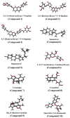
Figure 1
Figure 1
3D structure of phytochemicals (compounds 110) from Saudi medicinal plants.
Figure 2
Figure 2
Active site in the SARS-CoV-2 target protein (RdRp). (A) Ribbon structure of RdRp (PDB ID: 7BV2) with ligand (remdesivir, RTP) indicating the active pocket in the target protein. (B) Magnified view of the active site of RdRp showing the catalytic dyad of residues interacting with RTP (green color). Black color (H-bond), dark red (H-π bond), dark blue (Van der Waals clashes), element color (atoms), residues are labeled as blue texts.
Figure 3
Figure 3
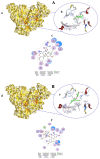
Surface representation showing the non-covalent docking of compound 1 (A(a)) and compound 2 (B(d)) with RdRp. Compounds 1 and 2 are green in color. Solvent-exposed regions of RdRp are dark yellow, hydrophobic regions are yellow, and polar regions are red in color. Magnified view showing the binding of compound 1 (A(b)) and compound 2 (B(e)) with the template primer nucleotides and residues in the active pocket of RdRp. Black color (H-bond), dark red (H-π bond), dark blue (Van der Waals clashes), element color (atoms), residues, and nucleotides are labeled as blue texts. Two-dimensional view of RdRp showing non-covalent binding with compound 1 (A(c)) and compound 2 (B(f)). Bonds and color descriptions are shown in the inset of the 2D image.
Figure 4
Figure 4
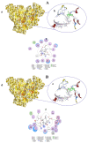
Surface representation showing the non-covalent docking of compound 3 (A(a)) and compound 4 (B(d)) with RdRp. Compounds 3 and 4 are in green color. Magnified view showing the binding of compound 3 (A(b)) and compound 4 (B(e)) with the template primer nucleotides and residues in the active pocket of RdRp. Two-dimensional view of RdRp showing non-covalent binding with compound 3 (A(c)) and compound 4 (B(f)).
Figure 5
Figure 5
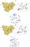
Surface representation showing the non-covalent docking of compound 5 (A(a)) and compound 6 (B(d)) with RdRp. Compounds 5 and 6 are in green color. Magnified view showing the binding of compound 5 (A(b)) and compound 6 (B(e)) with the template primer nucleotides and residues in the active pocket of RdRp. Two-dimensional view of RdRp showing non-covalent binding with compound 5 (A(c)) and compound 6 (B(f)).
Figure 6
Figure 6
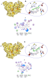
Surface representation showing the non-covalent docking of compound 7 (A(a)) and compound 8 (B(d)) with RdRp. Compounds 7 and 8 are in green color. Magnified view showing the binding of compound 7 (A(b)) and compound 8 (B(e)) with the template primer nucleotides and residues in the active pocket of RdRp. Two-dimensional view of RdRp showing non-covalent binding with compound 7 (A(c)) and compound 8 (B(f)).
Figure 7
Figure 7
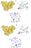
Surface representation showing the non-covalent docking of compound 9 (A(a)) and compound 10 (B(d)) with RdRp. Compounds 9 and 10 are in green color. Magnified view showing the binding of compound 9 (A(b)) and compound 10 (B(e)) with the template primer nucleotides and residues in the active pocket of RdRp. Two-dimensional view of RdRp showing non-covalent binding with compound 9 (A(c)) and compound 10 (B(f)).
Figure 8
Figure 8
Covalent docking of Vernolepin (compound 7) to SARS-CoV-2 target protein (RdRp) (A). (a) Surface representation of RdRp docked with compound 7 (green color). Solvent exposed region of RdRp is dark yellow, hydrophobic regions are yellow, and polar regions are red color. (b) Magnified view of the active pocket occupied by compound 7 showing interaction with residues in RdRp. H-bond (black color), H–π bond (dark red), Van der Waals clashes (dark blue), atoms (element color), and residues are labeled as blue texts. (c) Two-dimensional view of RdRp showing the interaction of amino acid residues with compound 7. Descriptions of bonds and color of 2D are shown in the inset of (c).
Figure 9
Figure 9
(A) Vernadolol (compound 8) covalently bound to SARS-CoV-2 target protein (RdRp). (a) Surface representation of RdRp docked with compound 8 (green color). Solvent exposed region of RdRp is dark yellow, hydrophobic regions are yellow, and polar regions are red in color. (b) Magnified view of the active pocket occupied by compound 8 showing interaction with residues in RdRp. H-bond (black color), H–π bond (dark red), Van der Waals clashes (dark blue), atoms (element color), and residues are labeled as blue texts. (c) Two-dimensional view of RdRp showing the interaction of amino acid residues with compound 8. Descriptions of bonds and color of 2D are shown in the inset of (c).
Figure 10
Figure 10
(A) 11β,13-Dihydrovernodalin (compound 9) covalently bound to SARS-CoV-2 target protein (RdRp). (a) Surface representation of RdRp docked with compound 9 (green color). Solvent exposed region of RdRp is dark yellow, hydrophobic regions are yellow, and polar regions are red in color. (b) Magnified view of the active pocket occupied by compound 9 showing interaction with residues in RdRp. H-bond (black color), H–π bond (dark red), Van der Waals clashes (dark blue), atoms (element color), and residues are labeled as blue texts. (c) Two-dimensional view of RdRp showing the interaction of amino acid residues with compound 9. Descriptions of bonds and color in 2D are shown in the inset of (c).
Figure 11
Figure 11
MD simulation analysis of RdRp and its complexation with compounds 7, 8, and 9. (A) RMSD of RdRp native structure and post-complexation with compounds 7, 8, and 9 at 50 ns. (B) RMSF of RdRp alone and after their complexation with compounds 7, 8, and 9. (C) Rg of RdRP showing the amino acid compactness in the absence and presence of compounds 7, 8, and 9 as a function of time.

Similar articles

References

    1. Bi Q., Wu Y., Mei S., Ye C., Zou X., Zhang Z., Liu X., Wei L., Truelove S.A., Zhang T., et al. Epidemiology and transmission of COVID-19 in 391 cases and 1286 of their close contacts in Shenzhen, China: A retrospective cohort study. Lancet Infect. Dis. 2020;20:911–919. doi: 10.1016/S1473-3099(20)30287-5. - DOI - PMC - PubMed
    1. Zhou P., Yang X.-L., Wang X.-G., Hu B., Zhang L., Zhang W., Si H.-R., Zhu Y., Li B., Huang C.-L., et al. A pneumonia outbreak associated with a new coronavirus of probable bat origin. Nature. 2020;579:270–273. doi: 10.1038/s41586-020-2012-7. - DOI - PMC - PubMed
    1. Lu H. Drug treatment options for the 2019-new coronavirus (2019-nCoV) Biosci. Trends. 2020;14:69–71. doi: 10.5582/bst.2020.01020. - DOI - PubMed
    1. Fu D.-J., Li P., Song J., Zhang S.-Y., Xie H.-Z. Mechanisms of synergistic neurotoxicity induced by two high risk pesticide residues—Chlorpyrifos and Carbofuran via oxidative stress. Toxicol. Vitr. 2019;54:338–344. doi: 10.1016/j.tiv.2018.10.016. - DOI - PubMed
    1. WHO WHO Coronavirus (COVID-19) Dashboard. [(accessed on 9 October 2023)];2023 Available online: https://covid19.who.int.

MeSH terms