. 2023 Oct 25;15(11):2153.
doi: 10.3390/v15112153.

Evaluation of the Neutralizing Antibody STE90-C11 against SARS-CoV-2 Delta Infection and Its Recognition of Other Variants of Concerns

Affiliations

Evaluation of the Neutralizing Antibody STE90-C11 against SARS-CoV-2 Delta Infection and Its Recognition of Other Variants of Concerns

Leila Abassi et al. Viruses. .

Abstract

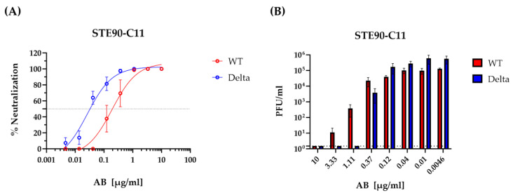
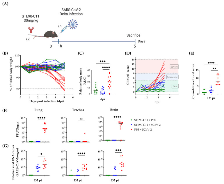
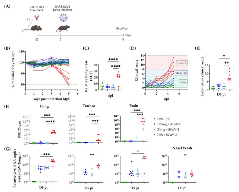
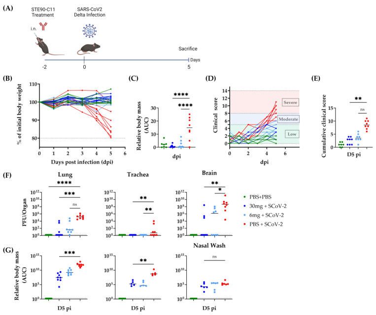
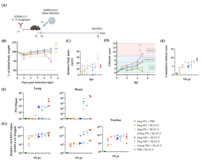
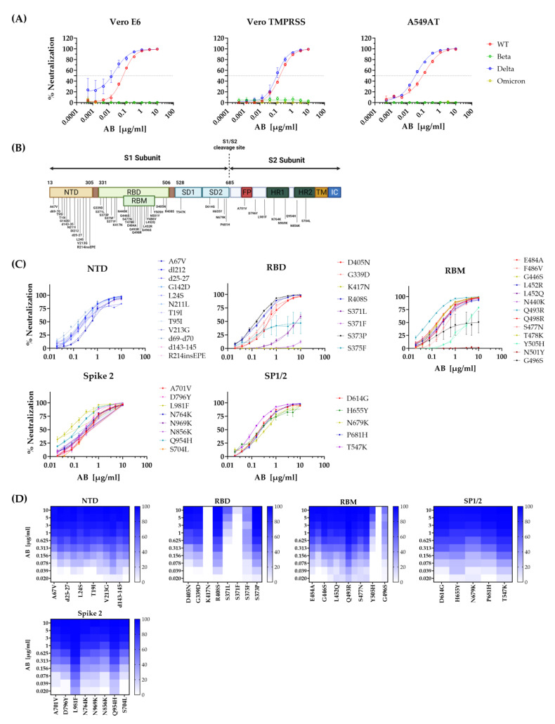
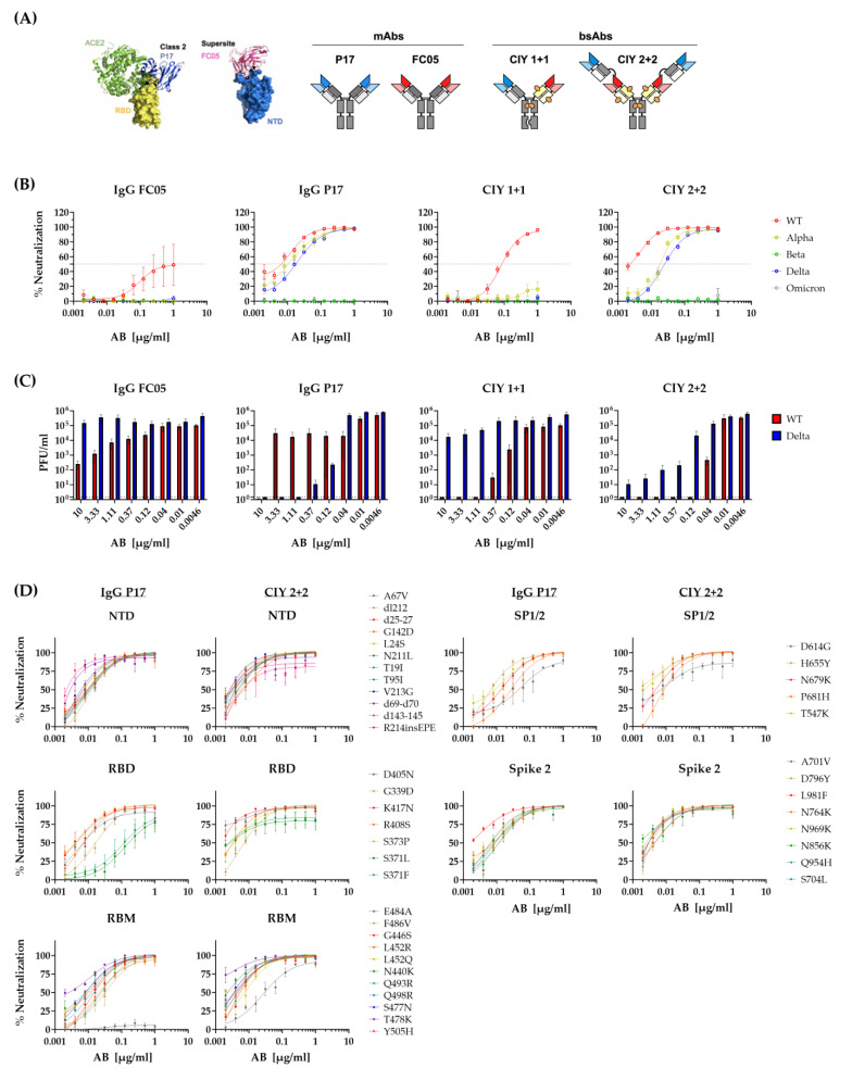
As of now, the COVID-19 pandemic has spread to over 770 million confirmed cases and caused approximately 7 million deaths. While several vaccines and monoclonal antibodies (mAb) have been developed and deployed, natural selection against immune recognition of viral antigens by antibodies has fueled the evolution of new emerging variants and limited the immune protection by vaccines and mAb. To optimize the efficiency of mAb, it is imperative to understand how they neutralize the variants of concern (VoCs) and to investigate the mutations responsible for immune escape. In this study, we show the in vitro neutralizing effects of a previously described monoclonal antibody (STE90-C11) against the SARS-CoV-2 Delta variant (B.1.617.2) and its in vivo effects in therapeutic and prophylactic settings. We also show that the Omicron variant avoids recognition by this mAb. To define which mutations are responsible for the escape in the Omicron variant, we used a library of pseudovirus mutants carrying each of the mutations present in the Omicron VoC individually. We show that either 501Y or 417K point mutations were sufficient for the escape of Omicron recognition by STE90-C11. To test how escape mutations act against a combination of antibodies, we tested the same library against bispecific antibodies, recognizing two discrete regions of the spike antigen. While Omicron escaped the control by the bispecific antibodies, the same antibodies controlled all mutants with individual mutations.

Keywords: Delta variant; SARS-CoV-2; bispecific antibodies; intranasal administration; intravenous administration; mice experiments; monoclonal antibody; pseudovirus assay; single mutations.


Conflict of interest statement

The authors declare the following conflict of interest. L.Č.-Š. was a consultant to CORAT Therapeutics GmbH, a subsidiary of YUMAB GmbH. T.S., A.F., S.D. and M.H. are founders and shareholders of YUMAB GmbH. F.B., M.S., P.A.H., S.D. and M.H. are inventors on a patent application on blocking antibodies against SARS-CoV-2.

Figures

Figure 1
Figure 1
Neutralization of SARS-CoV-2 variant strains by the mAb STE90-C11. Authentic SARS-CoV-2 neutralization titration by STE90-C11 performed using Vero E6 cells against SARS-CoV-2 WT-D614G (red) and Delta (blue) are represented as percentage of neutralization (A) and plaque number per ml (B). Lines represent nonlinear regression fit and data were shown as mean ± SEM of two independent experiments with two to three technical replicates.
Figure 2
Figure 2
STE90-C11 semi-therapeutic application against SARS-CoV-2 Delta (A) Experimental setup: 9–18 week-old mice received 30 mg/kg of STE90-C11 by intravenous injection (i.v.) 1 h before intranasal infection (i.n.) with 2 × 10³ PFU SARS-CoV-2 Delta strain in 20 µL volume. Organs were collected 5 days later. (B) Weight change after infection with SARS-CoV-2. (C) Cumulative relative mass reduction in SARS-CoV-2 infected and with mAb treated mice until D5 pi are shown as area under the curve (AUC). (D) Daily clinical scores upon SARS-CoV-2 infection and mAb treatment. The indicated thresholds represent the clinical severity of mice; low (green), moderate (blue), and severe (red, humane end-point). (E) Cumulative clinical score on D5 pi. (F) SARS-CoV-2 viral load at D5 pi in the lung (left), trachea (middle), and brain (left). (G) Viral RNA levels in the lung (left), trachea (middle), and brain (left) were measured. The dotted line indicates the limit of detection of the assay. Pooled data (n = 9–12 per group) from four independent experiments are shown. Each symbol is an individual mouse, and horizontal lines indicate the median of biological replicates. Statistical significance versus the infected control group was calculated using (C) one-way analysis of variance (ANOVA) followed by Dunnett´s post-analysis, (E,F) Kruskal–Wallis test followed by Dunn´s post-analysis, and (G) Mann–Whitney test. * for p < 0.05, ** for p < 0.005, *** for p < 0.001 and **** for p < 0.0001. ns: non-significant.
Figure 3
Figure 3
STE90-C11 prophylaxis against SARS-CoV-2 Delta. (A) Experimental setup: 10–18 week-old female and male K18-hACE 2 transgenic mice received 120 mg/kg, 30 mg/kg of STE90-C11 or PBS by intravenous injection two days upon intranasal inoculation with 2 × 10³ PFU SARS-CoV-2 Delta strain in 20 µL volume. Organs were collected 5 days later. (B) Relative weight upon infection in single mice. (C) Cumulative relative mass reduction in individual mice represented as area under the curve (AUC). (D) Daily clinical scores upon infection and mAb treatment. Clinical severity thresholds are indicated as low (green), moderate (blue), and severe (red, humane end-point). (E) Cumulative clinical score on D5 pi. (F) SARS-CoV-2 viral loads at dpi 5 in lungs, trachea, and brain. (G) Viral RNA levels in the lung, trachea, brain, and nasal wash were measured. The dotted line indicates the limit of detection of the assay. Data from three independent experiments were pooled (n = 6–8 per group). Each symbol is an individual mouse, and horizontal lines indicate the median of biological replicates. Statistical significance versus the infected control group was calculated using (C) One-way analysis of variance (ANOVA) followed by Dunnett´s post-analysis or (E,F) Kruskal–Wallis test followed by Dunn´s post-analysis, and (G) Mann–Whitney test. * for p < 0.05, ** for p < 0.005, *** for p < 0.001 and **** for p < 0.0001. ns non-significant.
Figure 4
Figure 4
Intranasal application of STE90-C11 against SARS-CoV-2 Delta (A) Experimental setup: 10–18 week-old female and male K18-hACE 2 transgenic mice received 30 mg/kg, 6 mg/kg of STE90-C11 or PBS by intranasal injection two days upon infection with 2 × 10³ PFU SARS-CoV-2 Delta strain in 20 µL volume intranasal. Organs were collected at 5 days post-infection. (B) Weight change after infection with SARS-CoV-2. (C) Cumulative relative mass reduction in SARS-CoV-2 infected and with mAb treated mice until D5 pi are shown as area under the curve (AUC). (D) Daily clinical scores upon SARS-CoV-2 infection and mAb treatment. The indicated thresholds represent the clinical severity of mice; low (green), moderate (blue), and severe (red, humane end-point). (E) Cumulative clinical score on D5 pi. (F) SARS-CoV-2 viral load at D5 pi in the lung (left), trachea (middle), and brain (left). (G) Viral RNA levels in the lung, trachea, and nasal wash were measured. The dotted line indicates the limit of detection of the assay. Pooled data (n = 3–6 per group) from three independent experiments are shown. Each symbol is an individual mouse, and horizontal lines indicate the median of biological replicates. Statistical significance versus the infected control group was calculated using (C) one-way analysis of variance (ANOVA) followed by Dunnett´s post-analysis (E,F) Kruskal–Wallis test followed by Dunn’s post-analysis, and (G) Mann–Whitney test. * for p < 0.05, ** for p < 0.005, *** for p < 0.001 and **** for p < 0.0001. ns non-significant.
Figure 5
Figure 5
Contribution of functional Fc fragment for the function of STE90-C11 against SARS-CoV-2 Delta infection. (A) Experimental setup: 13–25 week-old female and male K18-hACE 2 transgenic mice received different concentrations of either the functional Fc STE90-C11 (orange) or the non-functional Fc STE90-C11 (blue) by intravenous injection one hour before intranasal inoculation with 2 × 10³ PFU SARS-CoV-2 Delta strain in 20 µL volume. Organs were collected at 5 days post-infection. (B) Weight change after infection with SARS-CoV-2. (C) Cumulative relative mass reduction in SARS-CoV-2 infected and with mAb treated mice until D5 pi are shown as area under the curve (AUC). (D) Daily clinical scores upon SARS-CoV-2 infection and mAb treatment. The indicated thresholds represent the clinical severity of mice; low (green), moderate (blue), and severe (red, humane end-point). (E) Cumulative clinical score on D5 pi. (F) SARS-CoV-2 viral load at D5 pi in lung (left) and brain (left). (G) Viral RNA levels in the lung (left), brain (middle), and trachea (left) were measured. The dotted line indicates the limit of detection of the assay. Pooled data (n = 4 per group) from two independent experiments are shown. Each symbol is an individual mouse, and horizontal lines indicate the median of biological replicates. * for p < 0.05, ** for p < 0.005, *** for p < 0.001 and **** for p < 0.0001. ns non-significant.
Figure 6
Figure 6
Single mutation mapping of STE90-C11 in vitro. (A) The neutralization capacity of STE90-C11 was assessed using pseudoviruses expressing SARS-CoV-2 spike glycoprotein of either the original WT strain (red), Beta (green), Delta (blue), or Omicron (yellow) at different antibody concentrations on different cell types. (B) Schematic overview of the Omicron single mutations tested. (C) STE90-C11 neutralization capacity against pseudoviruses harboring single mutations of the Omicron variant. Data were divided after the region single mutations were located: NTD, RBD, RBM, SP1/2, and Spike 2. Lines represent nonlinear regression fit and data were shown as mean ± SEM of two independent experiments with two to three technical replicates. (D) The Heatmap-like representation shows the neutralization of STE90-C11 against Omicron single mutations in a dose-dependent manner.
Figure 7
Figure 7
Neutralization of SARS-CoV-2 variants of concern by the bispecific monoclonal antibodies. (A) Bispecific mAbs structures and regions of binding on the SARS-CoV-2 spike protein. (B) Neutralization capacities were assessed using pseudoviruses expressing SARS-CoV-2 spike glycoprotein of either the original WT strain (red), Alpha (yellow), Beta (green), Delta (blue), or Omicron (gray) at different antibody concentrations. (C) Authentic SARS-CoV-2 neutralization titration by the bispecific mAbs performed using Vero E6 cells are represented as plaque number per ml. Lines represent nonlinear regression fit and data were shown as mean ± SEM of two independent experiments with two to three technical replicates. (D) Neutralization capacity of IgG P17 (left) and CIY 2 + 2 (right) against pseudoviruses harboring single mutations of the Omicron variant. Data were divided after the region single mutations were located: NTD, RBD, RBM, SP1/2, and Spike 2. Lines represent nonlinear regression fit and data were shown as mean ± SEM of two independent experiments with two to three technical replicates.

Similar articles

Cited by

References

    1. Hu B., Guo H., Zhou P., Shi Z.L. Characteristics of SARS-CoV-2 and COVID-19. Nat. Rev. Microbiol. 2021;19:141–154. doi: 10.1038/s41579-020-00459-7. - DOI - PMC - PubMed
    1. Yeo Y.H., Wang M., He X., Lv F., Zhang Y., Zu J., Li M., Jiao Y., Ebinger J.E., Patel J.K., et al. Excess risk for acute myocardial infarction mortality during the COVID-19 pandemic. J. Med. Virol. 2023;95:e28187. doi: 10.1002/jmv.28187. - DOI - PMC - PubMed
    1. Spudich S., Nath A. Nervous system consequences of COVID-19. Science. 2022;375:267–269. doi: 10.1126/science.abm2052. - DOI - PubMed
    1. Tao K., Tzou P.L., Nouhin J., Gupta R.K., de Oliveira T., Kosakovsky Pond S.L., Fera D., Shafer R.W. The biological and clinical significance of emerging SARS-CoV-2 variants. Nat. Rev. Genet. 2021;22:757–773. doi: 10.1038/s41576-021-00408-x. - DOI - PMC - PubMed
    1. Natekar J.P., Pathak H., Stone S., Kumari P., Sharma S., Auroni T.T., Arora K., Rothan H.A., Kumar M. Differential Pathogenesis of SARS-CoV-2 Variants of Concern in Human ACE2-Expressing Mice. Viruses. 2022;14:1139. doi: 10.3390/v14061139. - DOI - PMC - PubMed

Supplementary concepts