

Hepatoprotective Effects of Biochanin A on Thioacetamide-Induced Liver Cirrhosis in Experimental Rats
- PMID: 38005330
- PMCID: PMC10674479
- DOI: 10.3390/molecules28227608
Hepatoprotective Effects of Biochanin A on Thioacetamide-Induced Liver Cirrhosis in Experimental Rats
Abstract
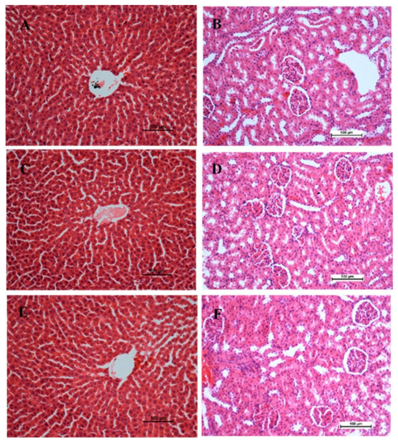
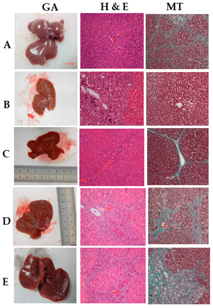
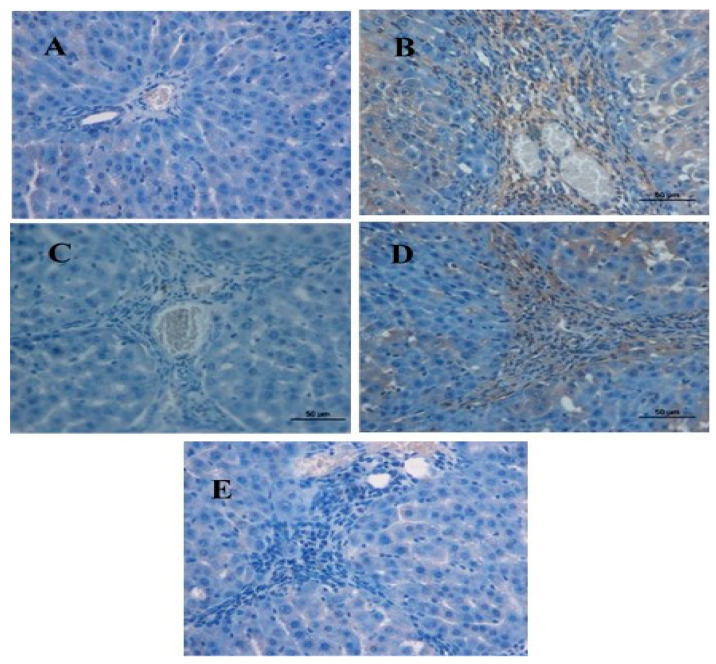
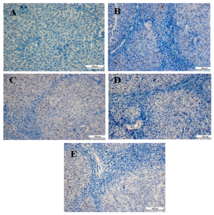
The protective effect of biochanin A (BCA) on the histopathology, immunohistochemistry, and biochemistry of thioacetamide (TAA)-induced liver cirrhosis in vivo was investigated. There was a significant reduction in liver weight and hepatocyte propagation, with much lower cell injury in rat groups treated with BCA (25 mg/kg and 50 mg/kg) following a TAA induction. These groups had significantly lower levels of proliferating cell nuclear antigen (PCNA) and α-smooth muscle actin (α-SMA). The liver homogenates showed increased antioxidant enzyme activity of superoxide dismutase (SOD), catalase (CAT), and glutathione peroxidase (GPx), as well as decreased malondialdehyde (MDA) levels. The serum biomarkers associated with liver function, namely alkaline phosphatase (ALP), alanine aminotransferase (ALT), aspartate aminotransferase (AST), and gamma glutamyl transaminase (GGT), returned to normal levels, comparable to those observed in both the normal control group and the reference control group. Taken together, the normal microanatomy of hepatocytes, the inhibition of PCNA and α-SMA, improved antioxidant enzymes (SOD, CAT, and GPx), and condensed MDA with repairs of liver biomarkers validated BCA's hepatoprotective effect.
Keywords: TAA; antioxidant enzymes; biochanin A; histopathology; immunohistochemistry; liver cirrhosis.
Conflict of interest statement
The authors declare no conflict of interest.
Figures
Similar articles
-
In vivo antioxidant and hepatoprotective activity of methanolic extracts of Daucus carota seeds in experimental animals.Asian Pac J Trop Biomed. 2012 May;2(5):385-8. doi: 10.1016/S2221-1691(12)60061-6. Asian Pac J Trop Biomed. 2012. PMID: 23569935 Free PMC article.
-
Role of quercetin in preventing thioacetamide-induced liver injury in rats.Toxicol Pathol. 2011 Oct;39(6):949-57. doi: 10.1177/0192623311418680. Epub 2011 Sep 1. Toxicol Pathol. 2011. Retraction in: Toxicol Pathol. 2024 May 20:1926233241256964. doi: 10.1177/01926233241256964. PMID: 21885874 Retracted.
-
Hepatoprotective effect of pinostrobin against thioacetamide-induced liver cirrhosis in rats.Saudi J Biol Sci. 2023 Jan;30(1):103506. doi: 10.1016/j.sjbs.2022.103506. Epub 2022 Nov 17. Saudi J Biol Sci. 2023. PMID: 36458098 Free PMC article.
-
Estrogen Deficiency Potentiates Thioacetamide-Induced Hepatic Fibrosis in Sprague-Dawley Rats.Int J Mol Sci. 2019 Jul 29;20(15):3709. doi: 10.3390/ijms20153709. Int J Mol Sci. 2019. PMID: 31362375 Free PMC article.
-
Effect of carnosine against thioacetamide-induced liver cirrhosis in rat.Peptides. 2010 Jan;31(1):67-71. doi: 10.1016/j.peptides.2009.11.028. Epub 2009 Dec 1. Peptides. 2010. PMID: 19958806
Cited by
-
Unveiling the Potential of Silymarin, Spirulina platensis, and Chlorella vulgaris towards Cardiotoxicity via Modulating Antioxidant Activity, Inflammation, and Apoptosis in Rats.Life (Basel). 2024 Oct 11;14(10):1289. doi: 10.3390/life14101289. Life (Basel). 2024. PMID: 39459589 Free PMC article.
-
Silymarin: Unveiling its pharmacological spectrum and therapeutic potential in liver diseases-A comprehensive narrative review.Food Sci Nutr. 2024 Feb 16;12(5):3097-3111. doi: 10.1002/fsn3.4010. eCollection 2024 May. Food Sci Nutr. 2024. PMID: 38726410 Free PMC article. Review.
References
-
- Bachar S.C., Bachar R., Jannat K., Jahan R., Rahmatullah M. Chapter Seven—Hepatoprotective natural products. In: Sarker S.D., Nahar L., editors. Annual Reports in Medicinal Chemistry. Academic Press; Cambridge, MA, USA: 2020. pp. 207–249.
-
- El-Fadaly A.A., Afifi N.A., El-Eraky W., Salama A., Abdelhameed M.F., El-Rahman S.S.A., Ramadan A. Fisetin alleviates thioacetamide-induced hepatic fibrosis in rats by inhibiting Wnt/β-catenin signaling pathway. Immunopharmacol. Immunotoxicol. 2022;44:355–366. doi: 10.1080/08923973.2022.2047198. - DOI - PubMed
MeSH terms
Substances
Grants and funding
LinkOut - more resources
Full Text Sources
Medical
Miscellaneous
