. 2023 Nov 17;24(22):16429.
doi: 10.3390/ijms242216429.

Role of CB1 Cannabinoid Receptors in Vascular Responses and Vascular Remodeling of the Aorta in Female Mice

Affiliations

Role of CB1 Cannabinoid Receptors in Vascular Responses and Vascular Remodeling of the Aorta in Female Mice

Bálint Bányai et al. Int J Mol Sci. .

Abstract

Both the endocannabinoid system (ECS) and estrogens have significant roles in cardiovascular control processes. Cannabinoid type 1 receptors (CB1Rs) mediate acute vasodilator and hypotensive effects, although their role in cardiovascular pathological conditions is still controversial. Estrogens exert cardiovascular protection in females. We aimed to study the impact of ECS on vascular functions. Experiments were performed on CB1R knockout (CB1R KO) and wild-type (WT) female mice. Plasma estrogen metabolite levels were determined. Abdominal aortas were isolated for myography and histology. Vascular effects of phenylephrine (Phe), angiotensin II, acetylcholine (Ach) and estradiol (E2) were obtained and repeated with inhibitors of nitric oxide synthase (NOS, Nω-nitro-L-arginine) and of cyclooxygenase (COX, indomethacin). Histological stainings (hematoxylin-eosin, resorcin-fuchsin) and immunostainings for endothelial NOS (eNOS), COX-2, estrogen receptors (ER-α, ER-β) were performed. Conjugated E2 levels were higher in CB1R KO compared to WT mice. Vasorelaxation responses to Ach and E2 were increased in CB1R KO mice, attenuated by NOS-inhibition. COX-inhibition decreased Phe-contractions, while it increased Ach-relaxation in the WT group but not in the CB1R KO. Effects of indomethacin on E2-relaxation in CB1R KO became opposite to that observed in WT. Histology revealed lower intima/media thickness and COX-2 density, higher eNOS and lower ER-β density in CB1R KO than in WT mice. CB1R KO female mice are characterized by increased vasorelaxation associated with increased utilization of endothelial NO and a decreased impact of constrictor prostanoids. Our results indicate that the absence or inhibition of CB1Rs may have beneficial vascular effects.

Keywords: cannabinoid type 1 receptor; endocannabinoid; endothelium; estrogen receptor; vascular remodeling.


Conflict of interest statement

The authors declare no conflict of interest. The funders had no role in the design of the study; in the collection, analyses or interpretation of data; in the writing of the manuscript; or in the decision to publish the results.

Figures
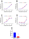
Figure 1
Figure 1
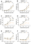
Contraction–relaxation vascular responses of aortic segments of wild-type (CB1R+/+) and CB1R knockout (CB1R−/−) female mice. Panel (A). Dose–response contraction curves to angiotensin II in wild-type (n = 14–25 rings from 9 animals) and in CB1R knockout (18–26 segments from 9 animals). Panel (B). Dose–response contraction curves to phenylephrine in CB1R+/+ (n = 28 segments from 9 animals) and in CB1R−/− (n = 27 segments from 9 animals). Panel (C). Dose–response relaxation curves to acetylcholine in CB1R+/+ (n = 26 segments from 9 animals) and in CB1R−/− (n = 27 segments from 8 animals). Panel (D). Dose–response relaxation curves to estradiol in CB1R+/+ (n = 8–10 segments from 8 animals) and CB1R−/− (n = 7–10 segments from 10 animals). Panel (E). Relaxation response of aortic segments to the CB1R agonist WIN 55,212-2 (10µM) in CB1R+/+ (n = 5) and CB1R−/− (n = 6) female mice. Mean ± SEM values. p < 0.05 values were considered significant. *: p < 0.05 and **: p < 0.01 between wild-type (CB1R+/+) and CB1R knockout (CB1R−/−) groups in two-way ANOVA with a Bonferroni post hoc test and unpaired t-test (panel E) (Contraction data were normalized to KCl contraction, relaxation data were calculated as percent values of precontraction level).
Figure 2
Figure 2
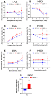
Effects of specific inhibitors Nω-nitro-L-arginine (LNA, inhibitor of nitric oxide synthase) and indomethacin (INDO, inhibitor of cyclooxygenase) on contraction–relaxation vascular responses in aortas of wild-type (CB1R+/+) and CB1R knockout (CB1R−/−) female mice. Panel (A). Effects of inhibitors on phenylephrine-induced vasoconstriction in CB1R+/+ female mice (n = 7–9, segments = 7–9). Panel (B). Effects of inhibitors on phenylephrine-induced vasoconstriction in CB1R−/− female mice (n = 8, segments = 8). Panel (C). Effects of inhibitors on acetylcholine-induced vasodilation in CB1R+/+ female mice (n = 8–9, segments = 8–9). Panel (D). Effects of inhibitors on acetylcholine-induced vasodilation in CB1R−/− female mice (n = 8–9, segments = 8–9). Panel (E). Effects of inhibitors on estradiol-induced vascular responses in CB1R+/+ female mice (n = 10, segments = 7–10). Panel (F). Effects of inhibitors on estradiol-induced vascular responses in CB1R−/− female mice (n = 10, segments = 7–10). Data are shown as mean ± SEM values. p < 0.05 values were considered significant. #: p < 0.05, ###: p < 0.001 between vehicle and LNA-treated segments, +: p < 0.05, +++: p < 0.001 between vehicle and INDO-treated segments (two-way ANOVA with a Bonferroni post hoc test). Abbreviations: INDO: indomethacin, LNA: Nω-nitro-L-arginine, CB1R: cannabinoid type 1 receptor, CB1+/+: CB1R wild-type, CB1−/−: CB1R knockout mice (Contraction data were normalized to KCl contraction, relaxation data were calculated as percent values of precontraction level).
Figure 3
Figure 3
Comparison of the effects of specific inhibitors Nω-nitro-L-arginine (LNA, inhibitor of nitric oxide synthase) and indomethacin (INDO, inhibitor of cyclooxygenase) on contraction-relaxation vascular responses in aortas of WT (CB1R+/+) and CB1R KO (CB1R−/−) female mice. Panel (A). Effects of inhibitor LNA on Phe-induced vasoconstriction in CB1R+/+ (n = 6, segments = 6) and CB1R−/− (n = 6, segments = 6) female mice. Panel (B). Effects of inhibitor INDO on Phe-induced vasoconstriction in CB1R+/+ (n = 5, segments = 5) and CB1R−/− (n = 6, segments = 6) female mice. Panel (C). Effects of inhibitor LNA on Ach-induced vasorelaxation in CB1R+/+ (n = 8, segments = 8) and CB1R−/− (n = 8, segments = 8) female mice. Panel (D). Effects of inhibitor INDO on Ach-induced vasodilation in CB1R+/+ (n = 8, segments = 8) and CB1R−/− (n = 8, segments = 8) female mice. Panel (E). Effects of inhibitor LNA on E2 induced vascular responses in CB1R+/+ (n = 9) and CB1R−/− (n = 8) female mice. Panel (F). Effects of inhibitor INDO on E2-induced vascular responses in CB1R+/+ (n = 9) and CB1R−/− (n = 9) female mice. Differences in contraction or relaxation values are plotted. Panel (G). Normalized effects of INDO on E2 relaxation responses (at 10 µmol/L). Values are plotted as percentage differences from control relaxation. Mean ± SEM values. p < 0.05 values were considered significant. * p < 0.05 between CB1R+/+ and CB1R−/− groups (two-way ANOVA with a Bonferroni post hoc test). Abbreviations: Ach: acetylcholine, Phe: phenylephrine, E2: estradiol, INDO: indomethacin, LNA: Nω-nitro-L-arginine, CB1R: cannabinoid type 1 receptor, WT: wild-type, KO: knockout, CB1R+/+: cannabinoid type 1 receptor wild-type mice, CB1R−/−: cannabinoid type 1 receptor knockout mice.
Figure 4
Figure 4
Free estradiol, conjugated estradiol and free 4-hydroxyestrone levels (pg/mL) in WT and CB1R KO female mice. Mean ± SEM values. p < 0.05 values were considered significant. *: p < 0.05 between CB1R+/+ (n = 14–24) and CB1R−/− (n = 11–17) groups. Abbreviations: fE2: free estradiol level, cE2: conjugated estradiol level, f4OH-E1: free 4-hydroxyestrone level, CB1R: cannabinoid type 1 receptor, WT: wild-type, KO: knockout, CB1R+/+: cannabinoid type 1 receptor wild-type mice, CB1R−/−: cannabinoid type 1 receptor knockout mice. *: p < 0.05 between groups (unpaired t-test). Note that sample takings were not adjusted to the estrus cycle, mean values reflect means during estrus cycle.
Figure 5
Figure 5
Morphological changes of the abdominal aorta wall. Panel (A). Intima–media ratio of the aorta wall with mean ± SEM, n = 6 (CB1R+/+) n = 10 (CB1R−/−) *: p < 0,05 CB1R+/+ vs. CB1R−/− group. Panel (B). Aorta wall thickness in micrometer with mean ± SEM n = 6 (CB1R+/+) n = 11 (CB1R−/−) *: p < 0.05 CB1R+/+ vs. CB1R−/− group. Panel (C). Representative photos of the hematoxylin-eosin staining photographed at 10× magnification. Panel (D). Optical density of elastic fiber on resorcin fuchsin stained sections. n = 7 (CB1R+/+), n = 10 (CB1R−/−). Panel (E). Representative photos of the resorcin fuchsin stained sections photographed at 10× magnification. Panel (F). Optical density of α-SMA stained sections. n = 5 (CB1R+/+) n = 14 (CB1R−/−) Panel (G). Representative photos of α-SMA stained aorta segments, visualization with diamino-benzidine (DAB) on hematoxylin counterstaining, photographed at 10× magnification. Statistical analysis executed with unpaired t-test. Data shown by noncalibrated optical density with mean ± SEM Abbreviations: CB1R: cannabinoid type 1 receptor, CB1R+/+: cannabinoid type 1 receptor wild-type mice, CB1R−/−: cannabinoid type 1 receptor knockout mice. RF: resorcin fuchsin, α-SMA: alpha smooth muscle actin, O.D.: optical density.
Figure 6
Figure 6
Vasoactive markers of the abdominal aorta wall. Panel (A). Results of eNOS immunostained sections. n = 9 (CB1R+/+) n = 14 (CB1R−/−). ***: p < 0.001 CB1R+/+ vs. CB1R−/− group. Panel (B). Representative photos of eNOS immunostained aorta segments, visualization with DAB on hematoxylin counterstaining, photographed at 20× magnification. Evaluation performed from the values of the endothelial layer Panel (C). Results of COX-2 immunostained sections. n = 8 (CB1R+/+), n = 12 (CB1R−/−). *: p < 0,05 CB1R+/+ vs. CB1R−/− group. Panel (D). Representative photos of COX-2 immunostained aorta segments, visualization with DAB on hematoxylin counterstaining, photographed by 20× magnification. Evaluation performed from the values of the endothelial layer Panel (E). Results of TP receptor immunostained sections. n = 6 (CB1R+/+), n = 13 (CB1R−/−). Panel (F). Representative photos of TP receptor immunostained aorta segments, visualization with DAB on hematoxylin counterstaining, photographed at 10× magnification. Evaluation performed from the values of the media layer. Statistical analysis performed with unpaired t-test. Data shown by noncalibrated optical density with mean ± SEM Abbreviations: CB1R: cannabinoid type 1 receptor, CB1R+/+: cannabinoid type 1 receptor wild-type mice, CB1R−/−: cannabinoid type 1 receptor knockout mice, eNOS: endothelial nitric oxide synthase, DAB: diamino-benzidine, COX: cyclooxygenase, TP: thromboxane-prostanoid receptor O.D.: optical density.
Figure 7
Figure 7
Estrogen receptor protein expression in the abdominal aorta wall. Panels (A, B) Results of ER-α immunostained sections. Panel (A). Results from the endothelial layer. n = 6 (CB1R+/+), n = 9 (CB1R−/−). Panel (B). Results from the media layer. n = 4 (CB1+/+) n = 9 (CB1R−/−). Panel (C). Representative photos of ER-α immunostained aorta segments, visualization with DAB on hematoxylin counterstaining, photographed at 20× magnification. Evaluation performed from the values of the endothelial and the media layer. Panels (D,E). Results of ER-β stained sections. Panel (D). Results from the endothelial layer. (CB1R+/+), n = 9 (CB1R−/−). *: p < 0,05 CB1R+/+ vs. CB1R−/− group. Panel (E). Results from the medial layer. n = 9 (CB1R+/+), n = 10 (CB1R−/−). ***: p < 0.001 CB1R+/+ vs. CB1R−/− group. Panel (F). Representative photos of ER-β immunostained aorta segments, visualization with DAB on hematoxylin counterstaining, photographed by 10× magnification. Evaluation performed from the values of the endothelial and the media layer. Statistical analysis performed with unpaired t-test. Data shown by noncallibrated optical density with mean ± SEM. Abbreviations: CB1R: cannabinoid type 1 receptor, CB1R+/+: cannabinoid type 1 receptor wild-type mice, CB1R−/−: cannabinoid type 1 receptor knockout mice, eNOS: endothelial nitric oxide synthase, DAB: diamino-benzidine, COX: cyclooxygenase, O.D.: optical density, ER: estrogen receptor.
Figure 8
Figure 8
Results of 3-nitrotyrosine density in the abdominal aorta wall. Panel (A). Noncalibrated optical density data are shown with mean ± SEM, n = 7 (CB1R+/+), n = 9 (CB1R−/−). Statistical analysis performed with unpaired t-test. Panel (B). Representative pictures of 3-nitrotyrosine staining. Visualization with DAB on hematoxylin counterstaining, photographed at 10× magnification. Evaluation performed from the values of the media layer. Abbreviations: CB1R: cannabinoid type 1 receptor, CB1R+/+: cannabinoid type 1 receptor wild-type mice, CB1R−/−: cannabinoid type 1 receptor knockout mice, DAB: diamino-benzidine, O.D.: optical density, NT: 3-nitrotyrosine staining.

Similar articles

Cited by

References

    1. Freund T.F., Katona I., Piomelli D. Role of endogenous cannabinoids in synaptic signaling. Physiol. Rev. 2003;83:1017–1066. doi: 10.1152/physrev.00004.2003. - DOI - PubMed
    1. Gyombolai P., Pap D., Turu G., Catt K.J., Bagdy G., Hunyady L. Regulation of endocannabinoid release by G proteins: A paracrine mechanism of G protein-coupled receptor action. Mol. Cell. Endocrinol. 2012;353:29–36. doi: 10.1016/j.mce.2011.10.011. - DOI - PMC - PubMed
    1. Pacher P., Bátkai S., Kunos G. Handbook of Experimental Pharmacology. Springer; Berlin/Heidelberg, Germany: 2005. Cardiovascular pharmacology of cannabinoids; pp. 599–625. - DOI - PMC - PubMed
    1. Pacher P., Bátkai S., Kunos G. The endocannabinoid system as an emerging target of pharmacotherapy. Pharmacol. Rev. 2006;58:389–462. doi: 10.1124/pr.58.3.2. - DOI - PMC - PubMed
    1. Szekeres M., Nádasy G.L., Turu G., Soltész-Katona E., Benyó Z., Offermanns S., Ruisanchez É., Szabó E., Takáts Z., Bátkai S., et al. Endocannabinoid-mediated modulation of Gq/11 protein-coupled receptor signaling-induced vasoconstriction and hypertension. Mol. Cell. Endocrinol. 2015;403:46–56. doi: 10.1016/j.mce.2015.01.012. - DOI - PubMed

LinkOut - more resources