

Rab Geranylgeranyltransferase Subunit Beta as a Potential Indicator to Assess the Progression of Amyotrophic Lateral Sclerosis
- PMID: 38002490
- PMCID: PMC10670085
- DOI: 10.3390/brainsci13111531
Rab Geranylgeranyltransferase Subunit Beta as a Potential Indicator to Assess the Progression of Amyotrophic Lateral Sclerosis
Abstract
Background: Currently, there is no effective treatment for amyotrophic lateral sclerosis (ALS), a devastating neurodegenerative disorder. Many biomarkers have been proposed, but because ALS is a clinically heterogeneous disease with an unclear etiology, biomarker discovery for ALS has been challenging due to the lack of specificity of these biomarkers. In recent years, the role of autophagy in the development and treatment of ALS has become a research hotspot. In our previous studies, we found that the expression of RabGGTase (low RABGGTB expression and no change in RABGGTA) is lower in the lumbar and thoracic regions of spinal cord motoneurons in SOD1G93A mice compared with WT (wild-type) mice groups, and upregulation of RABGGTB promoted prenylation modification of Rab7, which promoted autophagy to protect neurons by degrading SOD1. Given that RabGGTase is associated with autophagy and autophagy is associated with inflammation, and based on the above findings, since peripheral blood mononuclear cells are readily available from patients with ALS, we proposed to investigate the expression of RabGGTase in peripheral inflammatory cells.
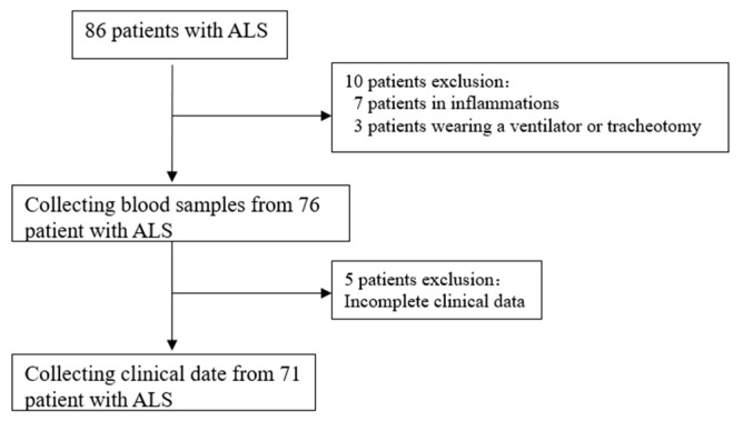
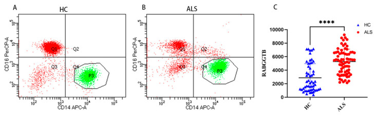
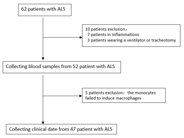
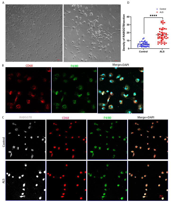
Methods: Information and venous blood were collected from 86 patients diagnosed with ALS between January 2021 and August 2023. Flow cytometry was used to detect the expression of RABGGTB in monocytes from peripheral blood samples collected from patients with ALS and healthy controls. Extracted peripheral blood mononuclear cells (PBMCs) were differentiated in vitro into macrophages, and then the expression of RABGGTB was detected by immunofluorescence. RABGGTB levels in patients with ALS were analyzed to determine their impact on disease progression.
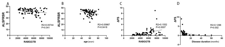
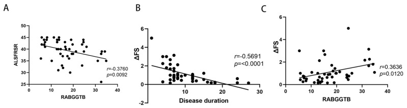
Results: Using flow cytometry in monocytes and immunofluorescence in macrophages, we found that RABGGTB expression in the ALS group was significantly higher than in the control group. Age, sex, original location, disease course, C-reactive protein (CRP), and interleukin-6 (IL-6) did not correlate with the ALS functional rating scale-revised (ALSFRS-R), whereas the RABGGTB level was significantly correlated with the ALSFRS-R. In addition, multivariate analysis revealed a significant correlation between RABGGTB and ALSFRS-R score. Further analysis revealed a significant correlation between RABGGTB expression levels and disease progression levels (ΔFS).
Conclusions: The RABGGTB level was significantly increased in patients with ALS compared with healthy controls. An elevated RABGGTB level in patients with ALS is associated with the rate of progression in ALS, suggesting that elevated RABGGTB levels in patients with ALS may serve as an indicator for tracking ALS progression.
Keywords: Rab geranylgeranyltransferase subunit beta (RABGGTB); amyotrophic lateral sclerosis (ALS); macrophages; monocyte.
Conflict of interest statement
The authors declare no conflict of interest.
Figures
Similar articles
-
Assessment of Rab geranylgeranyltransferase subunit beta in amyotrophic lateral sclerosis.Front Neurol. 2024 Aug 19;15:1447461. doi: 10.3389/fneur.2024.1447461. eCollection 2024. Front Neurol. 2024. PMID: 39224887 Free PMC article.
-
Protective effects of intrathecal injection of AAV9-RabGGTB-GFP+ in SOD1G93A mice.Front Aging Neurosci. 2023 Mar 14;15:1092607. doi: 10.3389/fnagi.2023.1092607. eCollection 2023. Front Aging Neurosci. 2023. PMID: 36967828 Free PMC article.
-
RABGGTB plays a critical role in ALS pathogenesis.Brain Res Bull. 2024 Jan;206:110833. doi: 10.1016/j.brainresbull.2023.110833. Epub 2023 Dec 1. Brain Res Bull. 2024. PMID: 38042502
-
C-reactive protein levels in patients with amyotrophic lateral sclerosis: A systematic review.Brain Behav. 2022 Mar;12(3):e2532. doi: 10.1002/brb3.2532. Epub 2022 Feb 24. Brain Behav. 2022. PMID: 35201675 Free PMC article. Review.
-
Creatine for amyotrophic lateral sclerosis/motor neuron disease.Cochrane Database Syst Rev. 2010 Jun 16;(6):CD005225. doi: 10.1002/14651858.CD005225.pub2. Cochrane Database Syst Rev. 2010. Update in: Cochrane Database Syst Rev. 2012 Dec 12;12:CD005225. doi: 10.1002/14651858.CD005225.pub3. PMID: 20556761 Updated. Review.
Cited by
-
Assessment of Rab geranylgeranyltransferase subunit beta in amyotrophic lateral sclerosis.Front Neurol. 2024 Aug 19;15:1447461. doi: 10.3389/fneur.2024.1447461. eCollection 2024. Front Neurol. 2024. PMID: 39224887 Free PMC article.
References
-
- Vacchiano V., Mastrangelo A., Zenesini C., Masullo M., Quadalti C., Avoni P., Polischi B., Cherici A., Capellari S., Salvi F., et al. Plasma and CSF Neurofilament Light Chain in Amyotrophic Lateral Sclerosis: A Cross-Sectional and Longitudinal Study. Front. Aging Neurosci. 2021;13:753242. doi: 10.3389/fnagi.2021.753242. - DOI - PMC - PubMed
-
- Thouvenot E., Demattei C., Lehmann S., Maceski-Maleska A., Hirtz C., Juntas-Morales R., Pageot N., Esselin F., Alphandéry S., Vincent T., et al. Serum neurofilament light chain at time of diagnosis is an independent prognostic factor of survival in amyotrophic lateral sclerosis. Eur. J. Neurol. 2020;27:251–257. doi: 10.1111/ene.14063. - DOI - PubMed
-
- De Schaepdryver M., Lunetta C., Tarlarini C., Mosca L., Chio A., Van Damme P., Poesen K. Neurofilament light chain and C reactive protein explored as predictors of survival in amyotrophic lateral sclerosis. J. Neurol. Neurosurg. Psychiatry. 2020;91:436–437. doi: 10.1136/jnnp-2019-322309. - DOI - PubMed
Grants and funding
LinkOut - more resources
Full Text Sources
Research Materials
Miscellaneous
