. 2023 Oct 25;11(11):2892.
doi: 10.3390/biomedicines11112892.

SARS-CoV-2 and Epstein-Barr Virus-like Particles Associate and Fuse with Extracellular Vesicles in Virus Neutralization Tests

Affiliations

SARS-CoV-2 and Epstein-Barr Virus-like Particles Associate and Fuse with Extracellular Vesicles in Virus Neutralization Tests

Johannes Roessler et al. Biomedicines. .

Abstract

The successful development of effective viral vaccines depends on well-known correlates of protection, high immunogenicity, acceptable safety criteria, low reactogenicity, and well-designed immune monitoring and serology. Virus-neutralizing antibodies are often a good correlate of protective immunity, and their serum concentration is a key parameter during the pre-clinical and clinical testing of vaccine candidates. Viruses are inherently infectious and potentially harmful, but we and others developed replication-defective SARS-CoV-2 virus-like-particles (VLPs) as surrogates for infection to quantitate neutralizing antibodies with appropriate target cells using a split enzyme-based approach. Here, we show that SARS-CoV-2 and Epstein-Barr virus (EBV)-derived VLPs associate and fuse with extracellular vesicles in a highly specific manner, mediated by the respective viral fusion proteins and their corresponding host receptors. We highlight the capacity of virus-neutralizing antibodies to interfere with this interaction and demonstrate a potent application using this technology. To overcome the common limitations of most virus neutralization tests, we developed a quick in vitro diagnostic assay based on the fusion of SARS-CoV-2 VLPs with susceptible vesicles to quantitate neutralizing antibodies without the need for infectious viruses or living cells. We validated this method by testing a set of COVID-19 patient serum samples, correlated the results with those of a conventional test, and found good sensitivity and specificity. Furthermore, we demonstrate that this serological assay can be adapted to a human herpesvirus, EBV, and possibly other enveloped viruses.

Keywords: EBV; Epstein–Barr virus; SARS-CoV-2; antibody; diagnostic test; extracellular vesicle; fusion; virus neutralization test; virus-like particle.


Conflict of interest statement

Authors D.P., R.Z. and W.H. are listed as inventors on a patent application relating to virus neutralization assays jointly filed by Helmholtz Munich and Eximmium Biotechlogies (Munich, Germany). All other authors declare that they have no competing interest.

Figures
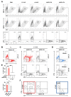
Figure 1
Figure 1
Association of SARS-CoV-2 VLP and ACE2 vesicles. (A) Purified SARS-CoV-2 virus-like particles (VLPs) generated by HEK293T cells and U251MG-derived extracellular vesicles (EVs) were stained with CTV or CTY, respectively, and analyzed via flow cytometry. After gating on side scatter height (SSC-H) and CTV or CTY, events in both gates as indicated were merged in the bottom set of panels. Particle-free diluent PBS was used as the control. (B,C) S+ or ∆S VLPs (CTV+) were incubated with ACE2+ or ACE2 EVs (CTY+) at a 1:1 ratio at 37 °C for 0 or 16 h and subjected to flow cytometric analysis. No double-positive CTV+ CTY+ events could be detected at t0 or at t = 16 h in the absence of spike (∆S) or the host receptor (ACE2), but co-association was observed exclusively between S+ VLPs and ACE2+ EVs. (D) The pre-incubation of S+ VLPs with the neutralizing monoclonal antibody (NmAb) 35B12 efficiently prevented co-association with ACE2+ EVs, while an isotype control IgG mAb did not. (E) Experiments from panels (BD) were carried out in triplicates, and the percentages of double-positive CTV+ CTY+ events were summarized. Results from independent t-tests are indicated; ns, not significant (p > 0.05); ** p ≤ 0.01. (F) The panels provide a schematic illustration of the experimental situations found in the top row of panels (C,D) above.
Figure 2
Figure 2
Fusion of SARS-CoV-2 VLP and ACE2 vesicles. (A) SARS-CoV-2 VLPs that comprise the viral structural proteins M, N, and E and trimeric, full-length (FL) S protein were engineered to carry CD63~HiBiT, a chimeric protein that anchors the activating peptide of split nanoluciferase (nLuc) inside the lumen. Susceptible EVs derived from U251MG cells and decorated with the host receptor ACE2 were engineered to bear CD63~LgBiT, the complementary entity of the split enzyme. Upon incubation, both particles associate via ACE2 and S, leading to the fusion of membranes and ultimately the reconstitution of functional nLuc. Neutralizing antibodies (NAbs) with specificity for spike inhibit this process and can be quantified accordingly via the reduction in luminescence intensity. (B) Preparations of CD63∼HiBiT+ and S+- or S-deficient (∆S) VLPs were incubated at 37 °C for 4 h with CD63∼LgBiT+ and ACE2+ or ACE2 EVs and subsequently analyzed for luminescence upon the addition of substrate. High relative light units (RLUs) and thus the fusion of particles were exclusively detected in the presence of viral fusion protein S and host receptor ACE2, confirming the specificity and tropism. Data are based on four independent experiments, and results from independent t-tests are indicated; **** p ≤ 0.0001. (C) To study the mechanism of S+ VLPs and ACE2+ EVs fusion, both classes of vesicles were incubated with increasing concentrations (c [M]) of protease inhibitors BB94, camostat-mesylate, and chloroquine or control diluents (DMSO and PBS) prior to luminescence analysis. Chloroquine, which prevents endosomal acidification, and camostat-mesylate, a TMPRSS2 inhibitor, did not prevent vesicle fusion while BB94 (batimastat), a broad inhibitor of matrix metalloproteinases (MMPs), effectively blocked fusion. Mean values of three biological replicates are displayed, with error bars indicating standard deviations.
Figure 3
Figure 3
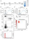
Characterization of purified particles. (A) S+ VLPs and ACE2+ EVs were purified from conditioned cell culture medium by differential centrifugation, sedimentation by ultracentrifugation, and fractionation in a flotation density gradient according to their specific weight. (B,C) Fractions were examined via BCA and NTA assays for total protein and total particle concentrations, respectively. Additionally, fractions were analyzed by reducing (red.) and non-reducing (non-red.) western blots, using anti-S antibodies specific for full-length (FL) spike, SFL or subunits S1 and S2, and antibodies specific for HiBiT or ACE2. (D) Combinations of S+ VLPs and ACE2+ EV fractions were incubated and analyzed for luminescence, and the resulting relative light units (RLU) were displayed in the heat map. Reconstitution of split nLuc reporter, i.e., VLP–EV fusion, was found to be limited to fraction F2 of S+ VLPs and fractions F2 and F3 of ACE2+ EVs. (E) S+ VLP fractions were further analyzed in a specific sandwich ELISA for SFL concentration and the cell-based assay with ACE2+, LgBiT+ U251MG recipient cells for fusion.
Figure 4
Figure 4
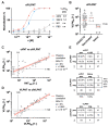
Validation of the cell-free SARS-CoV-2 virus neutralization test. (A) Neutralizing (42E2, 35B12) and non-neutralizing (5D1) anti-S specific monoclonal antibodies were analyzed in the cell-free VLP neutralization test (cfVLPNT) for the neutralization of S+ VLP fusion with ACE2+ EVs. Mean values of three biological replicates are displayed, and error bars indicate standard deviations. The 50% neutralization values, VLPN50, are listed or indicated as not applicable (n.a.). (B) Neutralization titers of 57 COVID-19 patients, 13 COVID-19 vaccinee, and 12 control serum specimens from healthy and immunologically naive donors tested in the cfVLPNT with S+ VLPs, are shown. Median values are indicated in red. Samples above the dotted line (1:≥140) scored positive according to the criteria applied. Samples in brackets indicate sera with a VLPN50 below the limit of detection (LOD). (C) VLPN50 titers from the cfVLPNT are plotted along VNT100 titers obtained in a conventional virus neutralization test (cVNT) with infectious SARS-CoV-2. Pearson correlation data (coefficient r, confidence interval CI, sample size n, and p-value) of 57 sera from COVID-19 patients are shown. Results left of the dotted vertical line denote sera, which scored below the LOD (1:<8) in the cVNT; these VNT100 values were defined as 1:4 and indicated in square brackets. (D) VLPN50 titers from the cell-free (vertical axis) and the cell-based (horizontal axis) VLPNT are plotted. As in panel (C), Pearson correlation data of 57 sera from COVID-19 patients are shown. Results left of the dotted vertical line denote sera, which scored negative (1:<25) in the VLPNT. (E) In the top table, the results of 57 COVID-19 samples tested in the cfVLPNT and a cVNT are compared. In the middle table, the sensitivity, specificity, positive predictive value (PPV), and negative predictive value (NPV) of the cfVLPNT were calculated based on 48 COVID-19 serum samples with neutralizing capacity according to the cVNT and 12 controls of immunologically naive donors. In the bottom table, the results of 57 COVID-19 samples tested in the cfVLPNT and the cell-based VLPNT are compared. Test results are indicated: pos., positive; neg., negative; <LOD, below the limit of detection; CP, concordant positive; CN, concordant negative; D, discrepant; TP, true positive; FN, false negative; FP, false positive; TN, true negative.
Figure 5
Figure 5
Fusion and neutralization of EB-VLP with vesicles and cells. (A) Purified samples of Epstein–Barr virus-like particles (EB-VLPs) and Daudi-cell-derived EVs were stained with CTV or CTY, respectively, and analyzed via flow cytometry. After gating on side scatter height (SSC-H) and CTV or CTY events, both gates were merged, as shown in the bottom row of panels. Particle-free PBS was used as a control. (B) EB-VLPs (CTV+) and Daudi cells were stained with glycoprotein gp350 and a CD21-specific antibody, respectively, and analyzed via flow cytometry. (C) EB-VLPs (CTV+) were incubated with Daudi-cell-derived EVs (CTY+) at 1:1 stoichiometry at 37 °C for 0 or 16 h and subjected to flow cytometric analyses. No double-positive CTV+ CTY+ events could be detected at t0, and the co-association of the two classes of vesicles was found after 16 h. (D) EB-VLPs (CD63~HiBiT+) were incubated with CD63~LgBiT+ Daudi cells or EVs at 37 °C for 4 h prior to the addition of substrate and luminescence analyses. Pre-incubation of EB-VLPs with 10 µg mL−1 monoclonal antibodies (mAb) specific for EBV antigens gp42 or gp350 effectively prevented fusion with Daudi cells and Daudi-cell-derived EVs while IgG isotype controls did not. Results from independent t-tests with biological triplicates are indicated; ** p ≤ 0.01; *** p ≤ 0.001. (E) EB-VLPs were pre-incubated with 1:50 diluted human sera from two EBV-seropositive and one EBV-negative donor one hour prior to incubation with Daudi cells or EVs. Signals were normalized to diluent PBS control and analyzed for neutralization. Results from independent t-tests based on triplicates are indicated; ** p ≤ 0.01; *** p ≤ 0.001; **** p ≤ 0.0001.

Similar articles

Cited by

  • Light-Induced Transformation of Virus-Like Particles on TiO2.
    Kohantorabi M, Ugolotti A, Sochor B, Roessler J, Wagstaffe M, Meinhardt A, Beck EE, Dolling DS, Garcia MB, Creutzburg M, Keller TF, Schwartzkopf M, Vayalil SK, Thuenauer R, Guédez G, Löw C, Ebert G, Protzer U, Hammerschmidt W, Zeidler R, Roth SV, Di Valentin C, Stierle A, Noei H. Kohantorabi M, et al. ACS Appl Mater Interfaces. 2024 Jul 17;16(28):37275-37287. doi: 10.1021/acsami.4c07151. Epub 2024 Jul 3. ACS Appl Mater Interfaces. 2024. PMID: 38959130 Free PMC article.

References

    1. Pollard A.J., Bijker E.M. A guide to vaccinology: From basic principles to new developments. Nat. Rev. Immunol. 2021;21:83–100. doi: 10.1038/s41577-020-00479-7. - DOI - PMC - PubMed
    1. Trovato M., Sartorius R., D’Apice L., Manco R., De Berardinis P. Viral Emerging Diseases: Challenges in Developing Vaccination Strategies. Front. Immunol. 2020;11:2130. doi: 10.3389/fimmu.2020.02130. - DOI - PMC - PubMed
    1. Khoury D.S., Cromer D., Reynaldi A., Schlub T.E., Wheatley A.K., Juno J.A., Subbarao K., Kent S.J., Triccas J.A., Davenport M.P. Neutralizing antibody levels are highly predictive of immune protection from symptomatic SARS-CoV-2 infection. Nat. Med. 2021;27:1205–1211. doi: 10.1038/s41591-021-01377-8. - DOI - PubMed
    1. Feng S., Phillips D.J., White T., Sayal H., Aley P.K., Bibi S., Dold C., Fuskova M., Gilbert S.C., Hirsch I., et al. Correlates of protection against symptomatic and asymptomatic SARS-CoV-2 infection. Nat. Med. 2021;27:2032–2040. doi: 10.1038/s41591-021-01540-1. - DOI - PMC - PubMed
    1. Earle K.A., Ambrosino D.M., Fiore-Gartland A., Goldblatt D., Gilbert P.B., Siber G.R., Dull P., Plotkin S.A. Evidence for antibody as a protective correlate for COVID-19 vaccines. Vaccine. 2021;39:4423–4428. doi: 10.1016/j.vaccine.2021.05.063. - DOI - PMC - PubMed

LinkOut - more resources