. 2024 Jan:190:106363.
doi: 10.1016/j.nbd.2023.106363. Epub 2023 Nov 22.

Characterisation and prion transmission study in mice with genetic reduction of sporadic Creutzfeldt-Jakob disease risk gene Stx6

Affiliations

Characterisation and prion transmission study in mice with genetic reduction of sporadic Creutzfeldt-Jakob disease risk gene Stx6

Emma Jones et al. Neurobiol Dis. 2024 Jan.

Abstract

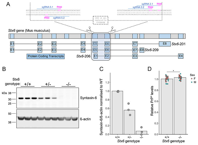
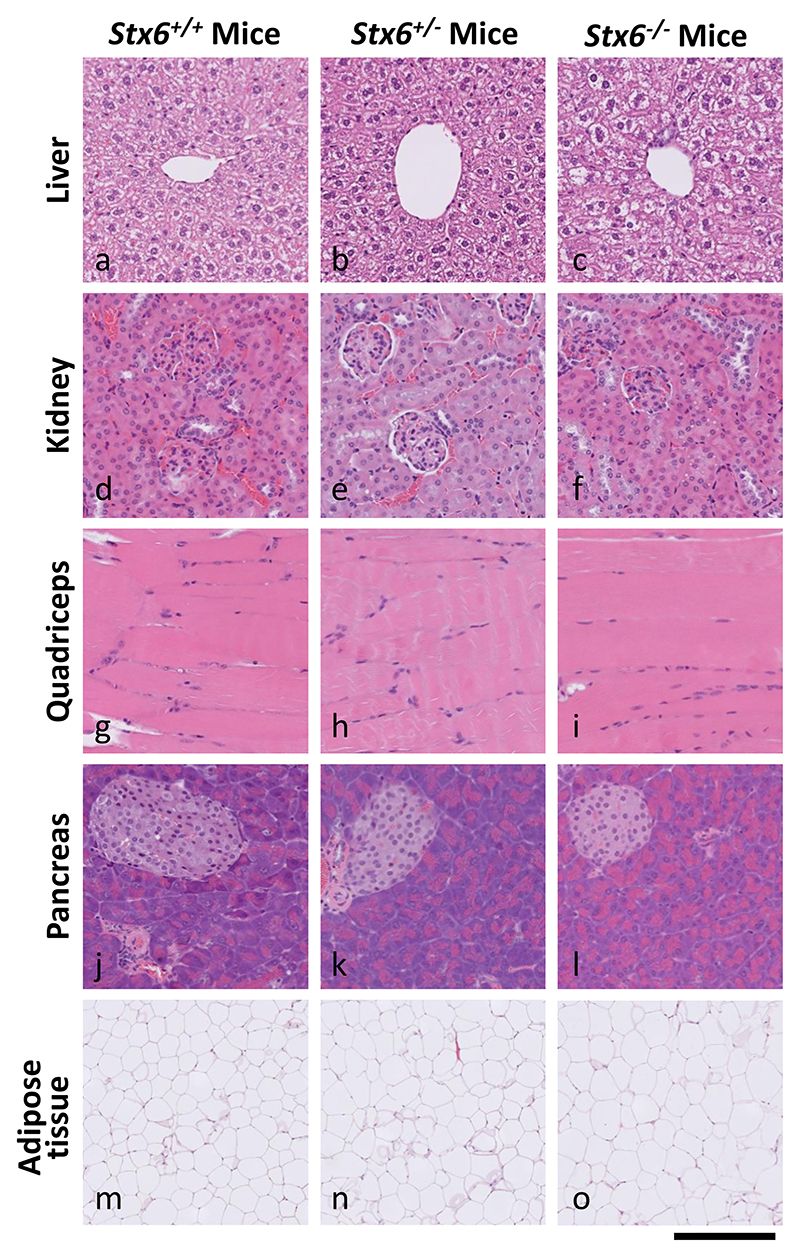
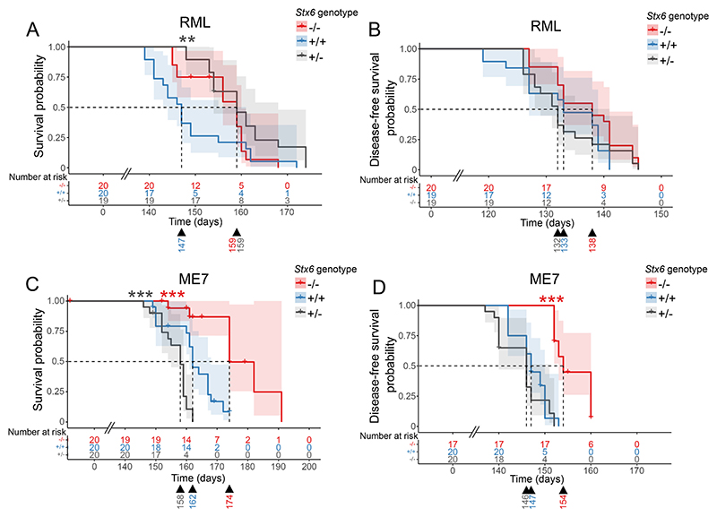
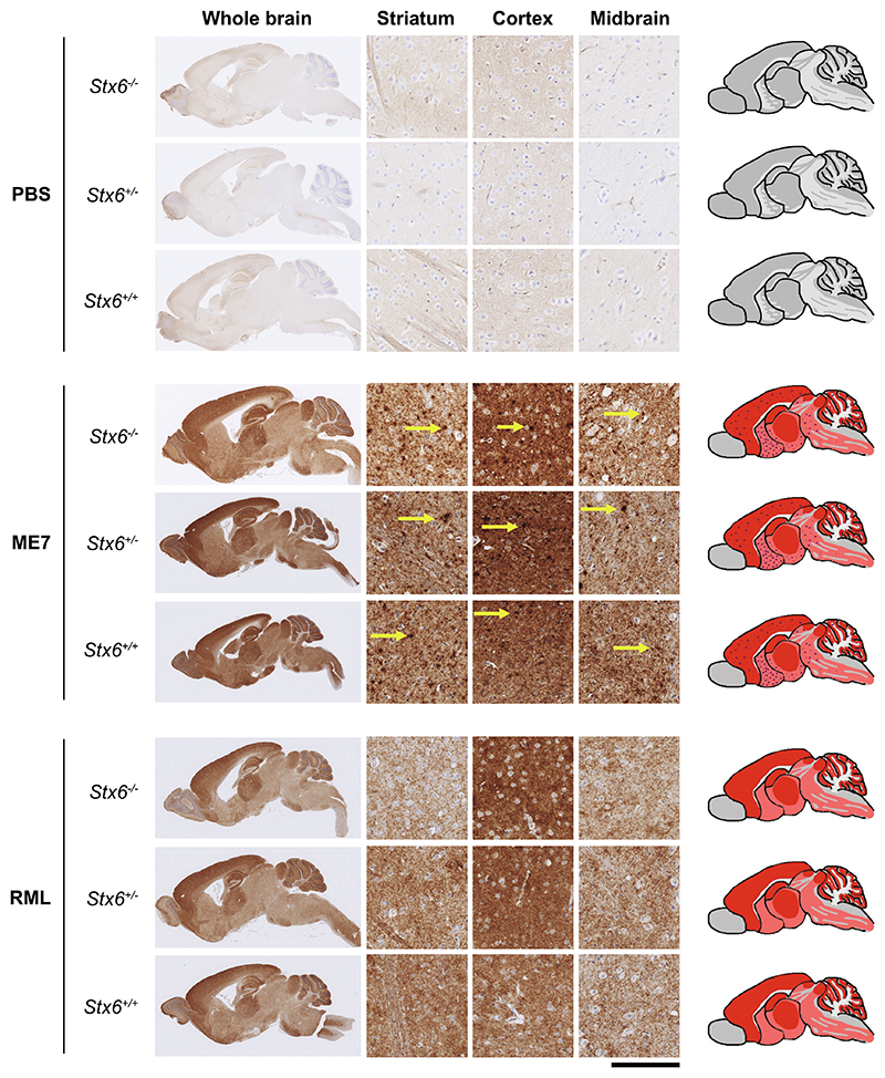
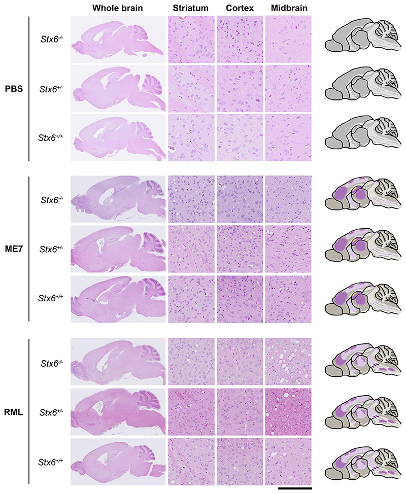
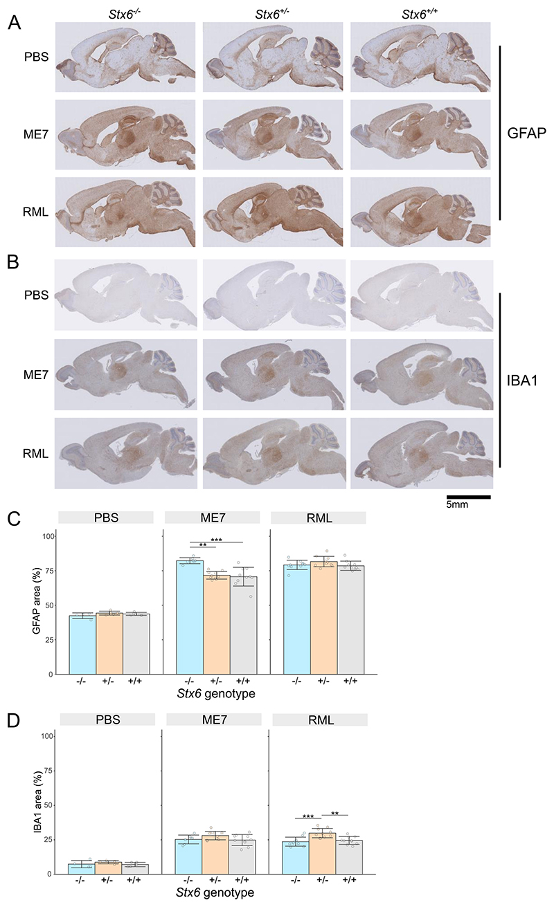
Sporadic Creutzfeldt-Jakob disease (sCJD), the most common human prion disease, is thought to occur when the cellular prion protein (PrPC) spontaneously misfolds and assembles into prion fibrils, culminating in fatal neurodegeneration. In a genome-wide association study of sCJD, we recently identified risk variants in and around the gene STX6, with evidence to suggest a causal increase of STX6 expression in disease-relevant brain regions. STX6 encodes syntaxin-6, a SNARE protein primarily involved in early endosome to trans-Golgi network retrograde transport. Here we developed and characterised a mouse model with genetic depletion of Stx6 and investigated a causal role of Stx6 expression in mouse prion disease through a classical prion transmission study, assessing the impact of homozygous and heterozygous syntaxin-6 knockout on disease incubation periods and prion-related neuropathology. Following inoculation with RML prions, incubation periods in Stx6-/- and Stx6+/- mice differed by 12 days relative to wildtype. Similarly, in Stx6-/- mice, disease incubation periods following inoculation with ME7 prions also differed by 12 days. Histopathological analysis revealed a modest increase in astrogliosis in ME7-inoculated Stx6-/- animals and a variable effect of Stx6 expression on microglia activation, however no differences in neuronal loss, spongiform change or PrP deposition were observed at endpoint. Importantly, Stx6-/- mice are viable and fertile with no gross impairments on a range of neurological, biochemical, histological and skeletal structure tests. Our results provide some support for a pathological role of Stx6 expression in prion disease, which warrants further investigation in the context of prion disease but also other neurodegenerative diseases considering syntaxin-6 appears to have pleiotropic risk effects in progressive supranuclear palsy and Alzheimer's disease.

Keywords: Creutzfeldt-Jakob disease; Incubation period; Prion disease; SNARE; Syntaxin-6.


Conflict of interest statement

Declaration of Competing Interest J.C. is a director and shareholder of D-Gen Limited, an academic spin-out company in the field of prion diagnosis, decontamination and therapeutics. There are no other competing interests.

Figures

Fig. 1
Fig. 1. Development and validation of Stx6 knockout mice.
(A) Strategy for CRISPR/Cas9-mediated knockout of Stx6 in C57BL/6N mice. Four single guide RNAs (sgRNAs) targeting the DNA sequences in exons 5 and 6 of the two main Stx6 protein-coding transcripts (Stx6–201 and Stx6–209), also encompassing the N-terminal portion of the shorter Stx6–206 transcript, were selected for gene editing in zygotes. This resulted in an 1808 nucleotide (nt) deletion replaced by an 8 nt insertion. Top: Full Stx6 genomic location. Bottom: protein coding transcripts. Inset: CRISPR/Cas9 genome editing at Stx6 locus. (B) Representative quantitative immunoblots with anti-syntaxin 6 and anti-β actin antibodies of whole brain homogenates from Stx6+/−, Stx6−/− and Stx6+/+ mice and (C) quantification of syntaxin-6 intensity relative to β-actin normalised to Stx6+/+ control demonstrates loss of the primary ~32 kDa protein isoform in Stx6−/− mice (0.0757% ± 0. 0.0652 (mean ± SD)) with ~50% expression (55.9% ± 0. 0.131) in Stx6+/− mice (n = 3/genotype, mean of 3 replicates per animal shown). (D) Analysis of PrPC expression in whole brain homogenates from Stx6−/− and Stx6+/+ early adult 9 week old mice (n = 30/genotype, mixed sex) by ELISA shows minor increase in relative PrPC expression in Stx6−/− mice relative to wildtype (Stx6+/+: 1.004 ± 0.0441; Stx6−/−: 1.030 ± 0.0337 (mean ± SD)). Values are relative to Stx6+/+. Relative differences were assessed by 2-way ANOVA with sex and genotype as factors.
Fig. 2
Fig. 2. Multi-organ H&E staining shows expected tissue architecture and appearance in Stx6−/− and Stx6+/− mice which lack consistent tissue-intrinsic pathologies.
The following organs were harvested from Stx6+/+ (n = 5), Stx6+/ - (n = 5) and Stx6−/− (n = 4) female mice at 56–61 weeks of age: liver, pancreas, kidney, skeletal muscle and white adipose. These were formalin fixed, processed to paraffin and underwent haematoxylin and eosin (H&E) staining, revealing no consistent differences between genotypes. Scale bar corresponds to 120 μm (a-l), and 500 μm (m-o).
Fig. 3
Fig. 3. Incubation periods in mice with Stx6 reduction following intracerebral inoculation with RML and ME7 mouse-adapted scrapie prions.
(A-D) Kaplan-Meier curve of survival probability following intracerebral inoculation of Stx6−/− (red), Stx6+/− (grey) and Stx6+/+ (blue) C57BL/6N mice (n = 20 per group) with 1% brain homogenate from (A,B) RML and (C,D) ME7 infected C57BL/6N mice. (A, C) Survival probability until animals were culled due to scrapie sickness shows a 12-day increase in median incubation period for (A) Stx6−/− and Stx6+/− mice following RML inoculation (significant only for Stx6+/− animals) and (C) for Stx6−/− mice only following ME7 inoculation relative to Stx6+/+ mice. (B,D) Disease-free survival probability (time until onset of the first scrapie symptom) (B) does not show a significant association with Stx6 genotype following RML-inoculation but (D) a 7-day increase in Stx6−/− mice inoculated with ME7, relative to Stx6+/+ mice. Significance shown from pair-wise log-rank test for Stx6−/− (red asterix) and Stx6+/− (black asterix) mice relative to wildtype controls (** P > 0.01; *** P > 0.001). Median incubation period shown below each plot. Crosses indicate censored animals. (For interpretation of the references to colour in this figure legend, the reader is referred to the web version of this article.)
Fig. 4
Fig. 4. Knockout of Stx6 does not alter PrP deposition in prion infected mice.
Immunohistochemistry using anti-PrP antibody ICSM35 in whole brain (left) with schematic depiction of PrP deposition (right; pink shading: moderate PrP deposition; red shading: intense PrP deposition; red dots: PrP micro-plaques) or representative images from striatum, cortex and midbrain (middle) from Stx6−/−, Stx6+/− and Stx6+/+ mice inoculated with PBS (top) or ME7 (middle) and RML (bottom) mouse-adapted scrapie prions, shows expected PrP deposition in all prion-inoculated samples at end-point, with widespread diffuse staining for both strains and additional presence of micro-plaques with ME7 (yellow arrows). There was no evidence of spontaneous PrP deposition in control animals or gross difference evident between genotypes. Scale bar corresponds to 100 μm in the high-power magnification and 1.8 mm for the overview images. (For interpretation of the references to colour in this figure legend, the reader is referred to the web version of this article.)
Fig. 5
Fig. 5. Neuronal loss and spongiform change were comparable in end-stage Stx6−/−, Stx6+/− and Stx6+/+ mice.
Haematoxylin and eosin (H&E) staining of whole brain (left) with schematic depiction of spongiosis (right; light purple shading: widely dispersed mild spongiosis; darker purple shading: moderate spongiosis; yellow shading: neuronal loss) and representative images from striatum, cortex and midbrain (middle) from Stx6−/−, Stx6+/− and Stx6+/+ mice inoculated with PBS (top) or ME7 (middle) and RML (bottom) mouse-adapted scrapie prions shows expected spongiform pathology in all samples at end-point, including mild hippocampal neuronal loss with ME7, with no gross differences evident between genotypes. No neuropathology was evident in PBS-inoculated controls. Scale bar corresponds to 100 μm in the high-power magnification and 1.8 mm for the overview images. (For interpretation of the references to colour in this figure legend, the reader is referred to the web version of this article.)
Fig. 6
Fig. 6. Stx6 genotype and neuroinflammation in prion disease.
(A, B) Example images of immunohistochemistry in Stx6−/−, Stx6+/− and Stx6+/+ mice inoculated PBS control or ME7 and RML mouse-adapted scrapie prions with (A) anti-GFAP antibody to measure reactive astrocytes and (B) anti-Iba1 antibody to measure microgliosis, shows expected neuroinflammation in prion-infected animals, including expected intense staining in the thalamus with both antibodies and intense GFAP staining in the hippocampus. (C, D) Quantification of percentage area stained with (C) anti-GFAP and (D) anti-Iba1 antibody demonstrates a ~ 10% increase in astrocyte area in ME7-inoculated Stx6−/− animals and a ~ 5% increase in microglia area in Stx6+/− mice in RML-inoculated animals (mean ± SD; linear regression model adjusted for age of death; ** P > 0.01, *** P > 0.001).

Similar articles

  • Genetic risk factors for Creutzfeldt-Jakob disease.
    Jones E, Mead S. Jones E, et al. Neurobiol Dis. 2020 Aug;142:104973. doi: 10.1016/j.nbd.2020.104973. Epub 2020 Jun 18. Neurobiol Dis. 2020. PMID: 32565065 Review.
  • Absence of spontaneous disease and comparative prion susceptibility of transgenic mice expressing mutant human prion proteins.
    Asante EA, Gowland I, Grimshaw A, Linehan JM, Smidak M, Houghton R, Osiguwa O, Tomlinson A, Joiner S, Brandner S, Wadsworth JDF, Collinge J. Asante EA, et al. J Gen Virol. 2009 Mar;90(Pt 3):546-558. doi: 10.1099/vir.0.007930-0. J Gen Virol. 2009. PMID: 19218199 Free PMC article.
  • Identification of novel risk loci and causal insights for sporadic Creutzfeldt-Jakob disease: a genome-wide association study.
    Jones E, Hummerich H, Viré E, Uphill J, Dimitriadis A, Speedy H, Campbell T, Norsworthy P, Quinn L, Whitfield J, Linehan J, Jaunmuktane Z, Brandner S, Jat P, Nihat A, How Mok T, Ahmed P, Collins S, Stehmann C, Sarros S, Kovacs GG, Geschwind MD, Golubjatnikov A, Frontzek K, Budka H, Aguzzi A, Karamujić-Čomić H, van der Lee SJ, Ibrahim-Verbaas CA, van Duijn CM, Sikorska B, Golanska E, Liberski PP, Calero M, Calero O, Sanchez-Juan P, Salas A, Martinón-Torres F, Bouaziz-Amar E, Haïk S, Laplanche JL, Brandel JP, Amouyel P, Lambert JC, Parchi P, Bartoletti-Stella A, Capellari S, Poleggi A, Ladogana A, Pocchiari M, Aneli S, Matullo G, Knight R, Zafar S, Zerr I, Booth S, Coulthart MB, Jansen GH, Glisic K, Blevins J, Gambetti P, Safar J, Appleby B, Collinge J, Mead S. Jones E, et al. Lancet Neurol. 2020 Oct;19(10):840-848. doi: 10.1016/S1474-4422(20)30273-8. Epub 2020 Sep 16. Lancet Neurol. 2020. PMID: 32949544 Free PMC article.
  • Prion Strain Characterization of a Novel Subtype of Creutzfeldt-Jakob Disease.
    Galeno R, Di Bari MA, Nonno R, Cardone F, Sbriccoli M, Graziano S, Ingrosso L, Fiorini M, Valanzano A, Pasini G, Poleggi A, Vinci R, Ladogana A, Puopolo M, Monaco S, Agrimi U, Zanusso G, Pocchiari M. Galeno R, et al. J Virol. 2017 May 12;91(11):e02390-16. doi: 10.1128/JVI.02390-16. Print 2017 Jun 1. J Virol. 2017. PMID: 28298604 Free PMC article.
  • Recent advances in the histo-molecular pathology of human prion disease.
    Baiardi S, Rossi M, Capellari S, Parchi P. Baiardi S, et al. Brain Pathol. 2019 Mar;29(2):278-300. doi: 10.1111/bpa.12695. Epub 2019 Jan 22. Brain Pathol. 2019. PMID: 30588685 Free PMC article. Review.

References

    1. Aguzzi A, Barres BA, Bennett ML. Microglia: scapegoat, saboteur, or something else? Science (New York, NY) 2013;339(6116):156–161. - PMC - PubMed
    1. Akhtar S, Wenborn A, Brandner S, Collinge J, Lloyd SE. Sex effects in mouse prion disease incubation time. PLoS One. 2011;6(12):e28741. - PMC - PubMed
    1. Akhtar S, Grizenkova J, Wenborn A, Hummerich H, Fernandez de Marco M, Brandner S, et al. Sod1 deficiency reduces incubation time in mouse models of prion disease. PLoS One. 2013;8(1):e54454. - PMC - PubMed
    1. Bock JB, Lin RC, Scheller RH. A new syntaxin family member implicated in targeting of intracellular transport vesicles. J Biol Chem. 1996;271(30):17961–17965. - PubMed
    1. Bock JB, Klumperman J, Davanger S, Scheller RH. Syntaxin 6 functions in trans-Golgi network vesicle trafficking. Mol Biol Cell. 1997;8(7):1261–1271. - PMC - PubMed

Supplementary concepts