. 2023 Nov 17;23(1):415.
doi: 10.1186/s12906-023-04255-7.

Prediction and validation of common targets in atherosclerosis and non-small cell lung cancer influenced by atorvastatin

Affiliations

Prediction and validation of common targets in atherosclerosis and non-small cell lung cancer influenced by atorvastatin

Yu-Qian Li et al. BMC Complement Med Ther. .

Abstract

Background: Cardiovascular disease and cancer are the main causes of morbidity and mortality worldwide. Studies have shown that these two diseases may have some common risk factors. Atorvastatin is mainly used for the treatment of atherosclerosis in clinic. A large number of studies show that atorvastatin may produce anti-tumor activities. This study aimed to predict the common targets of atorvastatin against atherosclerosis and non-small cell lung cancer (NSCLC) based on network pharmacology.

Methods: The target genes of atherosclerosis and NSCLC were obtained from The Cancer Genome Atlas (TCGA) and Gene Expression Omnibus (GEO) databases. The disease-target-component model map and the core network were obtained using Cytoscape 3.7.1. The MTS and wound healing assay were used to detect the effect of atorvastatin on cell viability and migration of A549 cells. The expression of potential common target genes of atorvastatin against atherosclerosis and NSCLC were confirmed in A549 cells and lung cancer tissues of patients.

Results: We identified 15 identical pathogenic genes, and four of which (MMP9, MMP12, CD36, and FABP4) were considered as the key target genes of atorvastatin in anti-atherosclerosis and NSCLC. The MTS and wound healing assays revealed that atorvastatin decreased A549 cells migration significantly. Atorvastatin markedly decreased the expression of MMP9, MMP12, CD36, and FABP4 in A549 cells and patients were treated with atorvastatin.

Conclusions: This study demonstrated 15 common pathogenic genes in both atherosclerosis and NSCLC. And verified that MMP 9, MMP 12, CD 36 and FABP 4 might be the common target genes of atorvastatin in anti-atherosclerosis and NSCLC.

Keywords: Atherosclerosis; Atorvastatin; Migration; Network pharmacology; Non-small cell lung cancer.


Conflict of interest statement

The authors declare no competing interests.

Figures

Fig. 1
Fig. 1
DEGs of atherosclerosis and DEGs of NSCLC, and potential common pathogenic genes of atherosclerosis and NSCLC. A Heat map of DEGs in atherosclerosis: red represents higher gene expression and blue represents lower gene expression. B Heat map of DEGs in NSCLC: red represents an over expressed gene and blue represents low gene expression. C Overlapping DEGs of atherosclerosis and DEGs of NSCLC. The network revealed the number of shared and unique genes of atherosclerosis and NSCLC
Fig. 2
Fig. 2
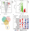
Key target genes of atorvastatin against atherosclerosis and NSCLC. A Volcano map of the expression of 15 identical pathogenic genes between normal tissues and atherosclerosis groups in the GEO database: the red represents the up-regulated genes in atherosclerosis tissues, and the green represents the down-regulated genes. B Volcano map of the expression of 15 same pathogenic genes in NSCLC; The red represents the upregulated genes in the NSCLC tissues and the green represents the down regulated genes. C Find 4 genes (MMP9, MMP12, CD36, and FABP4) were as potential therapeutic targets of atorvastatin for atherosclerosis and NSCLC through overlapped the genes of atorvastatin related targets, DEGs of atherosclerosis and DEGs of NSCLC. D Expression of four common genes associated with atorvastatin against atherosclerosis and NSCLC in various cancers
Fig. 3
Fig. 3
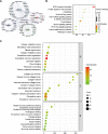
Topological properties, KEGG enrichment analysis and GO enrichment analysis. A The protein–protein interaction networks of MMP9, MMP12, CD36, and FABP4 of common genes were constructed by using Bisogenet in Cytoscape software. B KEGG analysis of top 10 enriched signaling pathways with p value < 0.05. C Top 10 signaling pathways of biological process, cell function, and molecular function in GO enrichment analysis with p < 0.05
Fig. 4
Fig. 4
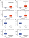
Expression of MMP12, MMP9, CD36, and FABP4 in NSCLC. A MMP12 was overexpressed in LUAD. B MMP12 was overexpressed in LUSC. C MMP9 was overexpressed in LUAD. D MMP9 was overexpressed in LUSC. E CD36 was underexpressed in LUAD. F CD36 was underexpressed in LUSC. G FABP4 was underexpressed in LUAD. H FABP4 was underexpressed in LUSC
Fig. 5
Fig. 5
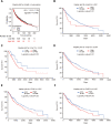
The relationship between overall survival and MMP9, MMP12, CD36, and FABP4 expression in patients with lung cancer. A MMP9 high expression was significantly correlated with lower overall survival (OS) rates in patients with lung cancer (p = 0.046). B The high expression of MMP9 was significantly correlated with the low incidence of OS in patients with LUSC (p = 0.01). C The high expression of MMP12 was significantly correlated with low OS rates in patients with LUAD (p = 0.03). D The high expression of MMP12 was significantly correlated with lower OS rates in patients with LUSC (p = 0.01). E The high expression of CD36 was significantly correlated with lower OS rates in patients with LUSC (p = 0.05). F The high expression of FABP4 was significantly correlated with lower OS rates in patients with LUSC (p = 0.03)
Fig. 6
Fig. 6
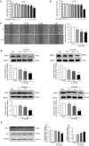
Effects of atorvastatin on the proliferation and migration of A549 cells. A A549 cells were exposed to atorvastatin at concentrations of 0, 0.1, 0.3, 1, 3, 10, 30, and 100 µM for 24 h, and then MTS assays was performed to determine the viability of the cell. Atorvastatin inhibited the proliferation of A549 cells in a dose-dependent manner. B  HepG2 cells were exposed to atorvastatin at concentrations of 0, 0.1, 0.3, 1, 3, 10, 30, and 100 µM for 24 h, and then CCK8 assays was performed to determine the viability of the cell. Atorvastatin inhibited the proliferation of HepG2 cells in a dose-dependent manner. C A549 cells were treated with atorvastatin at concentrations of 0, 0.1, 0.3, and 1 µM for 24 h. Results of the wound healing assay showed that cell healing over scratch was inhibited by the treatment of atorvastatin. The result indicated that atorvastatin therapy reduced the migration of A549 cells in a dose-dependent manner. Scale bar = 50 μm. D Effects of atorvastatin on the activities of MMP9, MMP12, CD36, and FABP4 in A549 cells. Western blot was used to evaluate the expression of MMP9, MMP12, CD36, and FABP4. The result indicated that atorvastatin significantly reduced the expression of MMP9, MMP12, CD36, and FABP4 in a concentration-dependent manner. E Atorvastatin treatment significantly upregulated the expression of pro-apoptosis protein Bax and downregulated the anti-apoptosis protein Bcl-2 protein. * p  < 0.05, ** p  < 0.01 vs. control, n  = 3. Scale bars = 20 μm
Fig. 7
Fig. 7
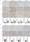
Immunohistochemistry staining determined MMP12, MMP9, CD36, and FABP4 expression in tissues of patients with NSCLC. A Compared with the adjacent non-cancerous lung tissues of patients taking atorvastatin, MMP12 and MMP9 were highly expressed in the tumor stroma of LUAD. CD36 presented a cell membrane pattern of staining, and the expression levels of CD36 in LUAD were higher compared with adjacent non-cancerous lung tissues in patients who took atorvastatin. FABP4 showed nuclear and cytoplasmic staining patterns, and the expression levels of CD36 in LUAD of patients taking atorvastatin was higher compared with adjacent noncancerous lung tissues. B Expression of MMP12, MMP9, CD36, and FABP4 expression in LUSC tissue samples and corresponding adjacent non-cancerous lung tissues. In patients taking atorvastatin, MMP12, MMP9, CD36, and FABP4 were highly expressed in the LUSC, butwere low expressed in the adjacent non-cancerous lung tissues. The intensity of immunohistochemical staining was analyzed using ImageJ 2.0 software. *p < 0.05, **p < 0.01 vs. control. ##p < 0.01 vs. ATV. Scale bars = 50 μm

Similar articles

References

    1. Raposeiras Roubín RS, Cordero A. The two-way relationship between Cancer and Atherosclerosis. Revista Esp De cardiologia (English ed) 2019;72(6):487–494. doi: 10.1016/j.recesp.2018.12.011. - DOI - PubMed
    1. Meijers WC, De B. Common risk factors for Heart Failure and cancer. Cardiovascular Res. 2019;5:844–853. doi: 10.1093/cvr/cvz035. - DOI - PMC - PubMed
    1. Bray F, Ferlay J, Soerjomataram I, et al. Global cancer statistics 2018: GLOBOCAN estimates of incidence and mortality worldwide for 36 cancers in 185 countries. Cancer J Clin. 2018;68(6):394–424. doi: 10.3322/caac.21492. - DOI - PubMed
    1. Matusewicz L, Czogalla A, Sikorski AF. Attempts to use statins in cancer therapy: an update. Tumor Biology. 2020;42(7):101042832094176. doi: 10.1177/1010428320941760. - DOI - PubMed
    1. Huang WY, Li CH, Lin CL, et al. Long-term statin use in patients with Lung cancer and dyslipidemia reduces the risk of death. Oncotarget. 2016;7(27):42208–42215. doi: 10.18632/oncotarget.9906. - DOI - PMC - PubMed

MeSH terms