. 2023 Nov 16;14(1):7431.
doi: 10.1038/s41467-023-43054-z.

Unconventional structure and mechanisms for membrane interaction and translocation of the NF-κB-targeting toxin AIP56

Affiliations

Unconventional structure and mechanisms for membrane interaction and translocation of the NF-κB-targeting toxin AIP56

Johnny Lisboa et al. Nat Commun. .

Abstract

Bacterial AB toxins are secreted key virulence factors that are internalized by target cells through receptor-mediated endocytosis, translocating their enzymatic domain to the cytosol from endosomes (short-trip) or the endoplasmic reticulum (long-trip). To accomplish this, bacterial AB toxins evolved a multidomain structure organized into either a single polypeptide chain or non-covalently associated polypeptide chains. The prototypical short-trip single-chain toxin is characterized by a receptor-binding domain that confers cellular specificity and a translocation domain responsible for pore formation whereby the catalytic domain translocates to the cytosol in an endosomal acidification-dependent way. In this work, the determination of the three-dimensional structure of AIP56 shows that, instead of a two-domain organization suggested by previous studies, AIP56 has three-domains: a non-LEE encoded effector C (NleC)-like catalytic domain associated with a small middle domain that contains the linker-peptide, followed by the receptor-binding domain. In contrast to prototypical single-chain AB toxins, AIP56 does not comprise a typical structurally complex translocation domain; instead, the elements involved in translocation are scattered across its domains. Thus, the catalytic domain contains a helical hairpin that serves as a molecular switch for triggering the conformational changes necessary for membrane insertion only upon endosomal acidification, whereas the middle and receptor-binding domains are required for pore formation.


Conflict of interest statement

The authors declare no competing interests.

Figures
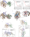
Fig. 1
Fig. 1. Three-dimensional structure of AIP56.
a Cartoon representation of the AIP56 monomer (Chain A), with domains colored wheat (catalytic domain), blue (middle domain) or green (receptor-binding domain). The active site residues are shown as red sticks, the disulfide bond (C262 and C298) as magenta sticks and the zinc ion as a black sphere. The regions not defined (ND) in the structure are indicated by arrows or represented by a dashed line. The N- (Nter) and C-termini (C-ter) are labeled. b SAXS profile showing adjustment of the curve (red line) calculated from the structural model of AIP56 obtained by the Modeller program (Supplementary Fig. 1a), as calculated in CRYSOL, to the experimental scattering curve (gray dots) of AIP56. c SAXS profile showing better fitting of the SREFLEX model (red line). The bottom panels in (b) and (c) show the residual plots for the respective fits, with the residuals defined as Δ/σ = [Iexp(q) – Icalc(q)]/σexp(q). χ2 scores were calculated in CRYSOL. d Complete SREFLEX simulated movement after the refinement stage. Vectors (red arrows) are drawn connecting the equivalent residues from the crystal structure (colored as in a) to the SREFLEX model (gray). e Cartoon representation of the superposed catalytic domains of AIP56 (wheat) and NleC (gray; PDB: 4Q3J). The modified Ψ-loop β-sheet motif (β1–β3) of AIP56 is colored blue. The N- (Nter) and C-termini (C-ter) and secondary structure elements are labeled. The inset shows a close-up of the active sites, including the zinc ion (Zn; black sphere) and the coordinating residues from AIP56 (red) and NleC (gray). f The linker peptide contacts both the catalytic and the receptor-binding domains. Top, cartoon representation highlighting the contact regions; Bottom, C-alpha trace of the contact region with contacts within a range of 4 Å is represented. Top and Bottom representations colored as in (a). g The catalytic and receptor-binding domains contact directly through helix α2 of the catalytic domain. Top and Bottom representations colored as in (a).
Fig. 2
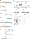
Fig. 2. Pore formation requires both the middle and receptor-binding domains.
a Schematic representation of AIP56 variants and chimeric proteins. AIP56 domains are colored as in Fig. 1a; β-lactamase (Bla) and diphtheria receptor-binding (DTR) domain, gray. b Only AIP56L258-N497 and BlaL19-W286AIP56L258-N497 interacted with artificial black lipid membranes. Single-channel record of DiPhPC/n‐decane membranes after addition of the indicated proteins to the cis-side of the black lipid bilayer at a final concentration indicated in the figure. Measurements were performed with 50 mV (AIP56L258-N497) or 150 mV (BlaL19-W286AIP56L258-N497) at room temperature. Membrane activity was induced by acidification (pH 4.8–5.0; arrows) of the aqueous phase at the cis‐side of the chamber with exception for BlaL19-W286AIP56L258-N497, which formed stable pores at pH 6. The average single-channel conductance was about 16 pS for 110 steps. Each result shown is representative of at least three (n = 3) independent measurements. c Pharmacological inhibition of vacuolar ATPase pump does not affect cytosolic delivery of Bla by BlaL19-W286AIP56L258-N497. The cleaved/uncleaved CCF4-AM ratios were determined by quantifying the indicated number of microscopic fields per condition. Representative images used for quantification are in Supplementary Fig. 4d. Results shown represent one out of three (n = 3) independent experiments. Statistical significance was tested by One-way ANOVA and p values for individual comparisons were calculated by Tukey’s HSD test and indicated on top of the brackets. Data are presented as mean values ± SD. Actual p values from left-to-right: p < 0.0001, p < 0.0001. Data of the three independent experiments are provided in the Source data file. CCF4, Fluorescence Resonance Energy Transfer (FRET) substrate. ConcA concanamycin A.
Fig. 3
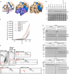
Fig. 3. Residues E218, H222, H231 and E234 in the D209-K247 hairpin control the low pH-triggered conformational changes required for AIP56 membrane interaction and translocation.
a Localization of the putative pH-sensing residues selected for replacement on the three-dimensional structure of the AIP56 catalytic domain. Cartoon (left) and surface representation of AIP56 catalytic domain with (middle) or without (right) the middle domain. The catalytic residues (H165, E166 and H169) are shown in red, the putative pH-sensing residues in orange and the D209-K247 hairpin in marine blue. The cysteine residues (pink sticks) forming the disulfide bridge (yellow) are also shown. b Analysis of NF-kB p65 cleavage in mouse bone marrow-derived macrophages (mBMDM) by V5 plus His-tagged AIP56 variants. Cleavage of p65 was assessed by western blotting (upper panel; chromogenic detection) and protein loading by staining the membranes with Ponceau S (lower panel). The result shown is representative of six (n = 6) independent experiments. c Peak ANS (8-Anilino-1-naphthalenesulfonic acid) fluorescence measured at 475 nm for the indicated pH, normalized by subtracting the corresponding values at pH 7 (fluorescence due to conformational changes caused by the mutation and not due to acidification). The measurement curves for each pH at different wavelengths are shown in Supplementary Fig. 7a. The results shown are representative of at least three (n = 3) independent experiments. AIP56, black; AIP56E214K, blue; AIP56E218K, purple; AIP56H222K, green; AIP56H231K, brown; AIP56E234K, pink; AIP56H231K/E234K, gray; AIP56E214K/E218K/H222K, orange; d Coomassie Blue-stained SDS-PAGE gels from limited proteolysis of AIP56 and AIP56H231K/E234K by Proteinase K. A and B mark the bands corresponding to the catalytic and receptor-binding domains, respectively. The results shown are representative of two (n = 2) independent experiments. e Interaction of AIP56 or AIP56 variants with black lipid bilayers. Single-channel recordings of DiPhPC/n-decane membranes after addition of the indicated proteins to one side of the black lipid bilayer at a final concentration of 14 nM. Membrane activity was induced by acidification (pH 4.8; red arrows) of the aqueous phase at the cis-side of the chamber. Each result shown is representative of at least three (n = 3) independent measurements. Source data for (b), (c) and (d) are provided in the Source data file.
Fig. 4
Fig. 4. Pharmacological inhibition of Hsp90 impairs the cytosolic delivery of Bla by BlaL19-W286AIP56P210-N497 but not of BlaL19-W286AIP56L258-N497.
a Schematic representation of chimera BlaL19-W286AIP56P210-N497. Bla, β-lactamase. b FRET-based assay to access the effect of Hsp90 inhibition on Bla delivery. The cleaved/uncleaved CCF4-AM ratios were determined by quantifying the indicated number of microscopic fields per condition. Results shown represent one out of three (n = 3) independent experiments. Statistical significance was tested by Kruskal–Wallis nonparametric test and the adjusted p values for individual comparisons were obtained by Bonferroni correction. Data are presented as mean values ± SD. Actual p values from left-to-right and upwards: p < 0.0001, p < 0.0001, p = 0.0993, p = 0.8533, p < 0.0001, p = 0.0051; ns non-significant, CCF4 Fluorescence Resonance Energy Transfer (FRET) substrate, DMAG 17-(dimethylaminoethylamino)−17-demethoxygeldanamycin (Hsp90 inhibitor), Bla β-lactamase. c Control of 17-DMAG activity by confirming its inhibitory effect on NF-kB p65 (nuclear factor kappa-light-chain-enhancer of activated B cells subunit p65) cleavage upon AIP56 intoxication of mBMDM. A representative blot of three (n = 3) independent experiments is shown. Loading correction was achieved by dividing the density of p65 by the respective density of the Ponceau S staining. Data are presented as mean values ± SD. Different symbols represent independent experiments. DMAG 17-(dimethylaminoethylamino)−17-demethoxygeldanamycin (Hsp90 inhibitor). Source data for (b) and (c) are provided in the Source data file.
Fig. 5
Fig. 5. An aspartate-rich patch located in the linker is important for AIP56 translocation but not for pore formation.
a V5 plus His-tagged AIP56 modified in the aspartate-rich motif (AIP56D274S/D276-278S and AIP56D274N/D276-278N) is unable to cleave p65 in intact cells. Cleavage of p65 was assessed by western blotting and protein loading by Ponceau S staining. The result shown is representative of six (n = 6) independent experiments. b V5 plus His-tagged AIP56D274S/D276-278S and AIP56D274N/D276-278N were unable to translocate across the host cell membrane in response to acidification. In all experiments, mock-treated cells were used as controls. NF-kB p65 cleavage was analyzed by western blotting. The result shown is representative of five (n = 5) independent experiments. The plot shows the quantification of intact NF-kB p65 normalized for Ponceau S. Statistical significance was tested by one-way ANOVA and p values for the individual comparisons were calculated using Tukey’s HSD test. Data are presented as mean values ± SD. Actual p values from left-to-right and upwards for pH 7.0: p < 0.001, p = 0.91, p > 0.99, p = 0.005, p = 0.90, p = 0.81; for pH 4.5: p = 0.02, p > 0.99, p > 0.99, p > 0.99, p = 0.78, p = 0.36; ns non-significant. Open (without concanamycin A) or closed (with concanamycin A) symbols as well as color coding have been added to facilitate the reading of the experimental conditions, as specified below the graph. Samples were derived from the same experiment and the blots processed in parallel. NF-kB p65, nuclear factor kappa-light-chain-enhancer of activated B cells subunit p65; ConcA, concanamycin A (black); AIP56, purple, AIP56D274S/D276-278S, red; AIP56D274N/D276-278N, green. c AIP56D274S/D276-278S and AIP56D274N/D276-278N retained the ability to interact with black lipid bilayers. Proteins were used at a final concentration of 14 nM. Membrane activity was induced by acidification (pH 4.8; red arrows) of the aqueous phase at the cis-side of the chamber. Each result shown is representative of at least three (n = 3) independent measurements. Source data for (b) and (c) are provided in the Source data file.

Similar articles

Cited by

References

    1. Viana F, Peringathara SS, Rizvi A, Schroeder GN. Host manipulation by bacterial type III and type IV secretion system effector proteases. Cell. Microbiol. 2021;23:e13384. doi: 10.1111/cmi.13384. - DOI - PubMed
    1. Falnes PO, Sandvig K. Penetration of protein toxins into cells. Curr. Opin. Cell Biol. 2000;12:407–413. doi: 10.1016/S0955-0674(00)00109-5. - DOI - PubMed
    1. Geny B, Popoff MR. Bacterial protein toxins and lipids: pore formation or toxin entry into cells. Biol. Cell. 2006;98:667–678. doi: 10.1042/BC20050082. - DOI - PubMed
    1. Geny B, Popoff MR. Bacterial protein toxins and lipids: role in toxin targeting and activity. Biol. Cell. 2006;98:633–651. doi: 10.1042/BC20060038. - DOI - PubMed
    1. Sandvig K, Skotland T, van Deurs B, Klokk TI. Retrograde transport of protein toxins through the Golgi apparatus. Histochem. Cell Biol. 2013;140:317–326. doi: 10.1007/s00418-013-1111-z. - DOI - PubMed

Publication types