. 2023 Nov 21;120(47):e2314440120.
doi: 10.1073/pnas.2314440120. Epub 2023 Nov 15.

rDNA magnification is a unique feature of germline stem cells

Affiliations

rDNA magnification is a unique feature of germline stem cells

Jonathan O Nelson et al. Proc Natl Acad Sci U S A. .

Abstract

Ribosomal DNA (rDNA) encodes ribosomal RNA and exists as tandem repeats of hundreds of copies in the eukaryotic genome to meet the high demand of ribosome biogenesis. Tandemly repeated DNA elements are inherently unstable; thus, mechanisms must exist to maintain rDNA copy number (CN), in particular in the germline that continues through generations. A phenomenon called rDNA magnification was discovered over 50 y ago in Drosophila as a process that recovers the rDNA CN on chromosomes that harbor minimal CN. Our recent studies indicated that rDNA magnification is the mechanism to maintain rDNA CN under physiological conditions to counteract spontaneous CN loss that occurs during aging. Our previous studies that explored the mechanism of rDNA magnification implied that asymmetric division of germline stem cells (GSCs) may be particularly suited to achieve rDNA magnification. However, it remains elusive whether GSCs are the unique cell type that undergoes rDNA magnification or differentiating germ cells are also capable of magnification. In this study, we provide empirical evidence that suggests that rDNA magnification operates uniquely in GSCs, but not in differentiating germ cells. We further provide computer simulation that suggests that rDNA magnification is only achievable through asymmetric GSC divisions. We propose that despite known plasticity and transcriptomic similarity between GSCs and differentiating germ cells, GSCs' unique ability to divide asymmetrically serves a critical role of maintaining rDNA CN through generations, supporting germline immortality.

Keywords: Drosophila germline; germline immortality; rDNA copy number maintenance; ribosomal DNA.


Conflict of interest statement

The authors declare no competing interest.

Figures

Fig. 1.
Fig. 1.
Model of rDNA magnification. (A) The rDNA locus is inherently unstable, requiring a mechanism to recover the CN for germline immortality. (B) USCE as a model of rDNA CN expansion (rDNA magnification). (C) Reintegration of extrachromosomal rDNA circle as a model of rDNA magnification. (D) Nonrandom sister chromatid segregation following USCE increases rDNA CN in GSCs (a model from ref. 3). (E) Schematic of germ cell differentiation in the Drosophila testis. GSCs reside at the apical tip of the testis, where they attach to somatic hub cells, which functions as the major component of the stem cell niche. GSCs divide asymmetrically to produce gonialblasts (GBs), which undergo mitotic proliferation to produce a cluster of interconnected spermatogonia (SGs). Upon 4 mitotic divisions, SGs enter meiotic prophase as spermatocytes.
Fig. 2.
Fig. 2.
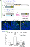
R2-dependent double-strand DNA breaks under low rDNA CN occur primarily in GSCs. (A) rDNA magnification occurs only when animals have insufficient rDNA CN (bbz9/Ybb0; magnifying condition), whereas animals with sufficient rDNA CN (bbz9/Y+ or X+/Y+, nonmagnifying conditions). (BD) Representative images of the apical tip of the testes from control (bbz9/Y+) (B), bbz9/Ybb0 (C), and bbz9/Ybb0 nos-gal4 > R2 RNAi (D) testes, stained for γH2Av (marker for DSBs, green) and Vasa (germ cells, blue). Asterisks (*) mark the hub. The dotted line indicates GSCs. White arrowheads indicate DSB-positive GSCs, and yellow arrows indicate DSB-positive SGs. (Bar: 10 µm.) (E) The frequency of γH2Av-positive GSCs vs. SGs in indicated genotypes. DSBs are most prominently elevated in GSCs under magnifying conditions (bbz9/Ybb0), which is repressed by RNAi-mediated knockdown of R2. P values displayed compared to the control condition in the same cell type using chi-squared test. Error bars = 95% CI.
Fig. 3.
Fig. 3.
Induction of R2 expression under magnifying condition is enriched in GSCs. (A and B) Representative images of the testis apical tip from animals under nonmagnifying (A, bbz9/Y+) vs. magnifying (B, bbz9/Ybb0) conditions. DAPI: blue, R2 RNA: green. (A’ and B’) R2 RNA in situ hybridization only channel in grayscale. Asterisks (*) indicate the hub. Broken blue circles indicate GSCs negative for R2 expression, broken yellow circles indicate GSCs positive for R2 expression (also indicated by arrowheads), and the white solid circle indicates SGs positive for R2 expression. (C) The frequency of R2-positive GSCs (white bars) and SGs (gray bars) under nonmagnifying (bbz9/Y+) vs. magnifying (bbz9/Ybb0) conditions. P values indicate comparison between genotypes among the same cell types using chi-squared test. Error bars = 95% CI.
Fig. 4.
Fig. 4.
Simulation of rDNA CN changes based on the modes of rDNA CN inheritance. (A) Schematic model of rDNA CN changes over successive GSC divisions when GBs consistently inherit increased rDNA CN (Top), or GSCs inherit increased rDNA CN (Bottom). Note that differentiating cells (GBs) will receive successively decreasing rDNA CN when they inherit higher rDNA CNs than GSCs at each division. (BE) Computer simulation of rDNA CN in sperm after 2 wk (or 28 GSC divisions). The initial rDNA CN was assumed to be 200 for nonmagnifying condition (B) and 100 for magnifying condition (CE). The starting CN is shown in blue (B) or red (CE). Other simulation parameters are also shown at the Top Left of each graph: the frequency of USCE in GSC and SG and the frequency of GSCs inheriting increased rDNA CN after UCSE (50%, 80%, and 20%). See the main text and Methods for details of parameter setting (some parameters are empirically determined based on previous studies).
Fig. 5.
Fig. 5.
Ectopic expression of R2 in differentiating germ cells (>4-cell SG) reduces the number of differentiating germ cells. (A and B) Example of γH2Av-positive GSC (A) and SG (B) stained for γH2Av (green), Vasa (germ cells, magenta), and Adducin-like (blue). (Bars: 10 µm.) The asterisk (*) marks the hub (A), and the dotted line indicates γH2Av-positive GSC (A) or γH2Av-positive SG (B). (C) The frequency of γH2Av-positive cells/testis for the indicated stage of germ cells (GSCs/GBs and 2-cell SGs (bam-negative germ cells)/4- to 16-cell SGs (bam-positive germ cells) upon ectopic expression of R2 by bam-gal4. This result demonstrates that bam-positive (i.e., bam-gal4-expressing) germ cells specifically induce DSBs monitored by γH2Av. P values indicate a comparison between genotypes among the same cell types using Student’s t test. Error bars = 95% CI. n = numbers of testes scored from three biological replicates. (DF) Representative images of the testis apical tip from control (D), bam>R2 (E), bam>R2ND (nuclease-dead) at the age of 2 wk. The boundary between the SG stage and the SC stage is indicated by the dotted line. (Bar: 50 µm.) The yellow double-headed arrow in E indicates fully differentiated sperm (which moved toward the apical side due to loss of earlier germ cells, such as SGs and SCs). (G) Number of SG cysts and SC cysts per testis upon expression of R2 or nuclease-dead R2 (R2ND). P values displayed from Tukey’s HSD multiple comparison test after one-way ANOVA. ANOVA P values are > 0.00001 for both SG and SC analyses. Error bars = 95% CI. Data are from three biological replicates.

Similar articles

Cited by

References

    1. Palazzo A. F., Lee E. S., Non-coding RNA: What is functional and what is junk? Front. Genet. 6, 2 (2015). - PMC - PubMed
    1. Kobayashi T., Strategies to maintain the stability of the ribosomal RNA gene repeats–collaboration of recombination, cohesion, and condensation. Genes. Genet. Syst. 81, 155–161 (2006). - PubMed
    1. Watase G. J., Nelson J. O., Yamashita Y. M., Nonrandom sister chromatid segregation mediates rDNA copy number maintenance in Drosophila. Sci. Adv. 8, eabo4443 (2022). - PMC - PubMed
    1. Ritossa F. M., Atwood K. C., Spiegelman S., A molecular explanation of the bobbed mutants of Drosophila as partial deficiencies of “ribosomal” DNA. Genetics 54, 819–834 (1966). - PMC - PubMed
    1. Henderson A. S., Warburton D., Atwood K. C., Letter: Ribosomal DNA connectives between human acrocentric chromosomes. Nature 245, 95–97 (1973). - PubMed

LinkOut - more resources