[Preprint]. 2023 Oct 31:2023.10.31.564884.
doi: 10.1101/2023.10.31.564884.

Legionella pneumophila exploits the endo-lysosomal network for phagosome biogenesis by co-opting SUMOylated Rab7

Affiliations

Legionella pneumophila exploits the endo-lysosomal network for phagosome biogenesis by co-opting SUMOylated Rab7

Chuang Li et al. bioRxiv. .

Update in

Abstract

L. pneumophila strains harboring wild-type rpsL such as Lp02rpsLWT cannot replicate in mouse bone marrow-derived macrophages (BMDMs) due to induction of extensive lysosome damage and apoptosis. The mechanism of this unique infection-induced cell death remains unknown. Using a genome-wide CRISPR/Cas9 screening, we identified Hmg20a and Nol9 as host factors important for restricting strain Lp02rpsLWT in BMDMs. Depletion of Hmg20a protects macrophages from infection-induced lysosomal damage and apoptosis, allowing productive bacterial replication. The restriction imposed by Hmg20a was mediated by repressing the expression of several endo-lysosomal proteins, including the small GTPase Rab7. We found that SUMOylated Rab7 is recruited to the bacterial phagosome via SulF, a Dot/Icm effector that harbors a SUMO-interacting motif (SIM). Moreover, overexpression of Rab7 rescues intracellular growth of strain Lp02rpsLWT in BMDMs. Our results establish that L. pneumophila exploits the lysosomal network for the biogenesis of its phagosome in BMDMs.

Keywords: CRISPR/Cas9; Caspases; Hmg20a; Lysosomes; Rab GTPase; SUMOylation; effectors; macrophages.


Conflict of interest statement

DECLARATION OF INTERESTS The authors declare no competing interests.

Figures

Figure 1
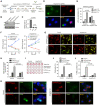
Figure 1. Identification of host factors involved in restricting intracellular growth of strain Lp02rpsLWT in BMDMs
(A) Intracellular growth of relevant L. penumophila strains in BMDMs from Cas9+/+ A/J mice. The indicated L. penumophila strains were used to infect BMDMs at an MOI of 0.05 and bacterial growth was determined by plating lysates of infected cells at the indicated time points. (B-C) The formation and size distribution of bacterial phagosome. BMDMs from Cas9+/+ A/J mice were infected with relevant L. pneumophila strains for 14 h and samples were processed for microscopic analysis for acquiring representative images of large LCVs (B) or their size distribution (C). Bar, 5 μm. LCVs containing more than 10 bacteria were defined as large; those containing 4 to 9 bacteria were classified as medium and those containing 1 to 3 bacteria were categorized as small vacuoles. (D) Schematic workflow of the genome-scale CRISPR/Cas9 screening. BMDMs from Cas9+/+ A/J mice were transduced with lentiviral particles carrying the CRISPR knockout sgRNA library for 6 d, cells were then infected with strain Lp02rpsLWT(pGFP) for 14 h. Fixed samples were used to isolate cells harboring large LCVs by flow cytometry and DNA was extracted and processed for analysis by next generation sequencing (NGS). (E) Gene enriched in BMDMs containing large LCVs vs those containing small LCVs shown by Dot plot. The significance of distribution of genes was determined based on the statistical significance (y axis) versus genomic position (x axis). The top 10 genes were highlighted. (F-H) Knockout of Hmg20 or Nol9 allowed strain Lp02rpsLWT to replicate in BMDMs. BMDMs were transduced by lentivirus particles carrying sgRNAs against Hmg20 or Nol9 and cells were infected with L. pneumophila for 14 h to assess intracellular replication by determining the distribution of bacterial phagosome in size (F and G) and total bacterial counts over 72 h at 24h intervals (H). In each case, for quantitative experiments, results (mean ± s.e.) were from three independent experiments each done in triplicate. Images shown were representatives from one experiment.
Figure 2
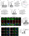
Figure 2. Deletion of Hmg20a allows strain Lp02rpsLWT to grow in mouse BMDMs
(A) Schematic of CRISPR/Cas9-mediated Hmg20a knockout in BMDMs and infection with strain Lp02rpsLWT(pGFP). BMDMs from Hmg20+/− Cas9+/+ A/J mice were transduced with lentiviruses carrying sgRNAs targeting Hmg20a for 3 d, non-transduced cells were eliminated by incubation in medium containing puromycin for 2 d. BMDMs seeded in medium without puromycin for an additional 3 d were infected with the bacteria for 14 h. (B-C) Hmg20a knockout efficiency in BMDMs. Lysates of cells treated as described in A were subjected to immunoblotting using anti-Hmg20a antibodies (B) and the rates of reduction were evaluated by measuring band density relative to bands of the loading control tubulin from three independent experiments (C). (D-F) Growth of strain Lp02rpsLWT in Hmg20a-deleted BMDMs. BMDMs treated as described in A were infected with strain Lp02rpsLWT at an MOI of 1 for 14 h and samples were processed for image acquisition (D), determining the distribution of the phagosome in size (E). Bar, 10 μm. Similar experiments with an MOI of 0.05 were used to analyze intracellular replication by determining total bacterial counts at the indicated time points (F). (G-L) Hmg20a knockout protected BMDMs from infection-induced lysosome damage and cell death. BMDMs received sgRNAs targeting Hmg20a or nontarget were infected with strain Lp02rpsLWT or Lp02rpsLK88R, samples loaded with AO were processed for image acquisition (G), for determining the ratios of cells with damaged lysosomes by AO staining (H) Cell death was measured by the release of lactate dehydrogenase (LDH) (I and J) and by TUNEL staining (K and L). In each case, quantitative results (mean ± s.e.) were from three independent experiments each done in triplicate. Images are representatives of one of the three experiments with similar results. Bar, 10 μm.
Figure 3
Figure 3. Hmg20a impacts the abundancy of proteins involved in lysosome function
(A) The protein abundancy of Hmg20a+/− and Hmg20a−/− BMDMs was analyzed by mass spectrometry and the comparative proteomic results were shown by a dot plot graph. Several representative proteins with marked changes were labeled in the graph. (B) Fold change of representative lysosome-related proteins in Hmg20a−/− cells relative to Hmg20+/− cells. (C-D) Evaluation of the change in protein abundancy by Immunoblotting (C), the changes were quantitated by measuring band intensity with tubulin as the reference (D). Results shown (mean ± s.e.) were from three independent experiments. (E) Comparison of mRNA levels of several Rab proteins between Hmg20a+/− and Hmg20a−/− BMDMs. Total mRNA was extracted and analyzed via RT-qPCR using primers specific for Hmg20a, Rab1, Rab7, Rab10, Rab12, Rab18, Rab33, LAMP1 or LAMP2. GAPDH was used as the control gene.
Figure 4
Figure 4. Overexpression of Rab7 permits productive intracellular replication of strain Lp02rpsLWT in BMDMs
(A) Knockout of Hmg20a but not several Rab genes allowed strain Lp02rpsLWT to grow in BMDMs. SgRNAs targeting Rab1, Rab7, Rab10, Rab12, Rab18, Rab33 or Hmg20a were introduced into BMDMs and the cells were infected with L. pneumophila at an MOI of 1 for 14 h prior to being processed for analysis of the formation of large LCVs. (B-E) Overexpression of Rab7 promotes growth of strain Lp02rpsLWT in BMDMs. Cells transduced to express Flag-Rab7 were used to determine bacterial growth by the formation and distribution of LCVs in size (B) and total bacterial counts in a 72-h experimental duration (D). The abundancy of ectopically expressed Flag-Rab7 was determined by immunoblotting with the Flag antibody (C and E). Note that Lp02rpsLWT infection started at 24 h post lentiviral transduction. (F-G) Only active Rab7 suppressed the restriction of the growth of strain Lp02rpsLWT by BMDMs. Cells transduced to express Flag-Rab7 or its mutants were infected with strain Lp02rpsLWT at an MOI of 1 for 14 h, samples were processed for analyzing the formation and distribution of LCVs (F). The expression of Rab7 and its mutant was detected by immunoblotting with the Flag antibody (G). (H-I) The association of Rab7 and its mutants with LCVs formed by Lp02rpsLWT in BMDMs. Cells transfected to express Flag-Rab7 or its mutants were infected with the common laboratory L. pneumophila strain Lp02rpsLK88R or strain Lp02rpsLWT for 14 h prior to immunostaining with antibodies specific for Rab7. Images were acquired using an Olympus X-81 microscope (H) and the distribution of LCVs was determined by counting at least 300 infected cells (I). (J-K) Rab7 recruitment by strains Lp02rpsLK88R and Lp02rpsLWT. Hmg20a+/− or Hmg20a−/− BMDMs were infected with the bacteria for 2 h and the recruitment of Rab7 to LCVs was analyzed by immunostaining (J) and the ratios of such association were determined (K). In all cases, quantitative results (mean ± s.e.) were from three independent experiments each done in triplicate. Images shown were a representative from three independent experiments with similar results. Bar, 10 μm.
Figure 5
Figure 5. SulF recruits SUMOylated Rab7 to LCVs through SUMO-SIM interaction
(A-C) SUMOylation is required for Rab7 to be recruited to the LCV Sketch of Rab7 with known post-translational modifications, including K38, S72, C83/84 and K175 that are modified by ubiquitination, phosphorylation, palmitoylation and SUMOylation, respectively (A) Mutations in the SUMOylation site of Rab7 abolished its recruitment to the LCV. BMDMs ectopically expressing Rab7 or its mutants were infected with strain Lp02rpsLWT, typical images of the association (B) and the ratios of Rab7-positive LCVs (C) were determined. (D-F) SUMOylation of Rab7 is required for its ability to support intracellular growth of strain Lp02rpsLWT in BMDMs. BMDMs were transduced to express Rab7 or its mutants and cells were infected with strain Lp02rpsLWT. Bacterial growth was determined by the formation of large LCVs (D) and total bacterial counts (E). The expression of transduced Rab7 or its alleles was probed by immunoblotting (F). (G-I) Identification of SulF as a SUMO-binding L. pneumophila effector. Comparison of the sequences of the predicted SUMO-binding motifs (SIM) of Dot/Icm substrates with SIM elements in established eukaryotic SUMO-binding proteins (G). The SIM motif is critical for the binding of SulF to SUMO. Recombinant GST-SulF or its SIM mutant SulFIV/AA and His6-SUMO was mixed for 2 h and the protein complexes were captured by GST or Ni2+ beads. After 3x washes, proteins associated with the beads resolved by SDS-PAGE were detected by CBB staining. Samples withdrawn prior to adding agarose beads were loaded as input controls (H). HEK293T cells co-transfected to express HA-SUMO2 and Flag-Rab7 or its mutants for 36 h were lysed and SUMOylated Rab7 or mutants were purified. SUMOylated Rab7 and unmodified Rab7 were used to determine their binding to SulF or SulFIV/AA by affinity pulldown. Tubulin was probed as a loading control (I). (J-L) Recruitment of Rab7 to LCVs by L. pneumophila required SulF with an intact SIM. BMDMs transfected to express Rab7 were infected with the indicated L. pneumophila strains for 2 h and the association of Rab7 with the LCVs was determined by acquiring images (J), quantitation (K) and the formation and ratio of large LCVs containing more than 10 bacteria (L) . (M-N) SulF is required for intracellular growth of strain Lp02rpsLWT in BMDMs overexpressing Rab7. BMDMs transduced to express Rab7 were infected with the indicated L. pneumophila strains, bacterial growth was analyzed by determining the total counts of bacteria at the indicated time points (M). The expression of SulF and its mutant in the complementation strains was detected by immunoblotting with the Flag-specific antibody (N). The metabolic enzyme ICDH was probed as a loading control.
Figure 6
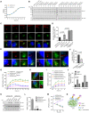
Figure 6. Overexpression of SulF increased Rab7 recruitment by strain Lp02rpsLK88R
(A-B) The abundancy of SulF in strain Lp02rpsLWT is higher than that in strain Lp02rpsLK88R in bacteria grown in bacteriological medium. Fresh cultures with OD600=0.3 established from saturated cultures of the indicated L. pneumophila strains were grown in a shaker and bacterial growth was monitored by measuring OD600 at 3-h intervals (left panel). Samples withdrawn at the indicated time points were resolved by SDS-PAGE and SulF was detected by immunoblotting. ICDH was probed as a loading control (middle and right panels). (C-D) The association of SulF with LCVs formed by relevant L. penumophila strains. BMDMs infected with the indicated L. penumophila strains were processed for immunostaining with SulF-specific antibodies, images were acquired using an Olympus IX-81 microscope (C). The density of SulF staining signals was measured from at least 300 phagosomes using Image J (D). Bar, 5 μm. (E-F) The recruitment of Rab7 by L. pneumophila requires an intact SIM motif. Strain Lp02rpsLWTΔsulF was complemented with sulF or its SIM-defective mutant. BMDMs infected with the indicated bacterial strains for 2 h were processed for immunostaining and imaging (E). Quantitation shown (mean ± s.e.) was from three independent experiments each done in triplicate (F). Bar, 5 μm. (G) The kinetics of Rab7 recruitment by relevant L. penumophila strains. BMDMs infected with the indicated bacterial stains were sampled at the indicated time points, cells were processed for immunostaining and analyzed using a fluorescence microscope. (H-K) Overexpression of SulF in strain Lp02rpsLK88R increased Rab7 recruitment. BMDMs infected with the indicated L. penumophila strains were processed for immunostaining. Representative images used for determining the ratios of Rab7-positive LCVs (H) and for measuring signaling intensity (J). The expression of the endogenous or plasmid-borne SulF was probed by immunoblotting with antibodies specific for Flag or SulF (K). (L-M) Overexpression of SulF does not endow strain Lp02rpsLWT the ability to grow in BMDMs. The indicated bacterial strains were used to infect BMDMs, the distribution of LCVs (L) and total bacterial counts for a 72-h experimental duration at 24-h intervals (M) were analyzed. In all cases, quantitative results (mean ± s.e.) were from three independent experiments each. (N) A model for SulF-mediated recruitment of SUMOylated Rab7 by LCVs.
Figure 7
Figure 7. SulF inhibits the GTPase activity of Rab7 by blocking the binding of a GAP protein
(A) Purification of SUMOylated Rab7. Flag-Rab7 and His6-SUMO2 were co-expressed in HEK293T cells, SUMO-Rab7 was purified by a two-step procedure using antibody specific for Flag and Ni2+ beads. Purified proteins separated by SDS-PAGE were detected by CBB staining. (B-C) SulF inhibits TBC1D15-induced GTPase activity of Rab7 in an SIM-dependent manner. SUMOylated Rab7 were co-incubated with TBC1D15 and SulF or its mutant for 15 min. Then the reactions were stopped and released Pi was measured. The image of a typical plate for the assay (B) and quantitative results (mean ± s.e.) from three independent experiments (C). (D) The impact of SulF on the binding of TBC1D15 to Rab7 requires SUMOylation. Lysates of cells transfected to express HA-SUMO2 and Rab7 variants were subjected to immunoprecipitation with Flag beads, and agarose beads coated with the indicated Rab7 or its mutants were used to evaluate their binding to TBC1D15 in reactions with or without SulF. Proteins retained by Flag beads were analyzed by immunoblotting with appropriate antibodies. (E-F) SulF interferes with the interactions between TBC1D15 and SUMOylated Rab7. His6-TBC1D15 was added to Flag beads coated with Flag-SUMO-Rab7 prepared as described in A. After 1 h incubation, GST-SulF or GST was added. His6-TBC1D15 retained by the beads was analyzed by immunoblotting (E). Reciprocal experiments were done in which SulF or its mutant was added prior to incubation with His6-TBC1D15 (F). (G) A model for the mechanism of action of SulF. SulF is anchored on the cytoplasmic surface of the LCV where it recruits SUMO-Rab7. The binding of SulF to the small GTPase prevents it from being accessed by the GAP protein TBC1D15.

Similar articles

References

    1. Jorgensen I, Rayamajhi M, Miao EA. Programmed cell death as a defence against infection. Nat Rev Immunol. 2017;17(3):151–164. doi:10.1038/nri.2016.147 - DOI - PMC - PubMed
    1. Kawai T, Akira S. The role of pattern-recognition receptors in innate immunity: update on Toll-like receptors. Nat Immunol. 2010;11(5):373–384. doi:10.1038/ni.1863 - DOI - PubMed
    1. Wu J, Chen ZJ. Innate immune sensing and signaling of cytosolic nucleic acids. Annu Rev Immunol. 2014;32:461–488. doi:10.1146/annurev-immunol-032713-120156 - DOI - PubMed
    1. Shi J, Gao W, Shao F. Pyroptosis: Gasdermin-Mediated Programmed Necrotic Cell Death. Trends Biochem Sci. 2017;42(4):245–254. doi:10.1016/j.tibs.2016.10.004 - DOI - PubMed
    1. Shi J, Zhao Y, Wang K, et al. Cleavage of GSDMD by inflammatory caspases determines pyroptotic cell death. Nature. 2015;526(7575):660–665. doi:10.1038/nature15514 - DOI - PubMed

Publication types