[Preprint]. 2023 Nov 4:2023.11.03.565564.
doi: 10.1101/2023.11.03.565564.

The membrane-associated ubiquitin ligase MARCHF8 stabilizes the human papillomavirus oncoprotein E7 by degrading CUL1 and UBE2L3 in head and neck cancer

Affiliations

The membrane-associated ubiquitin ligase MARCHF8 stabilizes the human papillomavirus oncoprotein E7 by degrading CUL1 and UBE2L3 in head and neck cancer

Mohamed I Khalil et al. bioRxiv. .

Update in

Abstract

The human papillomavirus (HPV) oncoprotein E7 is a relatively short-lived protein required for HPV-driven cancer development and maintenance. E7 is degraded through ubiquitination mediated by cullin 1 (CUL1) and the ubiquitin-conjugating enzyme E2 L3 (UBE2L3). However, E7 proteins are maintained at high levels in most HPV-positive cancer cells. A previous proteomics study has shown that UBE2L3 and CUL1 protein levels are increased by the knockdown of the E3 ubiquitin ligase membrane-associated ring-CH-type finger 8 (MARCHF8). We have recently demonstrated that HPV upregulates MARCHF8 expression in HPV-positive keratinocytes and head and neck cancer (HPV+ HNC) cells. Here, we report that MARCHF8 stabilizes the E7 protein by degrading the components of the SKP1-CUL1-F-box (SCF) ubiquitin ligase complex in HPV+ HNC cells. We found that MARCHF8 knockdown in HPV+ HNC cells drastically decreases the E7 protein level while increasing the CUL1 and UBE2L3 protein levels. We further revealed that the MARCHF8 protein binds to and ubiquitinates CUL1 and UBE2L3 proteins and that MARCHF8 knockdown enhances the ubiquitination of the E7 protein. Conversely, the overexpression of CUL1 and UBE2L3 in HPV+ HNC cells decreases E7 protein levels and suppresses tumor growth in vivo. Our findings suggest that HPV-induced MARCHF8 prevents the degradation of the E7 protein in HPV+ HNC cells by ubiquitinating and degrading CUL1 and UBE2L3 proteins.


Figures
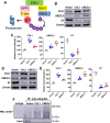
Fig. 1.
Fig. 1.. CUL1 and UBE2L3 overexpression increases the ubiquitination and decreases HPV16 E7 protein level in HPV+ HNC cells.
(A) The schematic model of E7 degradation through CUL1 and UBE2L3-mediated ubiquitination. CUL1 or UBE2L3 is overexpressed in HPV+ HNC (SCC152) (B-C) and SCC2 (D-E) cells using lentiviral transduction. CUL1, UBE2L3, HPV16 E7, and MARCHF8 proteins were detected by western blotting. The relative band intensities were quantified using NIH ImageJ in SCC152 (C) and SCC2 (E). β-actin was used as an internal control. (F) SCC152 cells with CUL1 or UBE2L3 overexpression were treated with MG132 (10 μM). Ubiquitinated proteins were pulled down from the cell lysate using anti-ubiquitin antibody-conjugated magnetic beads, and E7 protein was detected by western blotting. The data shown are means ± SD of three independent experiments. Student’s t-test was used to determine p values. *p < 0.05, **p < 0.01, ***p < 0.001.
Fig. 2.
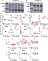
Fig. 2.. CUL1 and UBE2L3 protein levels are low in HPV+ HNC cells.
CUL1 and UBE2L3 protein levels in normal (N/Tert-1), HPV− HNC (SCC1, SCC9, and SCC19), and HPV+ HNC (SCC2, SCC90, and SCC152) cells were determined by western blotting (A). Relative band intensities were quantified using NIH ImageJ (B). HPV16 E7 and β-actin were used as viral and internal controls, respectively. CUL1 and UBE2L3 mRNA expression levels in normal (N/Tert-1), HPV+ HNC (SCC-2, SCC-90, and SCC-152), and HPV− HNC (SCC-1, SCC-9, and SCC-19) cells were quantified by RT-qPCR (C). The data shown are normalized by the GAPDH mRNA level as an internal control. CUL1, UBE2L3, and E7 protein levels in SCC152 cells treated with MG132 (10 μM) or DMSO (D). Relative band intensities were quantified using NIH ImageJ (E). CUL1 and UBE2L3 proteins were detected in N/Tert-1 cells expressing HPV16 E6, E7, or E6 and E7 by western blotting (F). An HA tag increases the size of HPV16 E7 in N/Tert-1 E7 cells to ~22 kDa from ~17 kDa. Relative band intensities were quantified using NIH ImageJ (G). Total RNA was extracted from N/Tert-1 containing an empty vector and N/Tert-1 cells expressing HPV16 E6, E7, or E6 and E7 (E6E7). The HPV16 E6 and E7 mRNA expression levels were quantified by RT-qPCR (H). The data shown are normalized by the GAPDH mRNA level as an internal control. All experiments were repeated at least three times, and the data shown are means ± SD of three independent experiments. Student’s t-test determined p values. *p < 0.05, **p < 0.01, ***p < 0.001.
Fig. 3.
Fig. 3.. Knockdown of MARCHF8 expression increases CUL1 and UBE2L3 protein levels and decreases HPV16 E7 protein levels in HPV+ HNC cells.
SCC152 (A & C) and SCC2 (B & D) cells were transduced with five and three lentiviral shRNAs against MARCHF8 (shR-MARCHF8), respectively, or scrambled shRNA (shR-scr) as a control. MARCHF8, CUL1, UBE2L3, HPV16 E7, and pRb proteins in SCC152 (A) and SCC2 (B) were detected by western blotting. The relative band intensities were quantified using NIH ImageJ (C and D). β-actin was used as an internal control. The mRNA levels of CUL1, UBE2L3, and E7 were quantified by RT-qPCR in SCC152 (E) and SCC2 (F) cells transduced with five and three lentiviral shRNAs against MARCHF8 (shR-MARCHF8), respectively, or scrambled shRNA (shR-scr) as a control. The data shown are normalized by the GAPDH mRNA level as an internal control. The data shown are means ± SD of three independent experiments. Student’s t-test determined p values. *p < 0.05, **p < 0.01, ***p < 0.001.
Fig. 4.
Fig. 4.. MARCHF8 protein interacts with and ubiquitinates CUL1 and UBE2L3 proteins.
MARCHF8 (A), CUL1 (B), and UBE2L3 (C) were pulled down from the lysate of SCC-152 cells treated with MG132 (10 μM) using anti-MARCHF8 (A), anti-CUL1 (B), and anti-UBE2L3 (C) antibodies, respectively. CUL1, UBE2L3, E7, and MARCHF8 were detected from the immunoprecipitated proteins by western blotting. (D-F) Ubiquitinated proteins were pulled down from the lysate of SCC152 cells with scrambled shRNA (shR-scr) or shRNA against MARCHF8 (shR-MARCHF8 clone 3) treated with MG132 (10 μM) using anti-ubiquitin antibody-conjugated magnetic beads. CUL1 (D), UBE2L3 (E), and HPV16 E7 (F) proteins were detected in the immunoprecipitated proteins by western blotting. All experiments were repeated at least three times. The data shown are means ± SD of three independent experiments. Student’s t-test determined p values. *p < 0.05, **p < 0.01, ***p < 0.001.
Fig. 5.
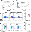
Fig. 5.. Knockdown of MARCHF8 expression suppresses proliferation of HPV+ HNC cells.
Cell proliferation rate was determined by cell counting using two HPV+ HNC cell lines, SCC152 (A) and SCC2 (B), with scrambled shRNA (shR-scr) or three shRNAs against MARCHF8 (shR-MARCHF8). 1 × 10⁵ cells per well were seeded in a 6-well plate. The cells were detached and counted at 24, 48, and 72 hours. BrdU incorporation assays were performed with SCC152 (shR-MARCHF8 clones 1 and 5) (C) and SCC2 (shR-MARCHF8 clones 4 and 5) (D) cells with MARCHF8 knockdown. 1 × 10⁶ cells were seeded in 10 cm Petri dishes and incubated for 16 hours with BrdU (10 mM). BrdU incorporation was analyzed by flow cytometry. Cell viability was determined by Zombie NIR staining as a control. All experiments were repeated at least three times, and the data shown are means ± SD. Student’s t-test determined p values. ***p < 0.001.
Fig. 6.
Fig. 6.. Marchf8 knockout restores CUL1 and UBE2L3 protein levels in HPV+ mouse oral cancer cells.
CUL1 and UBE2L3 protein levels in mouse normal immortalized (NiMOE), HPV− transformed (HPV− MOE), and HPV+ transformed (mEERL) oral epithelial cells were determined by western blotting (A). β-actin was used as an internal control. The relative band intensities were quantified using NIH ImageJ (B). mEERL cells were transduced with lentiviruses containing Cas9 and one of two sgRNAs against Marchf8 (sgR-Marchf8–2 and sgR-Marchf8–3) or scrambled sgRNA (sgR-scr). MARCHF8, CUL1, UBE2L3, and HPV16 E7 proteins were detected by western blotting (C). Relative band intensities were quantified using NIH ImageJ (D). The data shown are means ± SD of three independent experiments. All experiments were repeated at least three times, and the data shown are means ± SD. Student’s t-test determined p values. *p < 0.05, ***p < 0.001.
Fig. 7.
Fig. 7.. CUL1 and UBE2L3 overexpression suppresses HPV+ HNC tumor growth in vivo.
mEERL cells overexpressing Cul1 or Ube2l3 were generated by lentiviral transduction of the Cul1 or Ube2l3 genes, respectively, and blasticidin selection. CUL1, UBE2L3, and HPV16 E7 proteins were detected by western blotting (A). Relative band intensities were quantified using NIH ImageJ (B). β-actin was used as an internal control. The data shown are means ± SD of three independent experiments. Student’s t-test determined p values. *p < 0.05, **p < 0.01, ***p < 0.001. mEERL/vector (C), mEERL/Cul1 (D), or mEERL/Ube2l3 (E) cells were injected into the rear right flank of C57BL/6J mice (n = 10 per group). Tumor volume was measured twice a week (C-F). Survival rates of mice were analyzed using a Kaplan-Meier estimator (G). The time to event was determined for each group, with the event defined as a tumor size larger than 2000 mm3. The data shown are means ± SD. P values of mice injected with mEERL/Cul1 and mEERL/Ube2l3 cells compared with mice injected with mEERL/vector cells were determined for tumor growth (F) and survival (G) by two-way ANOVA analysis. Shown are representative of two independent experiments.
Fig. 8.
Fig. 8.. The schematic models of MARCHF8-mediated E7 stabilization by degrading CUL1 and UBE2L3.
The HPV oncoprotein E6 activates the MARCHF8 promoter activity through the MYC/MAX transcription factor complex and upregulates MARCHF8 in the HPV+ HNC cells (31). MARCHF8 protein ubiquitinates and degrades CUL1 and UBE2L3 proteins, leading to the prevention of E7 protein degradation.

Similar articles

References

    1. Burd EM. 2003. Human papillomavirus and cervical cancer. Clin Microbiol Rev 16:1–17. - PMC - PubMed
    1. Pyeon D, Newton MA, Lambert PF, den Boon JA, Sengupta S, Marsit CJ, Woodworth CD, Connor JP, Haugen TH, Smith EM, Kelsey KT, Turek LP, Ahlquist P. 2007. Fundamental differences in cell cycle deregulation in human papillomavirus-positive and human papillomavirus-negative head/neck and cervical cancers. Cancer Res 67:4605–19. - PMC - PubMed
    1. Griffin LM, Cicchini L, Pyeon D. 2013. Human papillomavirus infection is inhibited by host autophagy in primary human keratinocytes. Virology 437:12–9. - PMC - PubMed
    1. Mesri EA, Feitelson MA, Munger K. 2014. Human viral oncogenesis: a cancer hallmarks analysis. Cell Host Microbe 15:266–82. - PMC - PubMed
    1. Jabbar SF, Abrams L, Glick A, Lambert PF. 2009. Persistence of high-grade cervical dysplasia and cervical cancer requires the continuous expression of the human papillomavirus type 16 E7 oncogene. Cancer Res 69:4407–14. - PMC - PubMed

Publication types