Review
. 2023 Oct 27;23(21):8764.
doi: 10.3390/s23218764.

The Application of Graphene Field-Effect Transistor Biosensors in COVID-19 Detection Technology: A Review

Affiliations
Review

The Application of Graphene Field-Effect Transistor Biosensors in COVID-19 Detection Technology: A Review

Qin-Hong Liang et al. Sensors (Basel). .

Abstract

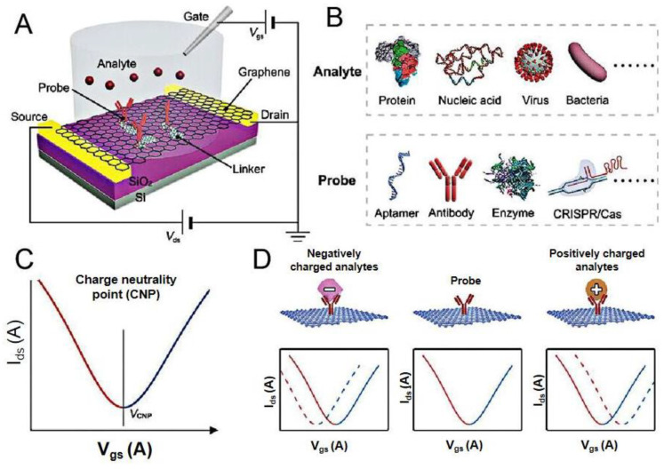
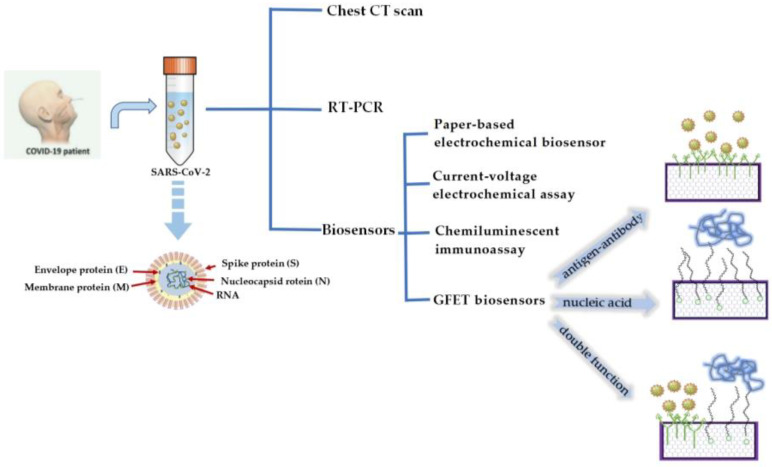
Coronavirus disease 2019 (COVID-19) is a disease caused by the infectious agent of severe acute respiratory syndrome coronavirus type 2 (SARS-CoV-2). The primary method of diagnosing SARS-CoV-2 is nucleic acid detection, but this method requires specialized equipment and is time consuming. Therefore, a sensitive, simple, rapid, and low-cost diagnostic test is needed. Graphene field-effect transistor (GFET) biosensors have become the most promising diagnostic technology for detecting SARS-CoV-2 due to their advantages of high sensitivity, fast-detection speed, label-free operation, and low detection limit. This review mainly focus on three types of GFET biosensors to detect SARS-CoV-2. GFET biosensors can quickly identify SARS-CoV-2 within ultra-low detection limits. Finally, we will outline the pros and cons of the diagnostic approaches as well as future directions.

Keywords: COVID-19; GFET biosensor; biological detection; biological sensors.


Conflict of interest statement

The authors declare no conflict of interest.

Figures

Figure 2
Figure 2
Working principles of GFET biosensors. (A) Schematic illustration of a liquid-gate GFET sensor. (B) The analytes include proteins, nucleic acids, viruses, and bacteria. The probes include aptamers, antibodies, enzymes, CRISPR/Cas. (C) Typical ambipolar transfer characteristics of graphene. (D) Sensing principle on the graphene surface: the binding of negatively (positively) charged analytes induced negative (positive) shifts in VCNP. Reprinted from [39], with permission from John Wiley and Sons.
Figure 1
Figure 1
Several detection methods for COVID-19 and the schematic diagram of a GFET biosensor for detecting SARS-CoV-2.
Figure 3
Figure 3
Schematic diagram of the GFET biosensor for detecting SARS-CoV-2 spike antibodies. Reprinted from [51], with permission from the American Chemical Society.
Figure 4
Figure 4
The virus detection performance of the laser-induced GFET. (A) Transfer characteristics of the laser-induced GFET biosensor responding to the complementary 1 pg/mL spike protein in PBS solution and (B) responding to the noncomplementary 1 pg/mL nucleocapsid protein in PBS solution. (C) Transfer characteristics of the laser-induced GFET biosensor responding to 1 pg/mL of complementary spike protein in human serum and (D) responding to 1 pg/mL noncomplementary nucleocapsid protein in human serum.
Figure 5
Figure 5
The kinetic response of the GFET device functionalized with the SARS-CoV-2 spike antibody at various concentrations of (A) SARS-CoV-2 spike protein added, ranging from 1 fM to 1 μM in 50 mM phosphate buffer (PB) (pH 7.2) and (B) MERS-CoV protein of various concentrations added (1 fM to 1 μM) in PB. Reprinted from [21], with permission from the American Chemical Society.
Figure 6
Figure 6
Schematic diagram of a Y-dual probe GFET biosensor. The dotted box is the structural schematic diagram of Y-shaped DNA dual probes. Reprinted from [54], with permission from the American Chemical Society.
Figure 7
Figure 7
Schematic diagram of the triple-probe TDF dimer GFET sensor for SARS-CoV-2 RNA testing. The dotted box is the structural schematic diagram of TDF dimer. Reprinted from [55], with permission from the American Chemical Society.
Figure 8
Figure 8
SARS-CoV-2 RNA testing. (A) Transfer curve measurement of adding different concentrations of target RNA (IdsVg response curve). (B) Real-time |ΔIds/Ids0| response upon different concentrations of target RNA (red line, modified with triple-probe TDF dimer; gray line, without immobilized probes). (C) |ΔIds/Ids0| responses of single- and triple-probe TDF dimer GFET sensors to different concentrations of target RNA. Reprinted from [55], with permission from the American Chemical Society.
Figure 9
Figure 9
The SARS-CoV-2 spike protein concentrations dependent transfer curves of (A) GO/Gr FET biosensor and (B) Gr FET biosensor. (C) The SARS-CoV-2 spike protein concentrations dependent ΔVDirac shifts for both GO/Gr FET (red line) and Gr FET (green line) biosensors. Reprinted from [37], with permission from Elsevier.
Figure 10
Figure 10
Excellent analytical performance of the COVID-19 GFET nanosensor. (A) Transfer curves upon incubation with PBS and nonspecific sequences including 1 nM non-complementary, SARS-CoV RdRp, and one-base mismatched RNA. (B) Variation of VCNP at detection of blank and three nonspecific sequences. Reprinted from [60], with permission from Elsevier.
Figure 11
Figure 11
The portable integrated platform developed by the research and development of 10-in-1 COVID-19 antigen detection, processing diagrams and photos. The red dashed box indicates one packaged multiantibody FET sensor using a printed circuit board substrate. A polydimethylsiloxane well was stamped above the graphene channel to hold the analyte solution. Reprinted from [62], with permission from the American Chemical Society.
Figure 12
Figure 12
Change in the electrical drain current for different types of viruses.

Similar articles

References

    1. Huang C., Wang Y., Li X., Ren L., Zhao J., Hu Y., Zhang L., Fan G., Xu J., Gu X., et al. Clinical Features of Patients Infected with 2019 Novel Coronavirus in Wuhan, China. Lancet. 2020;395:497–506. doi: 10.1016/S0140-6736(20)30183-5. - DOI - PMC - PubMed
    1. Wu J.T., Leung K., Leung G.M. Nowcasting and Forecasting the Potential Domestic and International Spread of the 2019-nCoV Outbreak Originating in Wuhan, China: A Modelling Study. Lancet. 2020;395:689–697. doi: 10.1016/S0140-6736(20)30260-9. - DOI - PMC - PubMed
    1. Cui F., Zhou H.S. Diagnostic Methods and Potential Portable Biosensors for Coronavirus Disease 2019. Biosens. Bioelectron. 2020;165:112349. doi: 10.1016/j.bios.2020.112349. - DOI - PMC - PubMed
    1. Van Doremalen N., Bushmaker T., Morris D.H., Holbrook M.G., Gamble A., Williamson B.N., Tamin A., Harcourt J.L., Thornburg N.J., Gerber S.I., et al. Aerosol and Surface Stability of SARS-CoV-2 as Compared with SARS-CoV-1. N. Engl. J. Med. 2020;382:1564–1567. doi: 10.1056/NEJMc2004973. - DOI - PMC - PubMed
    1. Tang Y.-N., Jiang D., Wang X., Liu Y., Wei D. Recent Progress on Rapid Diagnosis of COVID-19 by Point-of-Care Testing Platforms. Chin. Chem. Lett. 2023:108688. doi: 10.1016/j.cclet.2023.108688. - DOI - PMC - PubMed