Review
. 2023 Nov 4;24(21):15975.
doi: 10.3390/ijms242115975.

Multi-Scale Imaging of the Dynamic Organization of Chromatin

Affiliations
Review

Multi-Scale Imaging of the Dynamic Organization of Chromatin

Fabiola García Fernández et al. Int J Mol Sci. .

Abstract

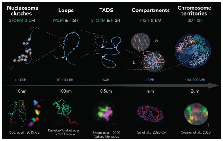
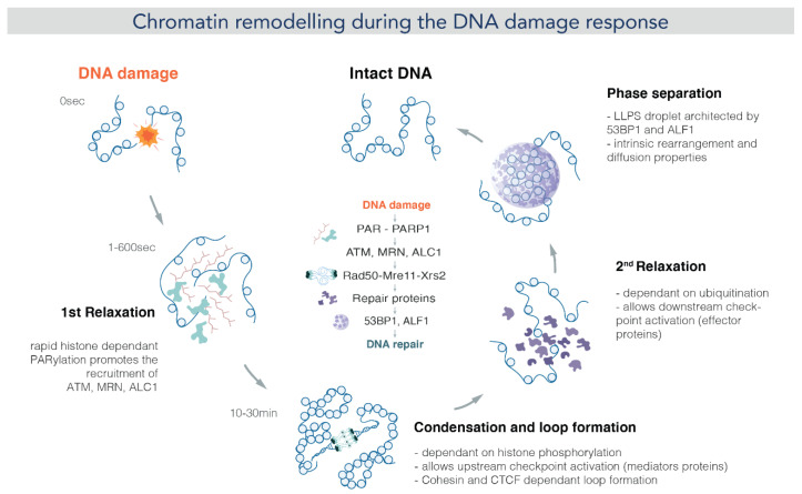
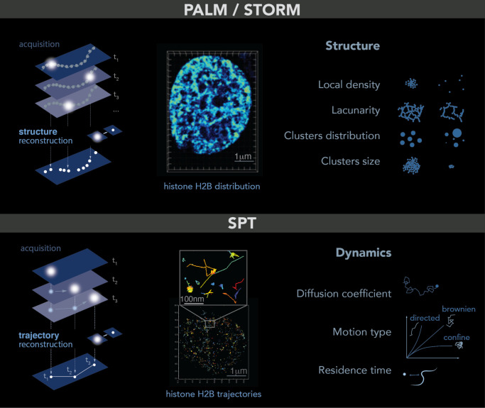
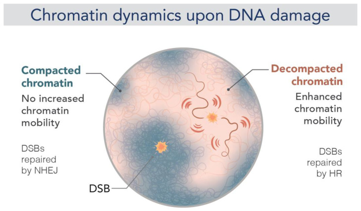
Chromatin is now regarded as a heterogeneous and dynamic structure occupying a non-random position within the cell nucleus, where it plays a key role in regulating various functions of the genome. This current view of chromatin has emerged thanks to high spatiotemporal resolution imaging, among other new technologies developed in the last decade. In addition to challenging early assumptions of chromatin being regular and static, high spatiotemporal resolution imaging made it possible to visualize and characterize different chromatin structures such as clutches, domains and compartments. More specifically, super-resolution microscopy facilitates the study of different cellular processes at a nucleosome scale, providing a multi-scale view of chromatin behavior within the nucleus in different environments. In this review, we describe recent imaging techniques to study the dynamic organization of chromatin at high spatiotemporal resolution. We also discuss recent findings, elucidated by these techniques, on the chromatin landscape during different cellular processes, with an emphasis on the DNA damage response.

Keywords: DNA repair; chromatin organization and dynamics; high resolution imaging.


Conflict of interest statement

The authors declare no conflict of interest.

Figures

Figure 2
Figure 2
Schematic representation of genome organization in mammals. From left to right: DNA wrapped around histones forms nucleosomes, which are organized into clutches. Each nucleosome clutch contains ~1–2 kb of DNA, as revealed by super-resolution image [32]. Genomic approaches and super-resolution imaging revealed the existence of chromatin loops, which are formed by loop extrusion and in a greater extent stabilized by CTCF and the cohesin ring [38]. At the scale of ~1 Mb, chromatin loops are the base of topologically associating domains (TADs), structures with delimited boundaries and high-rate interactions inside of these domains [45]. At a higher scale, up to several mega-bases, chromatin segregates into gene-active and gene-inactive compartments (A and B, respectively) [40]. Finally, as revealed by chromosome painting and fluorescence microscopy, individual chromosomes occupy specific regions, known as chromosome territories, that are several micrometers in length [82]. Top images are representations of each level of chromatin organization (art by Olga Markova), while the bottom images are real microscopy images taken from different studies (cited at the bottom of each image).
Figure 3
Figure 3
Chromatin remodeling during the DNA damage response: Upon DNA damage, PARP1 is activated and binds to the DNA damage site within seconds. Activated PARP1 catalyzes PAR chain formation and also PARylates the histone tails. This results in relaxation of the chromatin structure. Then, the PARylated PARP1 also activates and recruits other DDR factors at the DNA damage site. Among them, the ATM kinase is required to phosphorylate the H2AX histone. ATM signaling is performed through chromatin loops, which in turn triggers ƴ-H2AX spreading. The mega-base spreading of ƴ-H2AX transiently compacts chromatin around damage. This long period (10–30 min) of chromatin condensation allows the recruitment of upstream checkpoint factors. According to [124] Burgess et al., a second relaxation seems to be needed, since effector proteins cannot be recruited when chromatin is still condensed. Finally, certain repair proteins such as 53BP1 form condensates architecting chromatin in a way that repair is favored. (image by Olga Markova).
Figure 1
Figure 1
PALM and SPT: Principle, example and observables. Top: PALM/STORM principles. Left: a sparse subset of fluorescent probes is activated to produce single-particle images (represented by white circles) that do not overlap (left). After acquisition of images at a given time interval (t), a super-resolution image is reconstructed by plotting the measured positions of the fluorescent probes. Middle: example of histone H2B-mEOS distribution within a cell nucleus. (Right): further analysis of the final reconstructed images provides several parameters of the structure formed by the observed protein. Bottom: principle of SPT. (Left): during image acquisition, images are taken with a given exposure time (t) for the duration of several minutes. In each image, only a sparse number of emitters (white dots) are detected. Middle: using tracking and localization methods, it is possible to reconstruct the super-resolved trajectories of single molecules. Each histone H2B trajectory is represented within a cell nucleus by a colored trace. Right: different dynamic parameters can be extracted from SPT data by using mathematical approaches. (Image by Olga Markova).
Figure 4
Figure 4
Chromatin dynamics upon DNA damage. Representation of chromatin mobility after a single DSB in a mammalian nucleus under two different conditions. When a DSB is repaired by NHEJ (left, blue fiber), there is no change in chromatin mobility, and it thus remains compact. Repair by HR (right, red fiber), on the other hand, requires increased chromatin mobility (red flash), enabling a homology search within the nucleus. This increased mobility appears to be accompanied by chromatin decompaction and stiffening (image by Olga Markova).

Similar articles

References

    1. Valli J., Garcia-Burgos A., Rooney L.M., Vale de Melo e Oliveira B., Duncan R.R., Rickman C. Seeing beyond the Limit: A Guide to Choosing the Right Super-Resolution Microscopy Technique. J. Biol. Chem. 2021;297:100791. doi: 10.1016/j.jbc.2021.100791. - DOI - PMC - PubMed
    1. Chi K.R. Super-Resolution Microscopy: Breaking the Limits. Nat. Methods. 2009;6:15–18. doi: 10.1038/nmeth.f.234. - DOI
    1. Dupont C., Chahar D., Trullo A., Gostan T., Surcis C., Grimaud C., Fisher D., Feil R., Llères D. Evidence for Low Nanocompaction of Heterochromatin in Living Embryonic Stem Cells. EMBO J. 2023;42:e110286. doi: 10.15252/embj.2021110286. - DOI - PMC - PubMed
    1. Llères D., James J., Swift S., Norman D.G., Lamond A.I. Quantitative Analysis of Chromatin Compaction in Living Cells Using FLIM-FRET. J. Cell Biol. 2009;187:481–496. doi: 10.1083/jcb.200907029. - DOI - PMC - PubMed
    1. Audugé N., Padilla-Parra S., Tramier M., Borghi N., Coppey-Moisan M. Chromatin Condensation Fluctuations Rather than Steady-State Predict Chromatin Accessibility. Nucleic Acids Res. 2019;47:6184–6194. doi: 10.1093/nar/gkz373. - DOI - PMC - PubMed

LinkOut - more resources