Review
. 2023 Oct 27;24(21):15682.
doi: 10.3390/ijms242115682.

The Dysfunction of Ca2+ Channels in Hereditary and Chronic Human Heart Diseases and Experimental Animal Models

Affiliations
Review

The Dysfunction of Ca2+ Channels in Hereditary and Chronic Human Heart Diseases and Experimental Animal Models

Irina Shemarova. Int J Mol Sci. .

Abstract

Chronic heart diseases, such as coronary heart disease, heart failure, secondary arterial hypertension, and dilated and hypertrophic cardiomyopathies, are widespread and have a fairly high incidence of mortality and disability. Most of these diseases are characterized by cardiac arrhythmias, conduction, and contractility disorders. Additionally, interruption of the electrical activity of the heart, the appearance of extensive ectopic foci, and heart failure are all symptoms of a number of severe hereditary diseases. The molecular mechanisms leading to the development of heart diseases are associated with impaired permeability and excitability of cell membranes and are mainly caused by the dysfunction of cardiac Ca2+ channels. Over the past 50 years, more than 100 varieties of ion channels have been found in the cardiovascular cells. The relationship between the activity of these channels and cardiac pathology, as well as the general cellular biological function, has been intensively studied on several cell types and experimental animal models in vivo and in situ. In this review, I discuss the origin of genetic Ca2+ channelopathies of L- and T-type voltage-gated calcium channels in humans and the role of the non-genetic dysfunctions of Ca2+ channels of various types: L-, R-, and T-type voltage-gated calcium channels, RyR2, including Ca2+ permeable nonselective cation hyperpolarization-activated cyclic nucleotide-gated (HCN), and transient receptor potential (TRP) channels, in the development of cardiac pathology in humans, as well as various aspects of promising experimental studies of the dysfunctions of these channels performed on animal models or in vitro.

Keywords: HCN channels; RyR2; TRP channels; animal model; calcium channelopathies; cardiac arrhythmias; cardiac calcium channels; gene regulation; heart diseases; knockout model.


Conflict of interest statement

The author declares no conflict of interest.

Figures

Figure 1
Figure 1
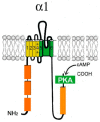
Schematic representation of the pore-forming channel α1-subunit Cav1. The α1-subunit includes four homologous domains (I–IV), each of which consists of six transmembrane segments (1–6). Segments 5 and 6 together with the linker peptide (linkers 5–6) form a selective pore permeable to Ca2+ ions. Segment 4 is a voltage-sensing module. The β-subunit is involved in the inactivation and closure of the channel. Both N- and C-termini are in the cytosol.
Figure 2
Figure 2
The cardiac CaV1.2 channel signaling complex. ABD, AKAP15 binding domain; DCRD, distal C-terminal regulatory domain; PCRD, proximal C-terminal regulatory domain; scissors, site of proteolytic processing.
Figure 3
Figure 3
Schematic representation of the Cav3 α1-subunit. The α1-subunit includes four homologous hydrophobic domains (I–IV), each of which consists of six transmembrane segments (1–6). Segments 5 and 6 together with linkers 5–6 form a highly conserved pore loop permeable to Ca2+ ions. Segment 4 is a voltage-sensing module. The amino (NH2 and carboxyl (COOH) termini and the cytoplasmic interdomain l-II-, II-III-, III-IV loops are in the cytosol.
Figure 4
Figure 4
Structural features of STIM1 (A) and Orai1 (B) proteins. Functional domains of proteins are enclosed in rectangles: EF—Ca2+-binding motif “EF-hands”; SAM—so-called “sterile”-α-motif (sterile-α-motif); TM—transmembrane domain; ERM—protein binding domain of the ERM complex; S/P—domain enriched with serine and proline; C-C—domain enriched with lysine; CAD—domain responsible for channel activation; SOAR, STIM1-Orai—activation region; PR—domain enriched with proline and arginine; TM1–4—transmembrane domains.
Figure 5
Figure 5
Schematic representation of the Trpm2 monomer structure. The Trpm2 monomer consists of six transmembrane domains (yellow and dark green rectangles). The pore-firming loop is located between segments 5 and 6 (black rectangle). Both N- and C-termini are in the cytosol. The N-terminus contains four modules of the Trpm subfamily melastatin homology domain (MHD) (orange rectangles). In the second MHD, there is an IQ-like motif that binds Ca2+-calmodulin. The C-terminus contains a Trp box (TRP) (black rectangle), a coiled-coil domain (CC) (red rectangle), and a adenosine diphosphate ribose (ADPR) pyrophosphatase homolog domain.
Figure 6
Figure 6
Schematic representation of the α1-subunit of the HCN channel. Each HCN monomer contains six TM segments (1–6), including a positively charged potential sensor (S4) and P-region located between the pore-forming segments 5 and 6. The C-terminus contains two conserved structured regions: the C-linker, contributing to tetramerization, and the CNBD, which allows for modulation by cAMP. A region at the end of the N-terminal domain (orange rectangles) is conserved among HCN channel subtypes and has been found to be important for channel trafficking.

Similar articles

  • TRP Channels in the Heart.
    Freichel M, Berlin M, Schürger A, Mathar I, Bacmeister L, Medert R, Frede W, Marx A, Segin S, Londoño JEC. Freichel M, et al. In: Emir TLR, editor. Neurobiology of TRP Channels. Boca Raton (FL): CRC Press/Taylor & Francis; 2017. Chapter 9. In: Emir TLR, editor. Neurobiology of TRP Channels. Boca Raton (FL): CRC Press/Taylor & Francis; 2017. Chapter 9. PMID: 29356479 Free Books & Documents. Review.
  • HCN-related channelopathies.
    Baruscotti M, Bottelli G, Milanesi R, DiFrancesco JC, DiFrancesco D. Baruscotti M, et al. Pflugers Arch. 2010 Jul;460(2):405-15. doi: 10.1007/s00424-010-0810-8. Epub 2010 Mar 8. Pflugers Arch. 2010. PMID: 20213494 Review.
  • Direct evidence for calcium conductance of hyperpolarization-activated cyclic nucleotide-gated channels and human native If at physiological calcium concentrations.
    Michels G, Brandt MC, Zagidullin N, Khan IF, Larbig R, van Aaken S, Wippermann J, Hoppe UC. Michels G, et al. Cardiovasc Res. 2008 Jun 1;78(3):466-75. doi: 10.1093/cvr/cvn032. Epub 2008 Feb 5. Cardiovasc Res. 2008. PMID: 18252758
  • Review: HCN Channels in the Heart.
    Depuydt AS, Peigneur S, Tytgat J. Depuydt AS, et al. Curr Cardiol Rev. 2022;18(4):e040222200836. doi: 10.2174/1573403X18666220204142436. Curr Cardiol Rev. 2022. PMID: 35125083 Free PMC article. Review.
  • Cardiac and neuronal HCN channelopathies.
    Rivolta I, Binda A, Masi A, DiFrancesco JC. Rivolta I, et al. Pflugers Arch. 2020 Jul;472(7):931-951. doi: 10.1007/s00424-020-02384-3. Epub 2020 May 18. Pflugers Arch. 2020. PMID: 32424620 Review.

References

    1. Shah K., Seeley S., Schulz C., Fisher J., Gururaja Rao S. Calcium Channels in the Heart: Disease States and Drugs. Cells. 2022;11:943. doi: 10.3390/cells11060943. - DOI - PMC - PubMed
    1. Bkaily G., Jacques D. Calcium Homeostasis, Transporters, and Blockers in Health and Diseases of the Cardiovascular System. Int. J. Mol. Sci. 2023;24:8803. doi: 10.3390/ijms24108803. - DOI - PMC - PubMed
    1. Catterall W.A., Lenaeus M.J., Gamal El-Din T.M. Structure and Pharmacology of Voltage-Gated Sodium and Calcium Channels. Annu. Rev. Pharmacol. Toxicol. 2020;60:133–154. doi: 10.1146/annurev-pharmtox-010818-021757. - DOI - PubMed
    1. Collins H.E., Zhang D., Chatham J.C. STIM and Orai Mediated Regulation of Calcium Signaling in Age-Related Diseases. Front. Aging. 2022;3:876785. doi: 10.3389/fragi.2022.876785. - DOI - PMC - PubMed
    1. Venkatachalamo K., Montell C. TRP channels. Annu Rev. Biochem. 2007;76:387–417. doi: 10.1146/annurev.biochem.75.103004.142819. - DOI - PMC - PubMed

MeSH terms

Substances

Grants and funding

The work is supported by the IEPhB Research Program 075-00967-23-00.