. 2024 Feb 23;115(3):511-524.
doi: 10.1093/jleuko/qiad141.

TIPE proteins control directed migration of human T cells by directing GPCR and lipid second messenger signaling

Affiliations

TIPE proteins control directed migration of human T cells by directing GPCR and lipid second messenger signaling

Jiyeon Yu et al. J Leukoc Biol. .

Abstract

Tissue infiltration by circulating leukocytes via directed migration (also referred to as chemotaxis) is a common pathogenic mechanism of inflammatory diseases. G protein-coupled receptors (GPCRs) are essential for sensing chemokine gradients and directing the movement of leukocytes during immune responses. The tumor necrosis factor α-induced protein 8-like (TIPE or TNFAIP8L) family of proteins are newly described pilot proteins that control directed migration of murine leukocytes. However, how leukocytes integrate site-specific directional cues, such as chemokine gradients, and utilize GPCR and TIPE proteins to make directional decisions are not well understood. Using both gene knockdown and biochemical methods, we demonstrated here that 2 human TIPE family members, TNFAIP8 and TIPE2, were essential for directed migration of human CD4+ T cells. T cells deficient in both of these proteins completely lost their directionality. TNFAIP8 interacted with the Gαi subunit of heterotrimeric (α, β, γ) G proteins, whereas TIPE2 bound to PIP2 and PIP3 to spatiotemporally control immune cell migration. Using deletion and site-directed mutagenesis, we established that Gαi interacted with TNFAIP8 through its C-terminal amino acids, and that TIPE2 protein interacted with PIP2 and PIP3 through its positively charged amino acids on the α0 helix and at the grip-like entrance. We also discovered that TIPE protein membrane translocation (i.e. crucial for sensing chemokine gradients) was dependent on PIP2. Collectively, our work describes a new mechanistic paradigm for how human T cells integrate GPCR and phospholipid signaling pathways to control directed migration. These findings have implications for therapeutically targeting TIPE proteins in human inflammatory and autoimmune diseases.

Keywords: PI3K signaling; TIPE proteins; chemoattractant sensing; directed migration; heterotrimeric g proteins.


Conflict of interest statement

Conflict of interest. The authors declare that the research was conducted in the absence of any commercial or financial relationships that could be construed as a potential conflict of interest.

Figures
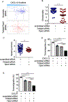
Figure 1.
Figure 1.. Complete loss of directionality in TNFAIP8/TIPE2 double-knockdown (DKD) human CD4+ T cells.
(A-C) Human CD4+ T cells were electroporated with individual or combined siRNA oligos specific for TNFAIP8 and/or TIPE2. Individual or combination knockdown samples together with controls (electroporated with scrambled siRNA) were loaded into a Collagen IV-coated Ibidi μ-slide and recorded using a Leica DMI4000 microscope with Yokogawa CSU-X1 spinning disk confocal attachment. Migration tracks (A), directionality (B), and speed (C) of CD4+ T control cells (scrambled siRNA) (n=41) and DKD cells (n=41) in response to CXCL12, as determined in the μ-slide migration assay. Loss of TNFAIP8 and TIPE2 impairs human CD4+ T cell transmigration induced with CXCL12 (D) or CCL21 (E). The transmigration assays were carried out using Neuro Probe ChemoTx transwell system. Bottom wells were filled with migration buffer with or without CXCL12 or CCL21 (100 ng/ml). Cells were loaded into top wells and allowed to migrate at 37°C for 4 hours. Transmigrated cells were collected from bottom wells and quantified using a hemacytometer. Data are presented as mean ± s.e.m. and pooled from at least three independent experiments. *P<0.05; **P<0.01; ***P<0.001 (Student’s t-test).
Figure 2.
Figure 2.. TNFAIP8 and TIPE2 promote chemotaxis of human CD4+ T cells by directing GPCR, PI3K, and Rac-GTPase signaling.
Pertussis toxin (PT) abolishes chemotaxis of human CD4+ T cells induced by CXCL12 (A) or CCL21(B) while PI3K and Rac1 inhibitors only partially block it. Human CD4+ T cells were pre-incubated with various inhibitors (pertussis toxin (100 ng/ml), LY294002 (50 μM) and NSC27366 (100 μM)) for 30 min prior to the transmigration assay. DKD human CD4+ T cells are defective in chemotaxis induced by CXCL12 (C) or CCL21(D) to a similar level of Gαi blockage by PT. Transfected human CD4+ T cells were pre-incubated with pertussis toxin (100 ng/ml) for 30 min before the start of the transmigration assay. Experiments were repeated independently at least three times and values are mean ± s.e.m. *P<0.05; **P<0.01; ***P<0.001 (Student’s t-test).
Figure 3.
Figure 3.. Gαi interacts with TNFAIP8 through its C-terminal domain.
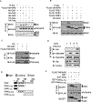
(A) TNFAIP8 specifically binds to Gαi subunits but not Gαq and Gαs subunits. 293T cells were co-transfected with HA-tagged Gαi isoform expression constructs and FLAG-tagged TNFAIP8 expression construct as indicated. Twenty-four hours after transfection, cell lysates were prepared and immunoprecipitated (IP) with anti-FLAG-M2 beads. Lysates and precipitates were Western blotted with anti-HA antibody. (B) TNFAIP8, but not other TIPE proteins, interacts with Gαi. 293T cells were co-transfected with HA-tagged Gαi2 expression construct and FLAG-tagged TNFAIP8, TIPE1, TIPE2, or TIPE3 expression construct as indicated. Twenty-four hours after transfection, cell lysates were immunoprecipitated and analyzed as described in (A). (C) Gαi2 is reversely co-immunoprecipitated with TNFAIP8. 293T cells were co-transfected with HA-tagged Gαi2 and FLAG-tagged TNFAIP8 expressing constructs, and cell lysates were prepared and IPed as described in (B) except using anti-HA-beads. Precipitates were blotted with anti-FLAG antibody. (D) TNFAIP8 and Gαi interaction occurs endogenously. Human CD4+ T cells were stimulated with CXCL12 for 30 min or left unstimulated. Total cell lysates were immunoprecipitated with anti-TNFAIP8 antibody or control Ig, then Western blotted with anti-Gαi antibody. (E) Schematic diagrams of the Gαi2 domain structure, full-length Gαi2, and truncated TNFAIP8 mutants. (F) The C-terminal domain of Gαi mediates interaction with TNFAIP8. 293T cells were transiently transfected with GST- Gαi2 or N-terminal truncated Gαi2 mutants, and full-length FLAG-TNFAIP8 construct. Twenty-four hours after transfection, the cells were immunoprecipitated (IP) with anti-FLAG antibody and Western blotted to detect the region of Gαi2 that bound to TNFAIP8. All experiments were repeated at least three times with similar results.
Figure 4.
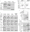
Figure 4.. CXCL12 promotes TNFAIP8 and TIPE2 membrane translocation and AKT activation whereas deficiency in TNFAIP8 and TIPE2 leads to dysregulated AKT and cofilin signaling.
(A) Increased membrane translocation of TNFAIP8 and TIPE2 upon CXCL12 stimulation. Human CD4+ T cells were treated with CXCL12 for the indicated times. The cytoplasmic, membrane, and nuclear fractions were then prepared and immunoblotted with anti- TNFAIP8 or anti-TIPE2 antibody. Anti-α-tubulin was used as the cytosolic loading control. Na+/K+ ATPase was used as the membrane loading control. Histone-H3 was used as the nuclear loading control. (B) Densitometric quantification of TNFAIP8 or TIPE2 membrane translocation shown in (A). (C) The effect of TNFAIP8 and TIPE2 knockdown on CXCL12-induced signaling in human CD4+ T cells. Control cells were treated with scrambled siRNA and knockdown cells were treated with siRNA against TNFAIP8 and TIPE2. Control, single and double knockdown human CD4+ T cells were treated with 100ng/ml CXCL12 for up to 15 min and lysed with RIPA buffer and resolved by SDS-PAGE, transferred to a membrane, and analyzed by immunoblotting with antibodies to pAKT (S473 or T308), total AKT, pCofilin, total cofilin, pLIMK1, TNFAIP8, TIPE2, and GAPDH. (D) TNFAIP8 competes with CXCR4 for Gαi binding. 293T cells were co-transfected with HA-tagged Gαi2, Myc-tagged CXCR4 and FLAG-tagged TNFAIP8 expression constructs as indicated. Twenty-four hours after transfection, cell lysates were prepared and IP with anti-HA antibody and protein A/G beads. Precipitates and lysates then were resolved by SDS-PAGE, transferred to a membrane, and Western blotted with anti-FLAG, Myc and HA antibody to detect TNFAIP8, CXCR4, and Gαi2, respectively. (E) Time-dependent interactions of endogenous Gαi with TNFAIP8 and CXCR4. Human CD4+ T cells were stimulated with 100ng/ml CXCL12 for 5 or 30 min or left unstimulated. Total cell lysates of human CD4+ T cells were immunoprecipitated with anti-Gαi or control Ig. IPs and cell lysates were prepared as described in (Fig. 4D) and blotted with anti-TNFAIP8 or CXCR4 antibodies to visualize the interactions among TNFAIP8, CXCR4, and Gαi. All the experiments were repeated two times with similar results.
Figure 5.
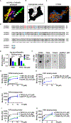
Figure 5.. Identification of TIPE2 amino acids that are crucial for PIP2 and PIP3 binding.
(A) Ribbon representation of the structure of mouse TIPE2 helices obtained from I-TASSER, containing the α0 helix missing from the crystal structure. (B) Representation of TIPE2 hydrophobic cavity size (≈ 1nm3). (C) Distribution of hydrophobic, positive, and negative charge amino acids on TIPE2 helices (positive (blue), negative (red) charges and (gold) hydrophobic). (D) Amino acids sequence similarity between mouse and human TIPE2 (94% sequence identity with identical aa marked by * and similar aa marked by.). (E and F) Positive charged amino acids in the α0 helix of TIPE2 (T2Δα) and at the entrance of the central hydrophobic cavity (T2Δent) are crucial for TIPE2 binding with PIP2 and PIP3. 1uM of TIPE2, T2Δα, T2Δent mutant, and Sumo proteins were added to immobilized lipid samples on a nitrocellulose membrane. Bound proteins were detected using anti-His primary antibody. The graphs shown are representative of at least two independent experiments. (G-K) Specificity of TIPE2 for phosphoinositide-containing SUVs. TIPE2, T2Δα, T2Δent, PLCD-PH, GRP1-PH solutions were injected over various SUV sensor surfaces. Representative binding curve and KD for the interaction between TIPE2 and liposome containing DOPC, PIP2 or PIP3 (G), comparison between binding levels of TIPE2 and PLCD1PH to PIP2 containing liposome (H), and comparison between binding levels of TIPE2 and GRP1PH to PIP3 containing liposome (I). Representative binding levels for the interaction between TIPE2, T2Δα, T2Δent with liposome containing PIP2 (J) or PIP3(K) (J and K). The sensorgrams were aligned and globally fit to a 1:1 binding model using BIAevaluation software as described under “Experimental Procedures.” The resulting binding curves are presented as the steady state binding response (R eq) as a function of respective protein concentration. The graphs shown are representative of at least two independent experiments.
Figure 6.
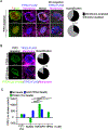
Figure 6.. TIPE protein membrane translocation is PIP2-dependent.
(A) TIPE2 localization and relocation is PIP2-dependent. 293 cells transfected with TIPE2 construct and the PJ/Lyn system (red/green, respectively), with the addition of 1 μM Rapamycin for 5 mins, results in recruitment of both components to the membrane and depletion of PIP2. The TIPE2 membrane translocation was analyzed by confocal microscopy. (B) PIP3 depletion does not affect TIPE2 localization and relocation. PTEN-Kras and PTEN-Lyn constructs, which deplete only PIP3, were used in lieu of the PJ/Lyn system and relocation of TIPE2 with loss of PIP3 was investigated. The pie charts show quantitative analysis of the localization of TIPE2, TIPE2 mutants, and TNFAIP8 isoforms ±PIP2 or ±PIP3. (C) Both TNFAIP8 and TIPE2 can extract PIP2 from lipid vesicle and convert it to PIP3 by PI3K while bound to the protein. All values shown were subtracted from no kinase control samples. N=12 technical replicates/group, representative of 2 independent experiments. For all graphs, **, p<0.01; ***, p<0.001; ****, p<0.0001 (Student’s t-test). The graphs shown are representative of at least two independent experiments.
Figure 7.
Figure 7.. TNFAIP8 functions as a pilot protein sensing chemokine gradient and guiding chemotaxis via heterotrimeric Gαi-dependent signaling.
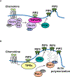
(A) Upon ligation of chemokines with GPCRs, TNFAIP8 is translocated to the plasma membrane where the receptors are activated via interacting with Gαi subunit of heterotrimeric G proteins, causing the dynamic change in PIP2 level at the site of receptor activation and facilitating the initial PIP3 generation, which defines the leading-edge location. (B) The dynamic change in PIP2/PIP3 level further accelerates the other TIPE proteins membrane translocation and facilitates a functional leading-edge formation in a Gαi- independent but PIP2-dependent manner. Collaboratively, they define the leading-edge location and promote cell migration.

Similar articles

References

    1. Cartier L, Hartley O, Dubois-Dauphin M, Krause KH. Chemokine receptors in the central nervous system: role in brain inflammation and neurodegenerative diseases. Brain Res Brain Res Rev. 2005;48(1):16–42. doi: 10.1016/j.brainresrev.2004.07.021 - DOI - PubMed
    1. Liehn EA, Zernecke A, Postea O, Weber C. Chemokines: inflammatory mediators of atherosclerosis. Arch Physiol Biochem. 2006;112(4–5):229–38. doi: 10.1080/13813450601093583 - DOI - PubMed
    1. Danese S, Gasbarrini A. Chemokines in inflammatory bowel disease. J Clin Pathol. 2005;58(10):1025–7. doi: 10.1136/jcp.2005.030916 - DOI - PMC - PubMed
    1. Muller WA. Getting leukocytes to the site of inflammation. Vet Pathol. 2013;50(1):7–22. doi: 10.1177/0300985812469883 - DOI - PMC - PubMed
    1. Nourshargh S, Alon R. Leukocyte migration into inflamed tissues. Immunity. 2014;41(5):694–707. doi: 10.1016/j.immuni.2014.10.008 - DOI - PubMed

Publication types