. 2023 Nov 10;14(1):7281.
doi: 10.1038/s41467-023-43145-x.

A closed translocation channel in the substrate-free AAA+ ClpXP protease diminishes rogue degradation

Affiliations

A closed translocation channel in the substrate-free AAA+ ClpXP protease diminishes rogue degradation

Alireza Ghanbarpour et al. Nat Commun. .

Abstract

AAA+ proteases degrade intracellular proteins in a highly specific manner. E. coli ClpXP, for example, relies on a C-terminal ssrA tag or other terminal degron sequences to recognize proteins, which are then unfolded by ClpX and subsequently translocated through its axial channel and into the degradation chamber of ClpP for proteolysis. Prior cryo-EM structures reveal that the ssrA tag initially binds to a ClpX conformation in which the axial channel is closed by a pore-2 loop. Here, we show that substrate-free ClpXP has a nearly identical closed-channel conformation. We destabilize this closed-channel conformation by deleting residues from the ClpX pore-2 loop. Strikingly, open-channel ClpXP variants degrade non-native proteins lacking degrons faster than the parental enzymes in vitro but degraded GFP-ssrA more slowly. When expressed in E. coli, these open channel variants behave similarly to the wild-type enzyme in assays of filamentation and phage-Mu plating but resulted in reduced growth phenotypes at elevated temperatures or when cells were exposed to sub-lethal antibiotic concentrations. Thus, channel closure is an important determinant of ClpXP degradation specificity.


Conflict of interest statement

The authors declare no competing interests.

Figures

Fig. 1
Fig. 1. Cryo-EM structure of substrate-free particles containing single-chain ClpX∆N and ClpP.
Structural side (a) and top (b) views. Cryo-EM density is colored light gray and fitted atomic models are displayed in cartoon representation and colored and labeled by ClpX subunit. Key structural features are noted. c Closeup of the axial ClpX pore, showing the locations of the six pore-1 loops and the pore-2 loop of subunit A.
Fig. 2
Fig. 2. The pore-1 and pore-2 loops of ClpX adopt similar conformations in the substrate-free and recognition complexes.
Side view of the complex of single-chain ClpX∆N and ClpP with semi-transparent surface representations of ClpX (purple) and ClpP (cyan). Inset highlights similar conformations of pore-1 (residues 150-156) and pore-2 loops from subunits A and C in the substrate free (purple) and recognition complex (yellow) structures. An atomic model of the ssrA degron from the recognition complex is depicted as a stick model and gray surface representation.
Fig. 3
Fig. 3. Flexible RKH loops assume altered conformations in the presence and absence of substrate.
The structure on the left shows a semi-transparent surface representation of the substrate-free complex of single-chain ClpX∆N and ClpP. The inset highlights the ssrA degron (stick representation with a transparent van der Waal’s surface) from the ClpXP recognition complex (10.2210/pdb6WRF/pdb) and RKH loops of chain C from the substrate-free and recognition structures (residues 220-238; cartoon representation). Note that the aromatic Tyr side chain in the ssrA tag would sterically impinge on the substrate-free conformation (purple) of this RKH loop.
Fig. 4
Fig. 4. Effects of the ∆NPS ClpX mutation on protein degradation.
a Loss of GFP fluorescence was used to assay the kinetics of ClpP (1.5 µM) degradation of different concentrations of GFP-ssrA* at 30 °C supported by ClpX∆N or ∆NPS ClpX∆N (0.5 µM). Symbols represent means ± 1 SD (n = 3 independent experiments). b After expression of GFP-ssrA* from a constitutive promoter at 37 °C in E. coli T7 Express ΔclpAΔclpPΔclpX cells, the bar chart depicts the fraction of GFP-ssrA* degraded 30 min after inducing co-expression of ClpX∆N/ClpP, ∆NPS ClpX∆N/ClpP, or an uninduced control as assessed by fluorescence microscopy. Values are means ± 1 SD (n = 3 independent experiments) with individual data points shown as symbols. c SDS-PAGE was used to assay degradation of unfolded AFtitin or AFtitin-ssrA (10 µM) at 30 °C by ClpX∆N or ∆NPS ClpX∆N (2.5 µM) and ClpP (2.5 µM). Symbols represent the average substrate remaining ± SD (n = 3 independent experiments). d FITC-casein (50 µM) degradation at 30 °C by purified variants of ClpX or ClpX∆N (0.5 µM) and ClpP (1.5 µM). Data points represent means of three technical replicates. Source data are provided as a Source Data file.
Fig. 5
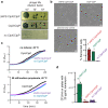
Fig. 5. Effects of ∆NPS ClpX on cellular phenotypes.
Assays were performed using E. coli W3110 ΔclpX cells expressing ClpP and different ClpX variants from a pPro33 plasmid under control of a sodium-propionate inducible promoter. a Dilutions of a phage Mucts stock were spotted onto lawns of cells expressing ∆NPS ClpX/ClpP, ClpX/ClpP, or an empty-vector control (37 °C; 12.5 mM sodium propionate). b Micrographs of cells (scale bar 6 µm) expressing ∆NPS ClpX/ClpP, ClpX/ClpP, or an empty-vector control 3 h after induction with 12.5 mM sodium propionate and growth at 37 °C. The graph quantifies the mean number of filamentous cells under these conditions (n = 3 technical replicates) with individual data points shown as symbols. c Growth curves at 42 °C with and without induction of ClpX variants using 50 mM sodium propionate. Data points are means (n = 3 of 3 technical replicates). d Equivalent numbers of cells expressing ∆NPS ClpX/ClpP, ClpX/ClpP, or an empty-vector control were applied to individual LB agar plates containing 20 µg/mL kanamycin and 12.5 mM sodium propionate and plates were incubated at 37 °C overnight. The graph shows mean colony forming units (CFU; n = 3 independent biological experiments) ± 1 SD with data points shown as symbols, determined by colony counting and correction for the dilution. Source data are provided as a Source Data file.
Fig. 6
Fig. 6. ClpP-mediated decapeptide cleavage.
a Rates of ClpP cleavage of the Abz-KASPVSLGYNO2D decapeptide (15 µM) were assayed by increased fluorescence in the presence/absence of ClpX variants (1 µM), ClpP (50 nM), and either 0 or 15 µM DHFR-gsylaalaa protein substrate. Values are means (n = 3 independent experiments) ± 1 SD with data points shown as symbols. b Degradation of fluorescent DHFR-gsylaalaa (15 µM) by ClpX∆N (0.5 µM) and ClpP (1.5 µM) assayed by SDS-PAGE proceeded at similar rates in the presence and absence of the nonspecific decapeptide (15 µM). Fluorescent DHFR-gsylaalaa (MR 18.8 kDa) comigrates with ClpP (MR 21.5 kDa) on the SDS gel. Source data, including a Coomassie-Blue stained gel for panel b and a fluorescent scan of the gel, are provided as a Source Data file.
Fig. 7
Fig. 7. A symmetry mismatch at the ClpX/ClpP interface could allow peptide-product release.
a Top-view surface representation of a ClpX∆N hexamer bound to ClpP. b Cartoon showing how the six IGF loops of ClpX (colored as in panel a) dock into six of the seven clefts of a heptameric ClpP ring. c Side-view surface representations of the single-chain ClpX∆N/ClpP complex depicting the presence of an empty cleft between the IGF loops of chains E and F. d Surface representation clipped in-plane at the ClpXP midline with key structural features noted.

Similar articles

Cited by

References

    1. Sauer RT, Baker TA. AAA+ proteases: ATP-fueled machines of protein destruction. Annu. Rev. Biochem. 2011;80:587–612. doi: 10.1146/annurev-biochem-060408-172623. - DOI - PubMed
    1. Baker TA, Sauer RT. ClpXP, an ATP-powered unfolding and protein degradation machine. Biochim. Biophys. Acta. 2012;1823:15–28. doi: 10.1016/j.bbamcr.2011.06.007. - DOI - PMC - PubMed
    1. Sauer RT, Fei X, Bell TA, Baker TA. Structure and function of ClpXP, a AAA+ proteolytic machine powered by probabilistic ATP hydrolysis. Crit. Rev. Biochem. Mol. Biol. 2021;57:188–204. doi: 10.1080/10409238.2021.1979461. - DOI - PMC - PubMed
    1. Moore SD, Sauer RT. The tmRNA system for translational surveillance and ribosome rescue. Annu. Rev. Biochem. 2007;76:101–124. doi: 10.1146/annurev.biochem.75.103004.142733. - DOI - PubMed
    1. Keiler KC. Mechanisms of ribosome rescue in bacteria. Nat. Rev. Microbiol. 2015;13:285–297. doi: 10.1038/nrmicro3438. - DOI - PubMed

Publication types

MeSH terms

Substances