. 2024 Jan;11(4):e2307182.
doi: 10.1002/advs.202307182. Epub 2023 Nov 10.

APP-C31: An Intracellular Promoter of Both Metal-Free and Metal-Bound Amyloid-β40 Aggregation and Toxicity in Alzheimer's Disease

Affiliations

APP-C31: An Intracellular Promoter of Both Metal-Free and Metal-Bound Amyloid-β40 Aggregation and Toxicity in Alzheimer's Disease

Eunju Nam et al. Adv Sci (Weinh). 2024 Jan.

Abstract

Intracellular C-terminal cleavage of the amyloid precursor protein (APP) is elevated in the brains of Alzheimer's disease (AD) patients and produces a peptide labeled APP-C31 that is suspected to be involved in the pathology of AD. But details about the role of APP-C31 in the development of the disease are not known. Here, this work reports that APP-C31 directly interacts with the N-terminal and self-recognition regions of amyloid-β40 (Aβ40 ) to form transient adducts, which facilitates the aggregation of both metal-free and metal-bound Aβ40 peptides and aggravates their toxicity. Specifically, APP-C31 increases the perinuclear and intranuclear generation of large Aβ40 deposits and, consequently, damages the nucleus leading to apoptosis. The Aβ40 -induced degeneration of neurites and inflammation are also intensified by APP-C31 in human neurons and murine brains. This study demonstrates a new function of APP-C31 as an intracellular promoter of Aβ40 amyloidogenesis in both metal-free and metal-present environments, and may offer an interesting alternative target for developing treatments for AD that have not been considered thus far.

Keywords: accelerator toward amyloidogenesis; amyloid precursor protein; amyloid-β; metal ions; protein-protein interaction.


Conflict of interest statement

The authors declare no conflict of interest.

Figures

Figure 1
Figure 1
Impact of APP‐C31 on the intracellular aggregation of Aβ40. a) Amino acid sequences of APP‐C31 (APP665–695) and Aβ40 that are generated from the proteolytic cleavage of APP. The self‐recognition site of Aβ is highlighted in bold. b) Scheme of the microinjection experiments. c) Mean size of intracellular Aβ40 aggregates in the absence and presence of APP‐C31 observed by confocal microscopy. Aggregates (red) with dimensions exceeding the resolution limit were analyzed from three or more randomly selected fields per condition. Conditions: [HF64740] = 10 µm; [APP‐C31] = 10 µm; 24 h incubation. Scale bars = 5 and 10 µm. Data are represented as mean ± s.e.m. n = 52 for the group of HF64740; n = 63 for the group of HF64740 with APP‐C31; *p < 0.05; Student's t‐test. The size distribution of Aβ40 aggregates detected in both groups was summarized in Figure S2, Supporting Information. d) Z‐stack images of Aβ40 aggregates (red) with and without APP‐C31. Nuclei were stained with SYTO16 (green). HF64740 aggregates observed from the nuclear region were indicated with white arrows. Individual green and red fluorescence images are presented in Figure S3, Supporting Information. Conditions: [HF64740] = 10 µm; [APP‐C31] = 10 µm; [SYTO16] = 2.5 µm; 24 h incubation. Scale bar = 10 µm.
Figure 2
Figure 2
Effect of APP‐C31 on the aggregation of Aβ40 and its interactions with Aβ40. a) Scheme of the aggregation experiments. b) Degree on the aggregation of Aβ40 upon incubation with or without APP‐C31 analyzed by the ThT assay. Experiments were carried out in triplicate. c) Morphology of the peptide aggregates produced by treatment of Aβ40 with or without APP‐C31 detected by TEM. White arrows indicate a mixture of globular aggregates and short fibrils. Scale bar = 200 nm. Conditions: [Aβ40] = 20 µm and [APP‐C31] = 20, 100, and 200 µm; 20 mm HEPES, pH 7.4, 150 mm NaCl; 37 °C; constant agitation (559 cpm). d) ESI–MS spectra of APP‐C31, Aβ40, and APP‐C31 incubated with Aβ40. Charge states are marked in the MS spectra. Conditions: [APP‐C31] = 100 µm; [Aβ40] = 100 µm; 20 mm ammonium acetate, pH 7.3; 37 °C; 2 h incubation; no agitation. The samples were diluted by tenfold prior to injection to the mass spectrometer. e) Interaction of APP‐C31 with monomeric Aβ40 analyzed by 2D 1H–15N HSQC NMR (900 MHz). The average of CSPs and the average + one standard deviation are indicated with solid and dashed lines, respectively. The zoomed‐in images of the regions where noticeable CSPs appear are depicted in Figure S11, Supporting Information. Conditions: [15N‐labeled Aβ40] = 40 µm; [APP‐C31] = 200 µm; 20 mm HEPES, pH 7.4; 5 °C. The amino acid residues indicated in blue or black asterisks represent the residues that were unresolved or not significantly shifted, respectively. f) Conformations of Aβ40 (PDB 2LFM)[ 55 ] and APP666–693 [PDB 3DXC; as a part of APP‐C31 (APP665–695)][ 56 ] used for metadynamics MD simulations. The self‐recognition site of Aβ shown in Figure 1a is highlighted in purple. g) A representative model of the APP666–693–Aβ40 interfaces from the trajectories of MD simulations. Possible hydrogen bonds within 3.0 Å and hydrophobic interactions observed within 4.0 Å are indicated with dashed black lines. The amino acid residues involved in hydrophobic interactions are presented in the space‐filling representation. The other representative model, Model II, is illustrated in Figure S16, Supporting Information.
Figure 3
Figure 3
Interactions of APP‐C31 with metal‐treated Aβ40 and its influence on metal‐induced Aβ40 aggregation. a) Interactions of APP‐C31 with metal‐treated Aβ40 observed by ESI–MS. The full MS spectra are depicted in Figures S21 and S22, Supporting Information. Conditions: [APP‐C31] = 100 µm; [Aβ40] = 100 µm; [CuCl2 or ZnCl2] = 100 µm; 20 mm ammonium acetate, pH 7.3; 37 °C; 2 h incubation; no agitation. The samples were diluted by tenfold prior to injection to the mass spectrometer. b) Interaction of APP‐C31 with Cu(II)‐treated Aβ40 analyzed by 2D 1H–15N HSQC NMR spectroscopy (700 MHz). The average of CSPs and the average + one standard deviation are indicated with solid and dashed lines, respectively. Conditions: [15N‐labeled Aβ40] = 40 µm; [APP‐C31] = 200 µm; [CuCl2] = 20 µm; 20 mm HEPES, pH 7.4; 10 °C. The amino acid residues indicated in blue or black asterisks represent the residues that were unresolved or not significantly shifted, respectively. c) Scheme of the aggregation experiments. d) Degree on the aggregation of metal‐added Aβ40 with or without APP‐C31 detected by the ThT assay. Experiments were conducted in triplicate. e) Morphology of the peptide aggregates generated by incubation of Cu(II) or Zn(II)‐treated Aβ40 with and without APP‐C31 for 60 or 40 h, respectively, monitored by TEM. Scale bar = 200 nm. Conditions: [APP‐C31] = 20 µm; [Aβ40] = 20 µm; [CuCl2] = 18 µm; [ZnCl2] = 20 µm; 20 mm HEPES, pH 7.4, 150 mm NaCl; 37 °C; constant agitation (559 cpm).
Figure 4
Figure 4
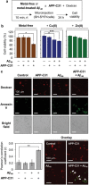
Influence of APP‐C31 on the toxicity of intracellular Aβ40. a) Scheme of the microinjection experiments. b) Cell viability of SH‐SY5Y cells microinjected with either APP‐C31, Aβ40, or both in the absence and presence of metal ions. Cell survival measurements were described in detail in the experimental section. Conditions: [Aβ40] = 10 µm; [APP‐C31] = 10 µm; [CuCl2 or ZnCl2] = 10 µm; [Texas Red‐labeled dextran] = 0.4 mg mL−1; 24 h incubation. All values denote mean ± s.e.m. *p < 0.05; ***p < 0.001; n = 3–5; Student's t‐test. c) Apoptosis monitored using Alexa Fluor 488‐tagged annexin V. Pearson's correlation coefficients were calculated to present the colocalization of the fluorophore‐tagged dextran and Annexin V (mean ± s.e.m, **p < 0.01; n = 3–5; Student's t‐test). White arrows indicate apoptotic cells microinjected with peptide samples. Conditions: [Aβ40] = 10 µm; [APP‐C31] = 10 µm; [Texas Red‐labeled dextran] = 0.4 mg mL−1; 24 h incubation. Scale bar = 100 µm.
Figure 5
Figure 5
Effects of APP‐C31 on the neurodegeneration and inflammatory response induced by Aβ40. a) Scheme of the experiments with human neuronal stem cells. b) Analysis of the average length of neurites as a function of APP‐C31's concentration. c) Representative images of neurons after 72 h incubation with either 20 µm of APP‐C31, Aβ40, or both. 15 neurons were analyzed per each condition. The average length of neurites in the presence of various concentrations of APP‐C31 only is reported in Figure S26, Supporting Information. Conditions: [Aβ40] = 20 µm; [APP‐C31] = 0, 10, 20, 40, and 100 µm; incubation for 24 and 72 h. Scale bar = 100 µm. Data are represented as mean ± s.e.m. *p < 0.05; ***p < 0.001; Student's t‐test. d) Scheme of in vivo experiments and injection sites in the brain. Hippocampus (hip), cortex (ctx), thalamus (th), caudate putamen (cp), and hypothalamus (hy) are shown. e) Microscopic images of the hippocampi of C57BL/6J mice injected with Aβ40, APP‐C31, or both, visualized by immunohistochemical analyses [primary antibodies, 6E10 (anti‐Aβ antibody; green), Y188 (anti‐APP antibody; green), Iba1 (anti‐microglial antibody; red)] or fluorescent dye staining (Hoechst for nucleus). Scale bar = 200 µm. f) Quantification results of the 6E10‐positive area and Iba1‐positive cells, calculated based on the fluorescence signals. All values denote mean ± s.e.m. *p < 0.05, **p < 0.01; n = 3 (for 6E10 staining), n = 5 (for Y188 staining), and n = 5 (for Iba1 staining); Student's t‐test.

Similar articles

References

    1. Müller U. C., Deller T., Korte M., Nat. Rev. Neurosci. 2017, 18, 281. - PubMed
    1. Hardy J. A., Higgins G. A., Science 1992, 256, 184. - PubMed
    1. Hamley I. W., Chem. Rev. 2012, 112, 5147. - PubMed
    1. Laferla F. M., Green K. N., Oddo S., Nat. Rev. Neurosci. 2007, 8, 499. - PubMed
    1. Lee J.‐H., Yang D.‐S., Goulbourne C. N., Im E., Stavrides P., Pensalfini A., Chan H., Bouchet‐Marquis C., Bleiwas C., Berg M. J., Huo C., Peddy J., Pawlik M., Levy E., Rao M., Staufenbiel M., Nixon R. A., Nat. Neurosci. 2022, 25, 688. - PMC - PubMed

Publication types

LinkOut - more resources