. 2023 Dec 21;30(12):1601-1616.e6.
doi: 10.1016/j.chembiol.2023.10.008. Epub 2023 Nov 7.

AGC kinase inhibitors regulate STING signaling through SGK-dependent and SGK-independent mechanisms

Affiliations

AGC kinase inhibitors regulate STING signaling through SGK-dependent and SGK-independent mechanisms

Johnny Castillo Cabrera et al. Cell Chem Biol. .

Abstract

Type 1 IFN expression is critical in the innate immune response, but aberrant expression is associated with autoimmunity and cancer. Here, we identify N-[4-(1H46 pyrazolo[3,4-b] pyrazin-6-yl)-phenyl]-sulfonamide (Sanofi-14h), a compound with preference for inhibition of the AGC family kinase SGK3, as an inhibitor of Ifnb1 gene expression in response to STING stimulation of macrophages. Sanofi-14h abrogated SGK activity and also impaired activation of the critical TBK1/IRF3 pathway downstream of STING activation, blocking interaction of STING with TBK1. Deletion of SGK1/3 in a macrophage cell line did not block TBK1/IRF3 activation but decreased expression of transcription factors, such as IRF7 and STAT1, required for the innate immune response. Other AGC kinase inhibitors blocked TBK1 and IRF3 activation suggesting common action on a critical regulatory node in the STING pathway. These studies reveal both SGK-dependent and SGK-independent mechanisms in the innate immune response and indicate an approach to block aberrant Ifnb1 expression.

Keywords: AGC kinases; IRF; SGK; STING; Sanofi-14h; TBK1; double strand DNA viral response; innate immunity; interferon; interferon regulatory factor; macrophages; serum and glucocorticoid kinase; stimulator of interferon genes; tank binding kinase.


Conflict of interest statement

Declaration of interests The authors declare no competing interests.

Figures
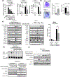
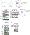
Figure 1:
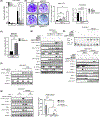
Figure 1:. SGK inhibitor Sanofi-14h impairs IFN expression and STING/TBK1/IRF3 signaling in macrophages.
A) RAW264.7 cells were pre-treated with Sanofi-14h at the indicated concentrations, whereas THP-1 and hMDM were pre-treated with 10 μM, for 2 hours and then treated with STING ligands DMXAA (10μg/mL, RAW cells) or diABZI (1μM) for an additional 2 hours. RT-qPCR of IFNβ mRNA expression was normalized to GAPDH for RAW264.7 and hMDM or 18S for THP1. B) RAW264.7 cells were pre-treated with Sanofi-14h (5 μM) for 24 h and then infected with HSV-1 KOS (MOI 0.1). Images shows representative plaque assays and graphs depict the HSV-1 PFU (left) and IFNβ mRNA (right). C) RAW264.7 macrophages (left panel) were pretreated with Sanofi-14h at the indicated doses for 2h prior to stimulation with DMXAA (10μg/mL) for 1h and 2h and lysates were immunoblotted. BMDM (right panel) were pretreated with Sanofi-14h (10 μM) for 2h and prior to DMXAA (10μg/mL) for 30 min, 1h and 2h. Graph shows IFNβ mRNA expression normalized to GAPDH in BMDM. pNDRG1(T346) was a used a control to measure Sanofi-14h activity in both cell lines. D) Dimerization (left panel) and nuclear localization (right panel) of IRF3 after pretreatment with Sanofi-14h at different doses for 2h and DMXAA for 1h. Lamin A/C and β-tubulin indicate nuclear and cytoplasmic fractions respectively. E) Effects of Sanofi-14h are upstream of IFNβ signaling. RAW cells were pretreated with Sanofi-14h (10 μM) or vehicle for 2h prior to treatment with DMXAA for 2h. A subset was treated with exogenous IFNβ (10 ng/mL) for the last 15 minutes. Immunoblots depict total and phosphorylated STAT3 and TBK1. BMDM: Bone Marrow Derived Macrophages, PFU: Plaque-Forming Unite, HSV: Herpes Simplex Virus. Data are representative of three different experiments (* p<0.05).
Figure 2:
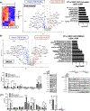
Figure 2:. Deletion of SGK1 and SGK3 impairs IFNβ production and STAT3 phosphorylation without affecting STING/TBK1/IRF3 phosphorylation.
A) IFNβ mRNA expression in single and double (dKO) RAW264.7 SGK1/3 KO cells with (top segment) and without (bottom segment) DMXAA treatment for 2h. B) RAW264.7 SGK KO cells infected with HSV-1 KOS. Images show plaque assay for single and dKO cells. Graphs indicated HSV-1 PFU and IFNβ mRNA production. C) HSV-1 PFU in SGK3 KO BMDMs. D) Cells were treated with DMXAA for 1h and 2h and lysates were immunoblotted for indicated proteins. SGK1 and SGK3 protein levels confirm knockout and pNDRG1 indicates SGK activity. E) Dimerization (left panel) and nuclear localization (right panel) of IRF3 after DMXAA treatment for 1h in SGK1 KO, SGK3 KO, and SGK1/3 dKO cells. Lamin A/C and β-tubulin indicate nuclear and cytoplasmic fractions, respectively. F) Immunoblot representing the treatment of SGK1/3 dKO cells with DMXAA (2 h total) with and without exogenous IFNβ (15 min). G) WT and SGK1/3 dKO cells were treated with Sanofi-14h for 2h prior to DMXAA stimulation for 2h. Lysates were immunoblotted for indicated proteins (left panel) and IFNβ mRNA was assayed by RT-PCR (right graph). Data are representative of three different experiments (* p<0.05).
Figure 3:
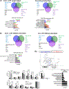
Figure 3:. SGK1 and SGK3 KO blocks expression of Interferon Stimulated Genes (ISGs) and innate immunity regulators.
A) Heatmap (left panel) depicts differentially expressed genes (DEGs) in unstimulated WT and SGK1/3 dKO RAW264.7 cells. Blue indicates downregulated and red indicates upregulated DEGs. Volcano plot (middle graph) depicts the expression and statistical significance of DEGs in unstimulated cells with highlighting of immune-related or Interferon Stimulated Genes (ISGs). Right graph shows Gene Set Enrichment Analysis (GSEA) representing significantly altered pathways in unstimulated SGK1/3 dKO cells relative to WT. B) Volcano plot (left graph) of gene expression in SGK1/3 dKO cells compared to WT after 2h DMXAA treatment. Significance was determined by an adjusted p value of <0.05 and a fold change >2 in either direction. GSEA (right graph) of the most affected pathways after DMXAA treatment in SGK1/3 dKO cells. C) IRF and STAT mRNA expression in Transcripts Per Million (TPM) (*p<0.05) in SGK1/3 dKO cells with and without DMXAA (2h). Immunoblot of IRFs and STATs protein expression at basal conditions (left panel) and their nuclear localization (right panel) after DMXAA treatment for 1h.
Figure 4:
Figure 4:. SGK1 and SGK3 individually control different subsets of genes under basal and STING stimulation:
A) Venn diagrams depicting the overlap of down- and up-regulated genes among unstimulated SGK1 KO, SGK3 KO, and SGK1/3 dKO cells relative to WT. GSEA showing affected pathways in individual and double knockouts relative to WT (lower panel). E) As in (D), comparison of gene expression changes in single and double knockouts after DMXAA stimulation. F) Volcano plot (left graph) indicating DEGs between vehicle- or Sanofi-14h-treated cells after DMXAA stimulation. GSEA (right graph) indicating pathway effects of Sanofi-14h on DMXAA-stimulated gene expression. (Bottom panel) Expression of individual IRF and STAT mRNAs with and without Sanofi-14h after DMXAA treatment. All experiments were repeated at least three times (* p<0.05).
Figure 5:
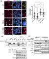
Figure 5:. Sanofi-14h disrupts TBK1/STING interaction.
A) Immunofluorescence (left) of RAW264.7 and TBK1 puncta quantification (right) after co-treatment with 10 mM of Sanofi-14h (2h) and 10μg/mL DMXAA (1h). Arrows indicate TBK1 puncta. B) Effects of Sanofi-14h on STING and TBK1 interaction after DMXAA treatment for 1h. STING was immunoprecipitated and the interaction was detected by TBK1 immunoblot. Immunoprecipitation with non-specific IgG serves as a negative control and inputs show the levels of both proteins in total lysate. C) RAW264.7 cells were fractionated and immunoblotted for TBK1 and IRF3 translocation into the ER-Golgi after Sanofi-14h (2h) and DMXAA treatment for 1h. Data is representative of 3 separate experiments (* p<0.05).
Figure 6:
Figure 6:. Other SGK inhibitors have variable effects on IRF DMXAA driven activation.
A) Structures of Sanofi-14h and Sanofi-17a. B) Effect of SGK inhibitors on DMXAA-stimulated IRF luciferase activity. Raw-Dual Luciferase cells were seeded in 96 well plates overnight and pretreated with indicated inhibitors for 2h prior to DMXAA stimulation for 24h. Luciferase signal indicates type I IFN production. X axis depicts log10 inhibitor concentration. Immunoblot (right panel) shows phosphorylation of indicated proteins after 2h DMXAA treatment. C) Immunoblot of RAW264.7 cells pre-treated with 10 μM Sanofi-14h followed by treatment with LPS at the indicated times. Data is representative of 3 separate experiments. D) Immunoblot (right panel) depicting effects of p70S6K (LY258702) and Akt (MK2206) inhibition on markers of the DMXAA response compared to Sanofi-14h. IRF activation luciferase assay (left graph) after co-treatment with DMXAA and indicated doses of LY258702 and MK2206. E) Co-immunoprecipitation (left panel) showing the effects of LY258702 (2 h pre-treatment) on TBK1/STING interaction after DMXAA treatment for 1 h. Data is representative of 3 separate experiments.
Figure 7:
Figure 7:. Model of SGK regulation of STING/TBK1 signaling.
Sanofi-14h inhibits phosphorylation of TBK1 through an unknown mechanism preventing STING and IRF3 phosphorylation, dimerization and nuclear translocation in the presence of 2’3’-cGAMP and DMXAA. SGK1/3 promote IRF7 expression and stabilization under basal and DMXAA stimulated conditions in macrophages. Adapted from “cGAS detects Cytosolic dsDNA”, BioRender.com (2022), https://app.biorender.com/biorender-templates

Similar articles

References

    1. McNab F, Mayer-Barber K, Sher A, Wack A, and O’Garra A (2015). Type I interferons in infectious disease. Nat Rev Immunol 15, 87–103. 10.1038/nri3787. - DOI - PMC - PubMed
    1. Choubey D, and Moudgil KD (2011). Interferons in autoimmune and inflammatory diseases: Regulation and roles. Journal of Interferon and Cytokine Research 31, 857–865. 10.1089/jir.2011.0101. - DOI - PMC - PubMed
    1. Davidson S, Crotta S, McCabe TM, and Wack A (2014). Pathogenic potential of interferon αβ in acute influenza infection. Nat Commun 5. 10.1038/ncomms4864. - DOI - PMC - PubMed
    1. Biron CA (2001). Interferons α and β as immune regulators - A new look. Immunity 14, 661–664. 10.1016/S1074-7613(01)00154-6. - DOI - PubMed
    1. Kim YM, and Shin EC (2021). Type I and III interferon responses in SARS-CoV-2 infection. Exp Mol Med 53, 750–760. 10.1038/s12276-021-00592-0. - DOI - PMC - PubMed

Publication types

MeSH terms

LinkOut - more resources