. 2023 Nov 7;6(1):1127.
doi: 10.1038/s42003-023-05511-9.

Synaptic proteasome is inhibited in Alzheimer's disease models and associates with memory impairment in mice

Affiliations

Synaptic proteasome is inhibited in Alzheimer's disease models and associates with memory impairment in mice

Felipe C Ribeiro et al. Commun Biol. .

Abstract

The proteasome plays key roles in synaptic plasticity and memory by regulating protein turnover, quality control, and elimination of oxidized/misfolded proteins. Here, we investigate proteasome function and localization at synapses in Alzheimer's disease (AD) post-mortem brain tissue and in experimental models. We found a marked increase in ubiquitinylated proteins in post-mortem AD hippocampi compared to controls. Using several experimental models, we show that amyloid-β oligomers (AβOs) inhibit synaptic proteasome activity and trigger a reduction in synaptic proteasome content. We further show proteasome inhibition specifically in hippocampal synaptic fractions derived from APPswePS1ΔE9 mice. Reduced synaptic proteasome activity instigated by AβOs is corrected by treatment with rolipram, a phosphodiesterase-4 inhibitor, in mice. Results further show that dynein inhibition blocks AβO-induced reduction in dendritic proteasome content in hippocampal neurons. Finally, proteasome inhibition induces AD-like pathological features, including reactive oxygen species and dendritic spine loss in hippocampal neurons, inhibition of hippocampal mRNA translation, and memory impairment in mice. Results suggest that proteasome inhibition may contribute to synaptic and memory deficits in AD.


Conflict of interest statement

The authors declare no competing interests.

Figures
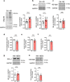
Fig. 1
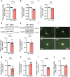
Fig. 1. Ubiquitinylated proteins accumulate in AD brains, and proteasome activity is impaired in human cortical slices exposed to AβOs in culture.
Immunoreactivities of polyubiquitin (n = 10 control, 11 AD) (a), 20S subunit α1 (n = 9 control, 11 AD) (b) and 19S subunit Rpt6 (n = 5 control, 9 AD) (c) were quantified in AD hippocampi compared to controls; two-tailed unpaired Student’s t test. df Chymotrypsin-, trypsin- and caspase-like proteasome activities in human adult cortical slices in culture exposed to vehicle or 0.5 µM AβOs for 24 h (n = 5 independent donors; two-tailed unpaired Student’s t test). g, h Proteasome 20S subunit α1 and 19S subunit Rpt6 immunoreactivities in human cortical slices in culture. GAPDH was used as loading control (n = 4 independent donors; two-tailed unpaired Student’s t test). Data are presented as mean ± SEM.
Fig. 2
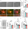
Fig. 2. AβOs induce proteasome inhibition in hippocampal cultures.
ac Chymotrypsin- (n = 5), trypsin- (n = 5) and caspase-like (n = 4) proteasome activities in primary hippocampal cultures exposed to vehicle or 0.5 µM AβOs for 24 h (n = 4–5 independent cultures; two-tailed unpaired Student’s t test). d, e Proteasome 20S subunit α1 (n = 7) and 19S subunit Rpt6 (n = 6) in hippocampal cultures exposed to vehicle or 0.5 µM AβOs (n = 6–7 independent cultures; two-tailed unpaired Student’s t test). fj Primary hippocampal cultures were exposed to vehicle or 0.5 µM AβOs for 24 h and were then labeled for proteasome 20S subunit α1 or 19S subunit Rpt6 (f). Quantification of dendritic (g, i) or cell body (h, j) immunoreactivities (n = 3 for cell body and 5 for dendrites in 20S subunit α1 and 3 for cell body and 5 for dendrites in Rpt6; symbols represent means from 30 images per experimental condition per culture; two-tailed unpaired Student’s t test). Data are presented as mean ± SEM. Scale bar: 20 µm.
Fig. 3
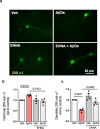
Fig. 3. Dynein inhibition prevents AβO-induced synaptic proteasome mislocalization in hippocampal cultures.
ac Primary hippocampal cultures were exposed to vehicle or 0.5 µM AβOs for 24 h in the presence or absence of erythro-9-[3-(2-hydroxynonyl)]adenine (EHNA; 100 µM), and were then immunolabeled for proteasome 20S subunit α1 (a). Quantification of dendritic (b) or cell body (c) immunoreactivities (n = 4 independent cultures; two-tailed two-way ANOVA with Holm-Sidak post hoc test). Data are presented as mean ± SEM. Scale bar: 20 µm.
Fig. 4
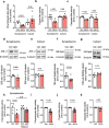
Fig. 4. AβOs inhibit synaptic proteasome activity in the mouse hippocampus.
Three-month-old Swiss mice received intracerebroventricular (i.c.v.) infusions of 10 pmol AβOs (or vehicle). ac Hippocampi were harvested 7 days after infusion, and tissue was fractionated for synaptosome preparation (see “Methods”). Proteasomal chymotrypsin-, (n = 4 per group) trypsin- (n = 5 per group), and caspase-like activities (n = 5 per group) were measured in synaptosomal preparations from independent mice; two-tailed unpaired Student’s t test). Proteasome 20S subunit α1 (d, e) (n = 5 veh and 4 AβOs in synaptosomal fraction and n = 5 vehicle and 4 AβOs in cytosol fraction) and 19S subunit Rpt6 (f, g) (n = 6 vehicle and 5 AβOs in synaptosomal fraction, 5 in cytosol vehicle and 5 in cytosol AβOs fractions) were determined by Western blotting in synaptosome or cytosolic fractions. h Proteasomal chymotrypsin-like activity was measured in synaptosomal preparations from veh-, AβO- and/or rolipram-treated mice (n = 5 mice in vehicle, AβOs and rolipram + AβOs; 4 mice in rolipram; two-way ANOVA with Holm-Sidak post hoc test). ik Hippocampi from naive mice were harvested and synaptosomes were isolated. Synaptosome preparations were then exposed to AβOs (or vehicle) for 1 h at 37 °C, and proteasomal chymotrypsin-, trypsin-, and caspase-like activities were measured (n = 9 synaptosomal preparations from independent mice in chymotrypsin and trypsin, 8 synaptosomal preparations from independent mice for caspase; two-tailed unpaired Student’s t test). Data are presented as mean ± SEM.
Fig. 5
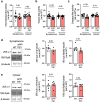
Fig. 5. Reduced synaptic proteasome activity in the APP/PS1 hippocampus.
Hippocampi from 12-month-old APP/PS1 mice or WT littermates were harvested for isolation of synaptosomes. ac Proteasomal chymotrypsin-, trypsin-, and caspase-like activities were measured (chymotrypsin: n = 14 WT and 12 APP/PS1 for synaptosome, n = 17 WT and 12 APP/PS1 for cytosol; trypsin: 19 WT synaptosome, 17 APP/PS1 synaptosome, 21 WT cytosol, 17 APP/PS1 cytosol; caspase-like: 15 WT synaptosome, 12 APP/PS1 synaptosome, 17 WT cytosol, 13 APP/PS1 cytosol); two-tailed unpaired Student’s t test). d, e Proteasome 20S subunit α1 and 19S subunit Rpt6 were determined by Western blotting in synaptosome (d) (n = 7 WT and 6 APP/PS1; two-tailed unpaired Student’s t test) or cytosolic fractions (e) (n = 6 for all; two-tailed unpaired Student’s t test). Data are presented as mean ± SEM.
Fig. 6
Fig. 6. Proteasome inhibition triggers AD-like features in neurons.
a, b Primary hippocampal cultures were exposed to vehicle, 0.5 µM AβOs or 0.5 µM lactacystin for 3 h and ROS were detected by DCF fluorescence (n = 4 independent cultures). Representative images in (a) show DCF fluorescence merged with brightfield images of cultures. Scale bar = 100 μm. c, d Primary hippocampal cultures were exposed to vehicle, 0.5 µM AβOs, or 0.5 µM lactacystin, and cells were double-labeled with neuronal marker MAP-2 (green) and F-actin probe phalloidin (red) for visualization of dendritic spines (n = 3 independent cultures). Images below the main panels are digital zoom images of selected dendrite segments. e 3-month-old C57/BL6 mice received intracerebroventricular infusions of vehicle, AβOs (10 pmol) or lactacystin (100 pmol). Hippocampi were harvested after 7 days, sliced, allowed to recover and incubated with puromycin for 45 min as described in “Methods”. SUnSET was performed by anti-puromycin immunolabeling (n = 8 mice for vehicle, 9 for AβOs; 5 mice for lactacystin). f, g 3-month-old mice received i.c.v. infusions of vehicle, AβOs (10 pmol) or lactacystin (100 pmol). Seven days after infusion, mice were tested in the novel object recognition (e) and novel object location (f) memory paradigms (n = 12 mice for vehicle, 13 mice for lactacystin, and 14 mice for AβOs). Symbols represent percentages of time of exploration of the novel object (or object at novel location) for individual mice. The dotted line at 50% corresponds to chance level. Unpaired two-tailed one-way ANOVA with Holm-Sidak post hoc test. Data are presented as mean ± SEM.

Similar articles

Cited by

References

    1. Murata S, Yashiroda H, Tanaka K. Molecular mechanisms of proteasome assembly. Nat. Rev. Mol. Cell Biol. 2009;10:104–115. doi: 10.1038/nrm2630. - DOI - PubMed
    1. Thibaudeau TA, Smith DM. A practical review of proteasome pharmacology. Pharmacol. Rev. 2019;71:170–197. doi: 10.1124/pr.117.015370. - DOI - PMC - PubMed
    1. Hamilton AM, et al. Activity-dependent growth of new dendritic spines is regulated by the proteasome. Neuron. 2012;74:1023–1030. doi: 10.1016/j.neuron.2012.04.031. - DOI - PMC - PubMed
    1. Dong C, Upadhya SC, Ding L, Smith TK, Hegde AN. Proteasome inhibition enhances the induction and impairs the maintenance of late-phase long-term potentiation. Learn. Mem. 2008;15:335–347. doi: 10.1101/lm.984508. - DOI - PMC - PubMed
    1. Lopez-Salon M, et al. The ubiquitin-proteasome cascade is required for mammalian long-term memory formation. Eur. J. Neurosci. 2001;14:1820–1826. doi: 10.1046/j.0953-816x.2001.01806.x. - DOI - PubMed

Publication types

Substances