[Preprint]. 2023 Oct 16:2023.10.11.561959.
doi: 10.1101/2023.10.11.561959.

Inflammatory stress-mediated chromatin changes underlie dysfunction in endothelial cells

Affiliations

Inflammatory stress-mediated chromatin changes underlie dysfunction in endothelial cells

Haibo Liu et al. bioRxiv. .

Abstract

Inflammatory stresses underlie endothelial dysfunction and contribute to the development of chronic cardiovascular disorders such as atherosclerosis and vascular fibrosis. The initial transcriptional response of endothelial cells to pro-inflammatory cytokines such as TNF-alpha is well established. However, very few studies uncover the effects of inflammatory stresses on chromatin architecture. We used integrative analysis of ATAC-seq and RNA-seq data to investigate chromatin alterations in human endothelial cells in response to TNF-alpha and febrile-range heat stress exposure. Multi-omics data analysis suggests a correlation between the transcription of stress-related genes and endothelial dysfunction drivers with chromatin regions exhibiting differential accessibility. Moreover, microscopy identified the dynamics in the nuclear organization, specifically, the changes in a subset of heterochromatic nucleoli-associated chromatin domains, the centromeres. Upon inflammatory stress exposure, the centromeres decreased association with nucleoli in a p38-dependent manner and increased the number of transcripts from pericentromeric regions. Overall, we provide two lines of evidence that suggest chromatin alterations in vascular endothelial cells during inflammatory stresses.

Keywords: TNF-alpha; centromeres; chromatin; cytokines; febrile; heat stress; heterochromatin; inflammation; stress; vascular endothelial cells.


Conflict of interest statement

DECLARATION OF INTEREST The authors declare that the research was conducted in the absence of any commercial or financial relationships that could be construed as a potential conflict of interest.

Figures
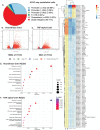
Figure 1.
Figure 1.. Chromatin accessibility assessment of febrile-like HS and TNF-alpha-treated endothelial cells revealed stress response and endothelial dysfunction drivers.
A. Distribution of peaks among genomic features. B. A volcano plot of HS-Cntrl differentially accessible regions (DARs) ATAC-seq regions. C. A volcano plot of TNF-Cntrl (DARs) ATAC-seq regions. D. ATAC-seq motif deviation from HUVEC cells treated with febrile-like HS (39°C for 2hrs) or TNF-alpha (10ng/ml for 6hrs) (C1, C2, T1, T2, and HS1, HS2 are biological replicates of control, TNF-alpha, and heat stress treated cells). E. HS-Cntrl up-regulated KEGG pathway overrepresentation analysis. F. TNF-Cntrl upregulated KEGG pathway overrepresentation analysis.
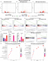
Figure 2.
Figure 2.. Febrile-like HS and TNF-alpha induce a profound transcriptional response, including EC dysfunction drivers.
A. A volcano plot of HS-Cntrl Differentially Expressed Genes, RNA-seq. B. A volcano plot of TNF-Cntrl Differentially Expressed Genes, RNA-seq. C. A volcano plot of TNF-HS Differentially Expressed Genes, RNA-seq. D. Gene Set Enrichment Analysis (GSEA) enrichment plot for cell adhesive molecules, CAMs. E. GSEA enrichment plot for mesenchymal transition genes. F. Q-PCR to confirm stress-induced upregulation of markers for EC dysfunction, CAMs, and anti-inflammatory protein KLF4. G. GSEA enrichment plots for cytokine signaling and chaperone-mediated protein folding for TNF-HS. H. KEGG analysis for HS-Cntrl and I. for TNF-Cntrl RNA-seq samples.
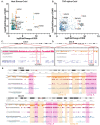
Figure 3.
Figure 3.. Transcriptional changes in multiple EC dysfunction markers also undergo alterations at the chromatin accessibility level.
A. Integrative analysis of ATAC-seq and RNA-seq for HS-Cntrl and B. for TNF-Cntrl (in orange color are examples of stress-specific genes, in red are examples of EC dysfunctional genes). C. An IGV browser snapshot of a region of chr.1 with human endothelial vein cells Hi-C track (ENCODE data accession number: ENCFF005ZBU), HS-T upregulated DARs, and TNF-C upregulated DARs at the site of VCAM1 gene. D. An IGV browser snapshot of a region of chr.5 with human endothelial vein cells Hi-C track (ENCODE data accession number: ENCFF005ZBU), HS-T upregulated DARs track and TNF-C upregulated DARs track at the site of VCAN gene. E. An IGV browser snapshot of a chr.9 and chr.11 with human endothelial vein cells Hi-C track (ENCODE data accession number: ENCFF005ZBU), HS-T upregulated and downregulated DARs tracks, and TNF-C upregulated and downregulated DARs tracks suggests more ATAC-seq DARs are within compartment A than compartment B.
Figure 4.
Figure 4.. Dynamics in centromere-nucleoli association during inflammatory stress exposure depends on p38 MAPK kinase.
A. Changes in centromere genes chromatin accessibility during exposure to inflammatory stresses. B. Confocal microscopy images of the centromere (CREST, magenta) -nucleoli (nucleoli, cyan) association in control and TNF-alpha treated HAEC cells (scale bar 10 μm). C. Quantification of nucleoli-centromere association: each dot represents the percentage of nucleoli-associated centromeres per nuclei. D. Quantification of alpha satellite (ASAT) transcripts per cell in control and TNF-alpha-treated cells. E. Q-PCR for endothelial dysfunction genes ICAM1, VCAM1, and IL-6 upregulation as a result of TNF-alpha exposure in HAEC cells. F. Quantification of nucleoli-centromere association in TNF-alpha and HS-treated HUVEC cells; each dot represents the percentage of nucleoli-associated centromeres per nuclei. When indicated, cells were exposed to stresses in the presence of p38MAPK inhibitors BIRB and SB. G. Confocal microscopy images of the centromere (CREST, magenta)-nucleoli (nucleoli, cyan) association in control and TNF-alpha treated HUVEC cells in the presence of p38 inhibitors, BIRB and SB (scale bar 10 μm). H. Model of the role of p38 in centromere-nucleoli dynamics during inflammatory stress. I. Nucleoli marker nucleophosmin (cyan, red arrow) disruption during HS recovery. Centrosome (pericentrin marker, PCNT, magenta, white arrow) is used as an intracellular marker for cellular recovery from stress in HUVEC cells, scale bar 10 μm.

Similar articles

References

    1. Evans S. S., Repasky E. A. & Fisher D. T. Fever and the thermal regulation of immunity: The immune system feels the heat. Nature Reviews Immunology (2015). doi:10.1038/nri3843 - DOI - PMC - PubMed
    1. Hotamisligil G. S. Inflammation, metaflammation and immunometabolic disorders. Nature (2017). doi:10.1038/nature21363 - DOI - PubMed
    1. Hsu T., Nguyen-Tran H. H. & Trojanowska M. Active roles of dysfunctional vascular endothelium in fibrosis and cancer. J. Biomed. Sci. 26, 1–12 (2019). - PMC - PubMed
    1. Steyers C. M. & Miller F. J. Endothelial dysfunction in chronic inflammatory diseases. International Journal of Molecular Sciences (2014). doi:10.3390/ijms150711324 - DOI - PMC - PubMed
    1. Pober J. S. & Sessa W. C. Evolving functions of endothelial cells in inflammation. Nature Reviews Immunology (2007). doi:10.1038/nri2171 - DOI - PubMed

Publication types