. 2023 Sep 27;15(10):2390.
doi: 10.3390/pharmaceutics15102390.

Combination of Phenethyl Isothiocyanate and Dasatinib Inhibits Hepatocellular Carcinoma Metastatic Potential through FAK/STAT3/Cadherin Signalling and Reduction of VEGF Secretion

Affiliations

Combination of Phenethyl Isothiocyanate and Dasatinib Inhibits Hepatocellular Carcinoma Metastatic Potential through FAK/STAT3/Cadherin Signalling and Reduction of VEGF Secretion

Gabriele Strusi et al. Pharmaceutics. .

Abstract

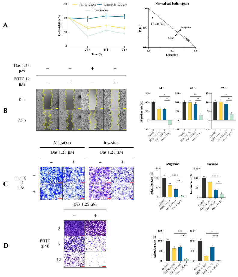
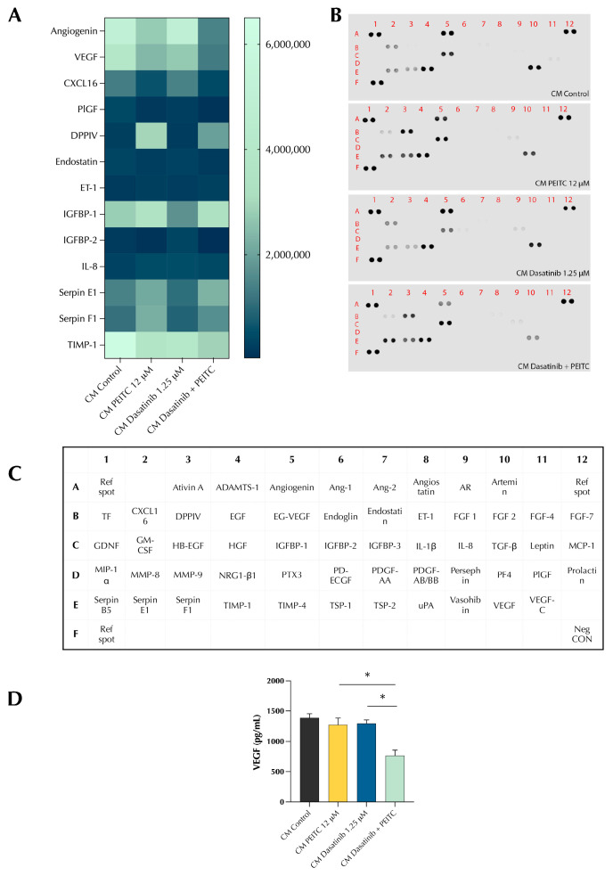
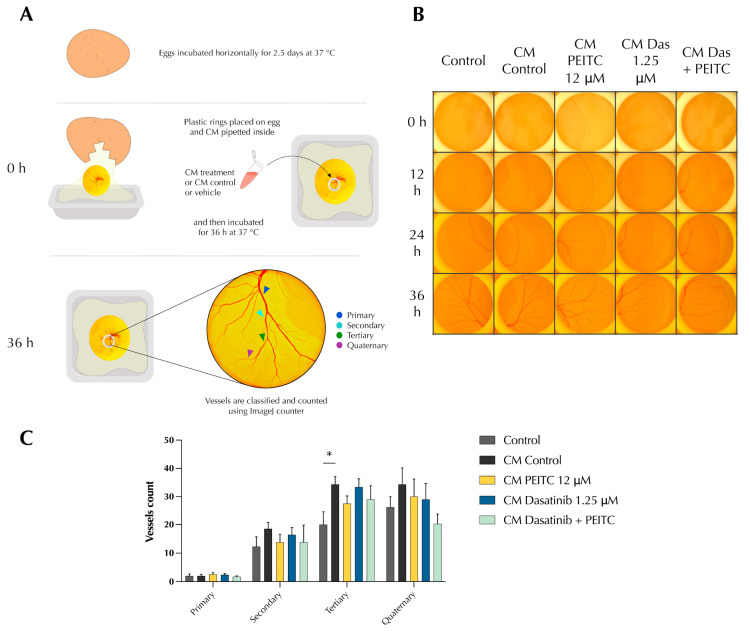
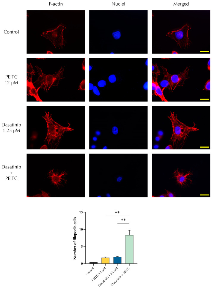
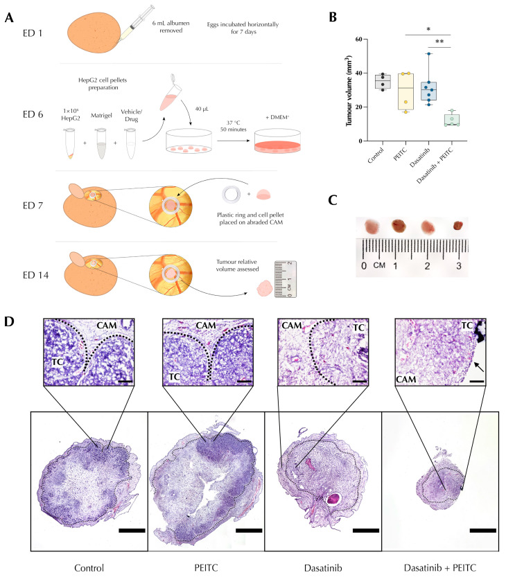
Cancerous cells are characterised by their ability to invade, metastasise, and induce angiogenesis. Tumour cells use various molecules that can be targeted to reverse these processes. Dasatinib, a potent Src inhibitor, has shown promising results in treating hepatocellular carcinoma (HCC) in vitro and in vivo. However, its effectiveness is limited by focal adhesion kinase (FAK) activation. Isothiocyanates, on the other hand, are phytochemicals with broad anticancer activity and FAK inhibition capabilities. This study evaluated the synergistic effect of dasatinib and phenethyl isothiocyanate (PEITC) on HCC. The combination was tested using various assays, including MTT, adhesion, scratch, Boyden chamber, chorioallantoic membrane (CAM), and yolk sac membrane (YSM) assays to evaluate the effect of the drug combination on HCC metastatic potential and angiogenesis in vitro and in vivo. The results showed that the combination inhibited the adhesion, migration, and invasion of HepG2 cells and reduced xenograft volume in the CAM assay. Additionally, the combination reduced angiogenesis in vitro, diminishing the growth of vessels in the tube formation assay. The inhibition of FAK/STAT3 signalling led to increased E-cadherin expression and reduced VEGF secretion, reducing HCC metastatic potential. Therefore, a combination of PEITC and dasatinib could be a potential therapeutic strategy for the treatment of HCC.

Keywords: CAM assay; FAK; HCC; PEITC; VEGF; cancer therapeutics; combination therapy; dasatinib; drug development; oncology.


Conflict of interest statement

The authors declare no conflict of interest.

Figures

Figure 1
Figure 1
Effect of PDc on cell proliferation, adhesion, migration, and invasion. (A) Left, HepG2 cell viability measurement via MTT assay (mean ± SEM, n = 3). Right, combination index value at 24 h calculated with CompuSyn software. (B) Scratch wound healing assay, representative images (5×, scale bar 200 µm) on the left, quantification of migration rate at 24 h, 48 h, 72 h employing ImageJ 1.55k plugin on the right (mean ± SEM, n = 3). (C) Migration and invasion assay using Boyden chambers. Left, representative images (20×, scale bar 100 µm); right, quantification of migration and invasion rate (mean ± SEM, n = 3). (D) Adhesion assay, representative images (10×, scale bar 200 µm) on the left, quantification of adhesion rate by ImageJ 1.55k on the right (mean ± SEM, n = 3). The degrees of significance are indicated as * p < 0.05, ** p < 0.01, *** p < 0.001, **** p < 0.0001, calculated with one-way ANOVA and Tukey’s post hoc multiple comparisons test.
Figure 2
Figure 2
Effect of PDc on tumour-induced angiogenesis in vitro. (A) Diagram of HepG2-conditioned medium (CM) model. (B) Effect of HepG2-conditioned medium on HUVECs adhesion and migration, representative images (10×, scale bar 20 µm) on the top; quantification of cell viability, adhesion, and migration rate with ImageJ 1.55k on the bottom. Effect of PDc HepG2-conditioned medium on HUVECs: (C) adhesion, representative images (10×, scale bar 20 µm) on the left, quantification of adhesion rate using ImageJ 1.55k on the right (mean ± SEM, n = 4); (D) migration, representative images (10×, scale bar 20 µm) on the left, quantification of migration rate with ImageJ (mean ± SEM, n = 4); (E) in vitro angiogenesis representative images (4×, scale bar 200 µm) on the left; quantification of segment length and number of meshes with ImageJ on the right (mean ± SEM, n = 4). The degrees of significance are indicated as * p < 0.05, ** p < 0.01, **** p < 0.0001 calculated with one-way ANOVA and Tukey’s post hoc multiple comparison.
Figure 3
Figure 3
Effect of PDc on HepG2-conditioned medium. (A) Heat map of angiogenesis proteome profiler. (B) Blots of Angiogenesis Proteome Profiler. (C) Legend of targets location on blot. (D) VEGF quantification of conditioned medium via ELISA. The degrees of significance are indicated as * p < 0.05, calculated with one-way ANOVA and Tukey’s post hoc multiple comparison.
Figure 4
Figure 4
Effect of PDc on HepG2-CM-induced angiogenesis in vivo. (A) Diagram of HepG2-conditioned medium model yolk sac membrane assay. (B) Representative images of YSM assay from three independent experiments (100×, scale bar 20 µm). (C) Quantification based on vessel size hierarchy using ImageJ 1.55k from three independent experiments (mean ± SEM, Control n = 4, CM Control n = 6, CM PEITC n = 4, CM Dasatinib n = 5, CM P + D n = 4). The degrees of significance are indicated as * p < 0.05, calculated with Student’s t-test.
Figure 5
Figure 5
Effect of PDc FAK/STAT3/Cadherin signalling (A) Western blot analysis of FAK and STAT3 expression; images are representative of three independent protein extractions. Data are depicted as fold change to control normalised to β-Actin expression (mean ± SEM, n ≥ 3) (B) Western blot analysis of E-Cadherin and N-Cadherin expression; images are representative of three independent protein extractions. Data are depicted as fold change to control normalised to β-Actin expression (mean ± SEM, n ≥ 3). The degrees of significance are indicated as * p < 0.05, ** p < 0.01, calculated using one-way ANOVA and Tukey’s post hoc multiple comparison.
Figure 6
Figure 6
Effect of PDc on HepG2 morphology. Top, phalloidin staining of f-actin in red, DAPI staining of nuclei in blue. Bottom, quantification of cells presenting filopodia structures. The degrees of significance are indicated as ** p < 0.01, calculated using one-way ANOVA and Tukey’s post hoc multiple comparison.
Figure 7
Figure 7
Effect of PDc on HepG2 chorioallantoic membrane (CAM) xenograft. (A) Diagram of HepG2 CAM xenograft method. (B) Quantification of tumour volume in three independent experiments (mean ± SEM, Control n = 4, PEITC n = 4, Dasatinib n = 6, P + D n = 5). The degrees of significance are indicated as * p < 0.05, ** p < 0.01, calculated using one-way ANOVA and Tukey’s post hoc multiple comparison. (C) Representative images of resected HepG2 xenografts (from the left: control, PEITC, dasatinib, PDc). (D) CAM xenograft histology H&E staining. TC = tumour cells, CAM = chorioallantoic membrane, dotted lines separation of tumour cells and CAM. Arrow indicate the edge of the tumour mass that is not surrounded by the CAM. Scale bar = 1 mm.

Similar articles

Cited by

References

    1. Calderon-Martinez E., Landazuri-Navas S., Vilchez E., Cantu-Hernandez R., Mosquera-Moscoso J., Encalada S., Al Lami Z., Zevallos-Delgado C., Cinicola J. Prognostic Scores and Survival Rates by Etiology of Hepatocellular Carcinoma: A Review. J. Clin. Med. Res. 2023;15:200–207. doi: 10.14740/jocmr4902. - DOI - PMC - PubMed
    1. Zhang H., Zhang W., Jiang L., Chen Y. Recent advances in systemic therapy for hepatocellular carcinoma. Biomark. Res. 2022;10:3. doi: 10.1186/s40364-021-00350-4. - DOI - PMC - PubMed
    1. Scott E.C., Baines A.C., Gong Y., Moore R., Pamuk G.E., Saber H., Subedee A., Thompson M.D., Xiao W., Pazdur R., et al. Trends in the approval of cancer therapies by the FDA in the twenty-first century. Nat. Rev. Drug Discov. 2023;22:625–640. doi: 10.1038/s41573-023-00723-4. - DOI - PubMed
    1. Araujo J., Logothetis C. Dasatinib: A potent SRC inhibitor in clinical development for the treatment of solid tumors. Cancer Treat. Rev. 2010;36:492–500. doi: 10.1016/j.ctrv.2010.02.015. - DOI - PMC - PubMed
    1. Chang A.Y., Wang M. In-vitro growth inhibition of chemotherapy and molecular targeted agents in hepatocellular carcinoma. Anticancer Drugs. 2013;24:251–259. doi: 10.1097/CAD.0b013e32835ba289. - DOI - PubMed

LinkOut - more resources