. 2023 Oct 22;24(20):15454.
doi: 10.3390/ijms242015454.

Activation of the TNF-α-Necroptosis Pathway in Parvalbumin-Expressing Interneurons of the Anterior Cingulate Cortex Contributes to Neuropathic Pain

Affiliations

Activation of the TNF-α-Necroptosis Pathway in Parvalbumin-Expressing Interneurons of the Anterior Cingulate Cortex Contributes to Neuropathic Pain

Yiwen Duan et al. Int J Mol Sci. .

Abstract

The hyperexcitability of the anterior cingulate cortex (ACC) has been implicated in the development of chronic pain. As one of the key causes of ACC hyperexcitation, disinhibition of the ACC may be closely related to the dysfunction of inhibitory parvalbumin (PV)-expressing interneurons (PV-INs). However, the molecular mechanism underlying the ACC PV-INs injury remains unclear. The present study demonstrates that spared sciatic nerve injury (SNI) induces an imbalance in the excitation and inhibition (E/I) of the ACC. To test whether tumor necrosis factor-α (TNF-α) upregulation in the ACC after SNI activates necroptosis and participates in PV-INs damage, we performed a differential analysis of transcriptome sequencing using data from neuropathic pain models and found that the expression of genes key to the TNF-α-necroptosis pathway were upregulated. TNF-α immunoreactivity (IR) signals in the ACCs of SNI rats were co-located with p-RIP3- and PV-IR, or p-MLKL- and PV-IR signals. We then systematically detected the expression and cell localization of necroptosis-related proteins, including kinase RIP1, RIP3, MLKL, and their phosphorylated states, in the ACC of SNI rats. Except for RIP1 and MLKL, the levels of these proteins were significantly elevated in the contralateral ACC and mainly expressed in PV-INs. Blocking the ACC TNF-α-necroptosis pathway by microinjecting TNF-α neutralizing antibody or using an siRNA knockdown to block expression of MLKL in the ACC alleviated SNI-induced pain hypersensitivity and inhibited the upregulation of TNF-α and p-MLKL. Targeting TNF-α-triggered necroptosis within ACC PV-INs may help to correct PV-INs injury and E/I imbalance in the ACC in neuropathic pain.

Keywords: anterior cingulate cortex; necroptosis; neuropathic pain; parvalbumin interneuron; tumor necrosis factor-α.


Conflict of interest statement

The authors declare no conflict of interest.

Figures
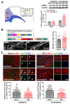
Figure 1
Figure 1
SNI induces an excitation/inhibition (E/I) imbalance in the ACC neural network structure. (A) SNI-induced upregulation of CaMKⅡ (a pyramidal neuron marker) in the contralateral ACC. The left part of the image shows that the ACC samples used to perform the Western blot include the Cg1 and Cg2 regions located between 2.2 and 0.5 mm on the anterior coronal plane of the bregma. A representative Western blot of CaMKⅡ expression in the bilateral ACC and the quantitative results of the Western blot is shown on the right. *** p < 0.001 versus the sham group (two-way ANOVA). (B) Supernova revealed a significant increase in the number of dendritic spines of CaMKⅡ-positive neurons in the ACC of mice subjected to SNI. Upper left: schematic for the Supernova vector set for sparsely labeled ACC neurons. Representative data and quantitative analysis are shown on the lower left and right, respectively. Scale bar = 12.5 μm. ** p < 0.01 versus the sham (unpaired t test). (C) SNI reduces inhibitory synaptic terminals on the surface of bilateral ACC pyramidal neurons. Upper, representative double immunofluorescence staining image showing CaMKⅡ-positive (red) somatic vGAT puncta-IR (inhibitory synaptic terminal marker, green) in the sham and SNI groups in the bilateral ACC. An enlarged image of the area enclosed by the white box is shown on the right. The white arrow indicates the colocalization (yellow) of vGAT puncta with CaMKⅡ. Blue fluorescence corresponds to DAPI, a nuclear counterstain. Below is the quantitative analysis of the number of vGAT-IR puncta on CaMKⅡ-positive neurons in the bilateral ACC of the sham and SNI groups. * p < 0.05, *** p < 0.001 versus the sham (unpaired t test).
Figure 2
Figure 2
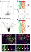
TNF-α-necroptosis pathway is activated in the ACC of neuropathic pain models. Volcano map (A) and heat map (B) showing the expression of TNF-α, RIP1, RIP3, and MLKL in the ACC of CCI model of neuropathic pain (n = 3/group) and controls (n = 3/group) (data from GEO dataset GSE212311). The Hub gene expression data of CCI model (red dots) and control (green dots) for TNF-α, RIP1, RIP3, and MLKL are shown in (C). * p < 0.05 versus the control (unpaired t test). Volcano map (D) and heat map (E) showing the expression of TNF-α, IL-6, RIP1, and MLKL in the ACC of SNI model (n = 3/group) and controls (n = 3/group) (data from GEO dataset GSE228065). (F) Representative triple staining shows the overlap of TNF-α (green) with p-RIP3 (red) and PV (magenta), or p-MLKL (red) and PV (magenta) on PO day 7. Enlarged and color-split images of the area enclosed in white boxes are shown below. White arrows indicate co-localization. Blue fluorescence corresponds to DAPI.
Figure 3
Figure 3
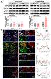
SNI increases the expression of necroptosis-related protein p-RIP1 in the contralateral ACC (A) Representative Western blot of RIP1 and p-RIP1 expression in the bilateral ACC is shown in the top panel. The quantitative results of Western blotting protein are shown below. SNI induces the expression of p-RIP1in the contralateral ACC at postoperative (PO) day 7. ** p < 0.01 versus the sham group (two-way ANOVA). (B) Representative double staining shows the overlap (yellow) of p-RIP1 (red) with NeuN (neuronal marker, green) and PV (PV-IN marker, green), but not with GFAP (astrocyte marker, green) or Iba1 (microglia marker, green), on PO day 7. Enlarged and color-split images of the area enclosed in white boxes are shown in the middle. White arrows indicate co-localization (yellow). Blue fluorescence corresponds to DAPI. The fluorescence intensity curves for red and green from boxed areas are shown on the right side of each group.
Figure 4
Figure 4
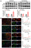
SNI upregulates RIP3/p-RIP3 proteins in the contralateral ACC (A) Representative Western blot of RIP3 and p-RIP3 expression in the bilateral ACC is shown in the top panel. Quantitative results of Western blot are shown below. SNI upregulates RIP3 and p-RIP3 in the contralateral ACC on PO day 7. *** p < 0.001 versus the sham group (two-way ANOVA). (B) Representative double staining showing the overlap (yellow) of p-RIP3 (red) with NeuN (green) and PV (green), but not with GFAP (green) or Iba1 (green), on PO day 7. Enlarged and color-split images of areas enclosed in white boxes are shown in the middle. White arrows indicate co-localization (yellow). Blue fluorescence corresponds to DAPI. The fluorescence intensity curves for red and green from boxed areas are shown on the right side of each group.
Figure 5
Figure 5
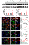
SNI increases the expression of necrotizing membrane-destroying proteins p-MLKL in the contralateral ACC (A) Representative Western blot of MLKL and p-MLKL expression in the bilateral ACC is shown in the top panel. Quantitative results of Western blot are shown below. SNI upregulates p-MLKL in the contralateral ACC on PO day 7. ** p < 0.01 versus the sham group (two-way ANOVA). (B) Representative double staining showing the overlap (yellow) of p-MLKL (red) with NeuN (green) and PV (green), but not with GFAP (green) or Iba1 (green), on PO day 7. Enlarged and color-split images of areas enclosed in white boxes are shown in the middle. White arrows indicate co-localization (yellow). Blue fluorescence corresponds to DAPI. The fluorescence intensity curves for red and green from boxed areas are shown on the right side of each group.
Figure 6
Figure 6
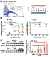
The effect of neutralizing SNI-induced elevation of TNF-α in the ACC on pain behavior and necroptosis activation (A) The injection site of anti-TNF-α antibodies (20 μg/mL, 10 μL) or control IgG (20 μg/mL, 10 μL) in the contralateral ACC is shown on the left, and behavioral test paradigms are shown on the right. (B) Changes in the bilateral paw withdrawal thresholds (PWTs) in the sham, IgG+SNI, and anti-TNF-α+SNI groups. SNI-induced ipsilateral mechanical allodynia in the vehicle control group could be significantly mitigated by microinjecting a TNF-α neutralization antibody on postoperative (PO) days 3, 5, and 7. ** p-value < 0.01, *** p-value < 0.001 versus PO day − 1 (Dunn’s multiple comparisons test) or ### p-value < 0.001 comparison among groups (multiple t test). (C) Representative Western blot showing the effect of the TNF-α neutralization antibody on the expression of TNF-α and p-MLKL in the contralateral ACC (left). Protein quantification results are shown right. * p-value < 0.05, ** p-value < 0.01, *** p-value < 0.001 (two-way ANOVA).
Figure 7
Figure 7
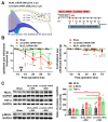
The effect of MLKL knockdown in the contralateral ACC on pain behavior and TNF-α expression. (A) The injection site of MLKL siRNA or control siRNA in the contralateral ACC is shown on the left, and the behavioral test paradigms are shown on the right. (B) Changes in the bilateral paw withdrawal thresholds (PWTs) in the sham, control siRNA+SNI, and MLKL siRNA+SNI groups. SNI-induced ipsilateral mechanical allodynia in the control siRNA group could be partially blocked by MLKL siRNA on postoperative (PO) days 3 and 5. ** p-value < 0.01, *** p-value < 0.001 versus PO day 1 (Dunn’s multiple comparisons test) or # p-value < 0.05, ## p-value < 0.01, ### p-value < 0.001 comparison among groups (multiple t test). (C) Representative Western blot showing the effect of the MLKL siRNA on the expression of MLKL, p-MLKL, and TNF-α in the contralateral ACC (left). Protein quantification results are shown right. * p-value < 0.05, *** p-value < 0.001 (two-way ANOVA).

Similar articles

Cited by

References

    1. Lançon K., Qu C., Navratilova E., Porreca F., Séguéla P. Decreased dopaminergic inhibition of pyramidal neurons in anterior cingulate cortex maintains chronic neuropathic pain. Cell Rep. 2021;37:109933. doi: 10.1016/j.celrep.2021.109933. - DOI - PMC - PubMed
    1. Smith M.L., Asada N., Malenka R.C. Anterior cingulate inputs to nucleus accumbens control the social transfer of pain and analgesia. Science. 2021;371:153–159. doi: 10.1126/science.abe3040. - DOI - PMC - PubMed
    1. Chen T., Taniguchi W., Chen Q.Y., Tozaki-Saitoh H., Song Q., Liu R.H., Koga K., Matsuda T., Kaito-Sugimura Y., Wang J., et al. Top-down descending facilitation of spinal sensory excitatory transmission from the anterior cingulate cortex. Nat. Commun. 2018;9:1886. doi: 10.1038/s41467-018-04309-2. - DOI - PMC - PubMed
    1. Chen T., Koga K., Descalzi G., Qiu S., Wang J., Zhang L.S., Zhang Z.J., He X.B., Qin X., Xu F.Q., et al. Postsynaptic potentiation of corticospinal projecting neurons in the anterior cingulate cortex after nerve injury. Mol. Pain. 2014;10:33. doi: 10.1186/1744-8069-10-33. - DOI - PMC - PubMed
    1. Li Q.Y., Chen S.X., Liu J.Y., Yao P.W., Duan Y.W., Li Y.Y., Zang Y. Neuroinflammation in the anterior cingulate cortex: The potential supraspinal mechanism underlying the mirror-image pain following motor fiber injury. J. Neuroinflamm. 2022;19:162. doi: 10.1186/s12974-022-02525-8. - DOI - PMC - PubMed

MeSH terms

Substances