Review
. 2023 Oct 13;24(20):15162.
doi: 10.3390/ijms242015162.

Interaction of Calmodulin with TRPM: An Initiator of Channel Modulation

Affiliations
Review

Interaction of Calmodulin with TRPM: An Initiator of Channel Modulation

Kristyna Vydra Bousova et al. Int J Mol Sci. .

Abstract

Transient receptor potential melastatin (TRPM) channels, a subfamily of the TRP superfamily, constitute a diverse group of ion channels involved in mediating crucial cellular processes like calcium homeostasis. These channels exhibit complex regulation, and one of the key regulatory mechanisms involves their interaction with calmodulin (CaM), a cytosol ubiquitous calcium-binding protein. The association between TRPM channels and CaM relies on the presence of specific CaM-binding domains in the channel structure. Upon CaM binding, the channel undergoes direct and/or allosteric structural changes and triggers down- or up-stream signaling pathways. According to current knowledge, ion channel members TRPM2, TRPM3, TRPM4, and TRPM6 are directly modulated by CaM, resulting in their activation or inhibition. This review specifically focuses on the interplay between TRPM channels and CaM and summarizes the current known effects of CaM interactions and modulations on TRPM channels in cellular physiology.

Keywords: TRPM channels; calcium homeostasis; calmodulin; calmodulin binding site; regulation.


Conflict of interest statement

The authors declare no conflict of interest.

Figures

Figure 1
Figure 1
Common schematic and representative structure of the TRPM channel. (A) Common membrane topology of TRPM in a dimer visualization. The TRPM monomeric unit consists of six transmembrane helices (orange and pink helices), with a pore region between helices five and six (channel centre, pink part). The pore serves to transport monovalent and divalent ions. The intracellular N- and C-termini modulatory domains present in blue and green bulbs are key players in binding intracellular regulatory molecules and changing the structural conformation of the entire channel to open or close the pore region for ion transport. The pink ball in the TRPM scheme at the N-termini location represents a potential ligand binding site. (B) Side view of the structure of TRPM4 structure (PDB: 6BQR). Both representations show the transmembrane part of the TRPM/TRPM4 channel in yellow/orange/red; N-termini in green/blue; and C-termini intracellular domains in deep violet and red. (A) was adapted from the “TRPM 2 channel” template by BioRender.com, retrieved from “https://app.biorender.com/biorender-templates (accessed on 20 July 2023); (B) was generated using PyMol software, version 1.20 [38].
Figure 2
Figure 2
CaM complex formations with TRP channel binding sites. (A) Structure of apo-CaM (PDB: 1QX5) and (B) holo-CaM complexed with Ca2+ (PDB: 5A2A). (C) Side and (D) front view of the interface of the CaM/Ca2+—TRPV1 peptide (PDB: 3SUI) complex in the backbone representation. (E) Side and (F) front view of the CaM/Ca2+—TRPM6 peptide complex interface in sphere representation as a result of molecular modelling and molecular dynamics simulations (MDs) [72]. TRPM6 binding site in orange (sphere representation; red represents basic AA residues; LIGRAYRSNYTRKHFR (bold) confirmed to be involved in the salt bridge formations with their CaM-binding counterparts). Color convention: CaM backbone shown in rainbow colors according to *CA atoms; pink spheres represent Ca2+; TRPV1 peptide backbone is shown in brown; TRPM6 peptide in ball orange/red representation.
Figure 3
Figure 3
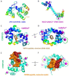
Localization of representative CaM binding sites at the TRPM4 and TRPM6 N-termini intracellular tails. (A,C) Side and (B,D) bottom view of TRPM4 structure ((A,B) PDB: 6BQR) and TRPM6 homology model (C,D) [22,72] with identified highlighted CaM binding sites accessible to CaM from intracellular cell environment [74,75]. Color convention: TRPM4 and TRPM6 backbones shown in rainbow ribbon according to *CA atoms; CaM binding sites of TRPM channels shown in pink and violet ball representations.
Figure 4
Figure 4
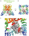
Structure of TRPV5 in the complex with CaM-Ca2+ (PDB: 6DMW) [87]. Cartoon representation of (A) side and (B) bottom views of CaM-bound TRPV5. (C) A detailed view of CaM bound in the complex with TRPV5 revealed two binding sites for N- and C-lobes of the CaM. Color convention: TRPV5 backbone in rainbow ribbon spectrum according to C-alpha; CaM in a surface representation, grey color; Ca2+ in the complex with CaM are shown as a magenta ball representation.

Similar articles

References

    1. Voets T., Talavera K., Owsianik G., Nilius B. Sensing with TRP channels. Nat. Chem. Biol. 2005;1:85–92. doi: 10.1038/nchembio0705-85. - DOI - PubMed
    1. Jimenez I., Prado Y., Marchant F., Otero C., Eltit F., Cabello-Verrugio C., Cerda O., Simon F. TRPM Channels in Human Diseases. Cells. 2020;9:2604. doi: 10.3390/cells9122604. - DOI - PMC - PubMed
    1. Lopez-Romero A.E., Hernandez-Araiza I., Torres-Quiroz F., Tovar Y.R.L.B., Islas L.D., Rosenbaum T. TRP ion channels: Proteins with conformational flexibility. Channels. 2019;13:207–226. doi: 10.1080/19336950.2019.1626793. - DOI - PMC - PubMed
    1. Samanta A., Hughes T.E.T., Moiseenkova-Bell V.Y. Transient Receptor Potential (TRP) Channels. Subcell Biochem. 2018;87:141–165. - PMC - PubMed
    1. Vangeel L., Voets T. Transient Receptor Potential Channels and Calcium Signaling. Cold Spring Harb. Perspect. Biol. 2019;11:a035048. doi: 10.1101/cshperspect.a035048. - DOI - PMC - PubMed