. 2023 Oct 26;14(1):6605.
doi: 10.1038/s41467-023-42330-2.

FHL1 promotes chikungunya and o'nyong-nyong virus infection and pathogenesis with implications for alphavirus vaccine design

Affiliations

FHL1 promotes chikungunya and o'nyong-nyong virus infection and pathogenesis with implications for alphavirus vaccine design

Wern Hann Ng et al. Nat Commun. .

Abstract

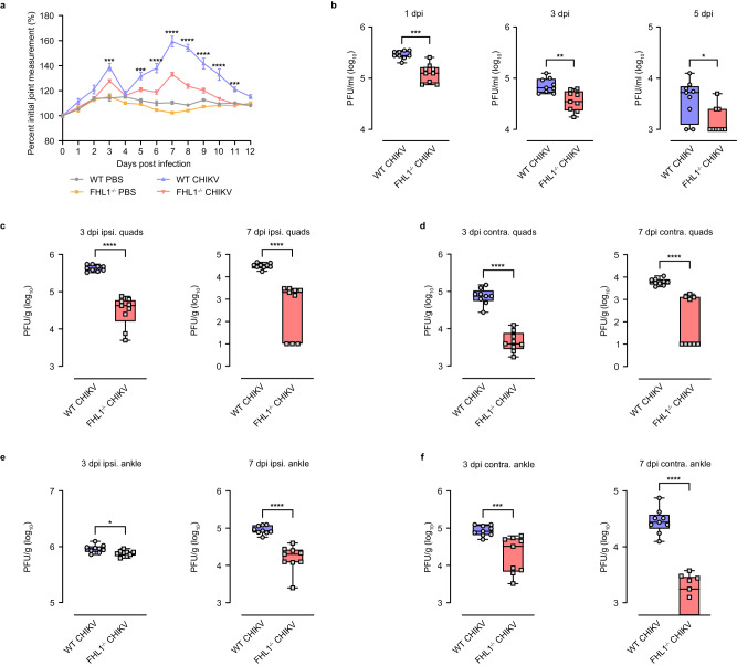
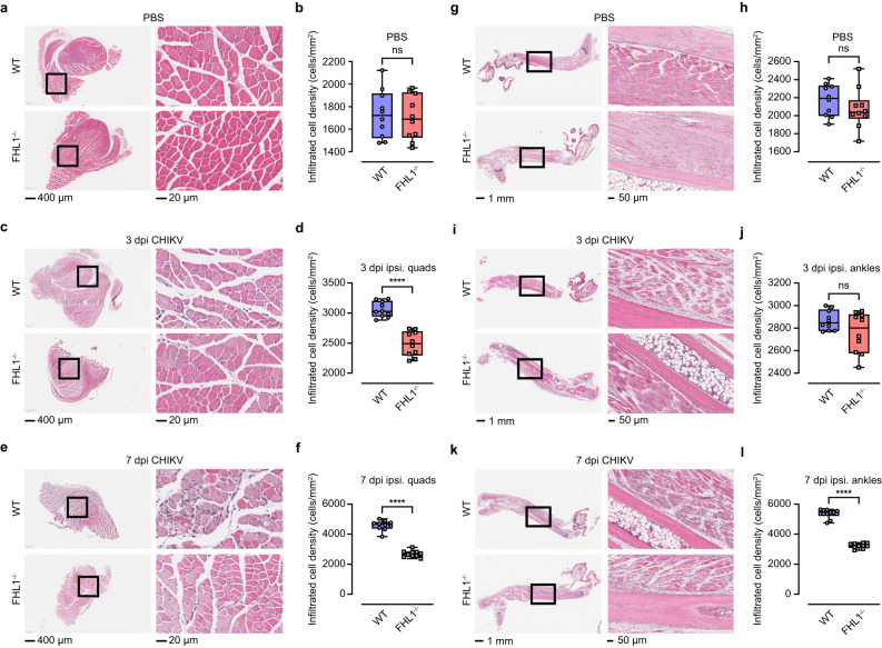
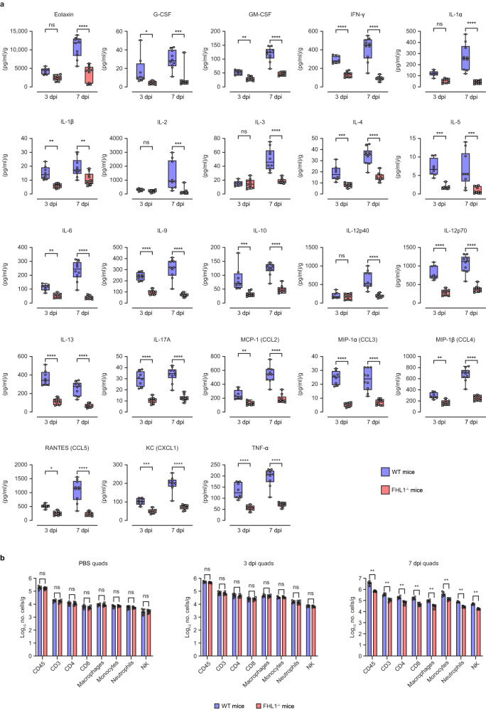
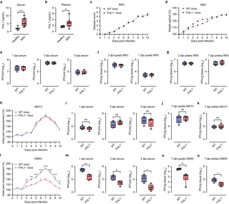
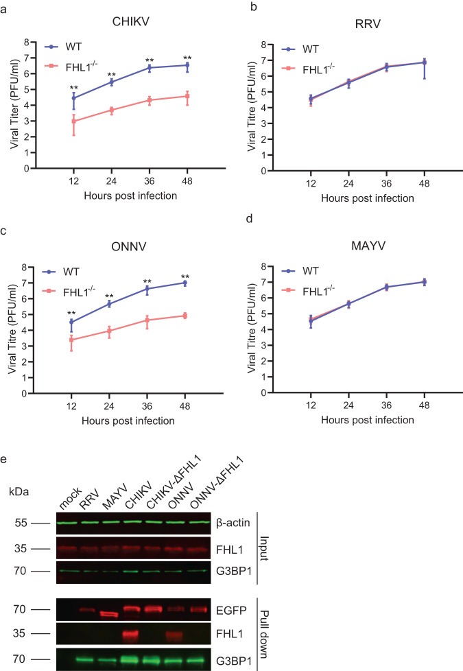
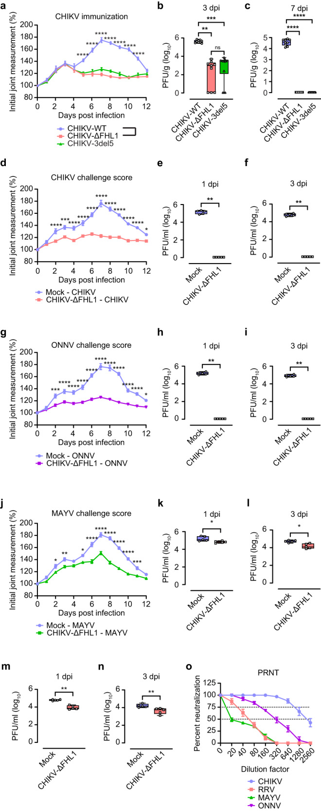
Arthritogenic alphaviruses are positive-strand RNA viruses that cause debilitating musculoskeletal diseases affecting millions worldwide. A recent discovery identified the four-and-a-half-LIM domain protein 1 splice variant A (FHL1A) as a crucial host factor interacting with the hypervariable domain (HVD) of chikungunya virus (CHIKV) nonstructural protein 3 (nsP3). Here, we show that acute and chronic chikungunya disease in humans correlates with elevated levels of FHL1. We generated FHL1-/- mice, which when infected with CHIKV or o'nyong-nyong virus (ONNV) displayed reduced arthritis and myositis, fewer immune infiltrates, and reduced proinflammatory cytokine/chemokine outputs, compared to infected wild-type (WT) mice. Interestingly, disease signs were comparable in FHL1-/- and WT mice infected with arthritogenic alphaviruses Ross River virus (RRV) or Mayaro virus (MAYV). This aligns with pull-down assay data, which showed the ability of CHIKV and ONNV nsP3 to interact with FHL1, while RRV and MAYV nsP3s did not. We engineered a CHIKV mutant unable to bind FHL1 (CHIKV-ΔFHL1), which was avirulent in vivo. Following inoculation with CHIKV-ΔFHL1, mice were protected from disease upon challenge with CHIKV and ONNV, and viraemia was significantly reduced in RRV- and MAYV-challenged mice. Targeting FHL1-binding as an approach to vaccine design could lead to breakthroughs in mitigating alphaviral disease.


Conflict of interest statement

The authors declare no competing interests.

Figures

Fig. 1
Fig. 1. FHL1 levels are increased in CHIKV infected humans and mice.
The levels of FHL1 in the serum from acute and chronic CHIKV disease patients were determined by ELISA (a). Data are presented as box and whisker ± SD with the mean indicated by a line across the box, maximum to minimum points. Dots represent individual participant (healthy individuals (control): n = 8; acute CHIKV patients: n = 39; chronic CHIKV patients: n = 17 (*P < 0.05; ****P < 0.0001; one-way ANOVA with the Kruskal–Wallis posttest). Mice were infected with CHIKV at 104 PFU or mock-infected with PBS. Serum was collected at 1, 3 and 5 dpi, and the ipsilateral quadriceps were harvested at 7 dpi. The quadriceps were processed for immunofluorescence staining (b) and qRT‒PCR analysis of FHL1 mRNA (c). The confocal immunofluorescence microscopy images shown are representative of n = 5 mice per group; the data are representative of two independent experiments. The serum was processed for ELISA analysis of FHL1 (d). Dots represent individual animals (n = 10). Data are representative of two independent experiments. Data are presented as box and whisker ± SD with the mean indicated by a line across the box, maximum to minimum points (*P < 0.05; ***P < 0.001; ****P < 0.0001; Mann–Whitney test) (c, d). Source data are provided as a Source Data file.
Fig. 2
Fig. 2. FHL1 facilitates CHIKV disease development and viral replication in mice.
WT and FHL1−/− mice were infected with CHIKV at 104 PFU or mock-infected with PBS. Disease was monitored daily and assessed by measuring the height and width of the perimetatarsal area of the ipsilateral hind foot (a). The data shown are representative of two independent experiments; n = 10 mice per group (***P < 0.001; ****P < 0.0001; two-way ANOVA with the Bonferroni posttest). Serum was collected at 1, 3 and 5 dpi and processed for plaque assays (b). Dots represent individual animals (n = 9). The data shown are representative of two independent experiments. Data are presented as box and whisker ± SD with the mean indicated by a line across the box, maximum to minimum points (*P < 0.05; **P < 0.01; ***P < 0.001; Mann–Whitney test). The contralateral and ipsilateral quadriceps and ankles were harvested at 3 and 7 dpi. Viral titers were determined by a plaque assay (cf). Dots represent individual animals (n = 9). The data shown are representative of two independent experiments. Data are presented as box and whisker ± SD with the mean indicated by a line across the box, maximum to minimum points (*P < 0.05; ***P < 0.001; ****P < 0.0001; Mann–Whitney test). Source data are provided as a Source Data file.
Fig. 3
Fig. 3. FHL1 facilitates cell infiltration in the quadriceps and ankles of mice with CHIKV disease.
WT and FHL1−/− mice were infected with CHIKV at 104 PFU or mock-infected with PBS. The ipsilateral quadriceps were collected at 3 and 7 dpi and processed for H&E staining (a, c, e). The microscopy images shown are representative of n = 10 mice per group. The numbers of infiltrated cells were analyzed using ImageScope [Min Nuclear Size (µm2) = 20; Max Nuclear Size (µm2) = 200; Min Roundness = 0.4; Min Compactness = 0.4; Min Elongation = 0.2]. Dots represent individual animals (n = 10); the data shown are representative of two independent experiments (b, d, f). Data are presented as box and whisker ± SD with the mean indicated by a line across the box, maximum to minimum points (ns, nonsignificant; ****P < 0.0001; Mann–Whitney test). The ipsilateral ankles were collected at 3 and 7 dpi and processed for H&E staining (g, i, k). The microscopy images shown are representative of n = 10 mice per group. The numbers of infiltrated cells were analyzed using ImageScope [Min Nuclear Size (µm2) = 20; Max Nuclear Size (µm2) = 200; Min Roundness = 0.4; Min Compactness = 0.4; Min Elongation = 0.2]. Dots represent individual animals (n = 10); the data shown are representative of two independent experiments (h, j, l). Data are presented as box and whisker ± SD with the mean indicated by a line across the box, maximum to minimum points (ns, nonsignificant; ****P < 0.0001; Mann–Whitney test). Source data are provided as a Source Data file.
Fig. 4
Fig. 4. Knock-out of FHL1 reduces levels of proinflammatory cytokines and chemokines in CHIKV-infected mice.
WT and FHL1−/− mice were infected with CHIKV at 104 PFU. The ipsilateral quadriceps were collected at 3 and 7 dpi and processed for a cytokine/chemokine multiplex assay (a). Dots represent individual animals (n = 8, WT mice; n = 10, FHL1−/− mice); the data shown are representative of two independent experiments. Data are presented as box and whisker ± SD with the mean indicated by a line across the box, maximum to minimum points (ns, nonsignificant; *P < 0.05; **P < 0.01; ***P < 0.001 ****P < 0.0001; two-way ANOVA with the Holm–Sidak posttest). WT and FHL1−/− mice were infected with 104 PFU CHIKV or mock-infected with PBS. The ipsilateral quadriceps were harvested at 3 and 7 dpi and processed for flow cytometry (b). Dots represent individual animals (n = 5); the data shown are representative of two independent experiments. Data are presented as the mean ± SEM from (ns nonsignificant; **P < 0.01; Mann–Whitney test). Source data are provided as a Source Data file.
Fig. 5
Fig. 5. Impact of FHL1 on RRV, MAYV and ONNV infection.
The levels of FHL1 in the serum and plasma from RRV disease patients were determined by ELISA (a, b). Dots represent individual participant (n = 10) (**P < 0.01; ****P < 0.0001; Mann-Whitney test). WT and FHL1−/− mice were infected with RRV at 104 PFU. Disease score and weight were monitored daily (c, d), (n = 5; two-way ANOVA with the Bonferroni posttest). Mouse serum was collected at 1, 3 and 5 dpi and processed for plaque assays (e). Mouse quadriceps and ankles were harvested at 3 and 7 dpi. Viral titers were determined by a plaque assay (f, g). For eg, dots represent individual animals; n = 5 mice per group. WT and FHL1−/− mice were infected with MAYV at 104 PFU. Mouse disease was monitored daily (h). (n = 5; two-way ANOVA with the Bonferroni posttest). Mouse serum was collected at 1, 3 and 5 dpi and processed for plaque assays (i). Mouse quadriceps and ankles were harvested at 3 and 7 dpi. Viral titers were determined by a plaque assay (j, k). For ik, dots represent individual animals; n = 5 mice per group (ns, nonsignificant). WT and FHL1−/− mice were infected with ONNV at 104 PFU. Mouse disease was monitored daily (l). (**P < 0.01; ***P < 0.001; ****P < 0.0001; n = 5; two-way ANOVA with the Bonferroni posttest). Mouse serum was collected at 1, 3 and 5 dpi and processed for plaque assays (m). Mouse quadriceps and ankles were harvested at 3 and 7 dpi. Viral titers were determined by a plaque assay (no). For panel m to o, dots represent individual animals; n = 5 mice per group (*P < 0.05; **P < 0.01; Mann–Whitney test). Data are presented as box and whisker ± SD with the mean indicated by a line across the box, maximum to minimum points for a, b, eg, ik and mo (Mann–Whitney test). Source data are provided as a Source Data file.
Fig. 6
Fig. 6. FHL1 is essential for the replication of CHIKV and ONNV.
Multi-step growth curves of CHIKV (a), RRV (b), ONNV (c) and MAYV (d). Primary murine fibroblasts derived from WT or FHL1−/− mice were infected at an MOI of 0.01. Data are presented as the mean ± SD from two experiments each performed in five technical replicates (**P < 0.05; Mann–Whitney test). EGFP pull-down experiment (e). U2OS cells were transfected with plasmids expressing EGFP fused with HVD of nsP3 of RRV, MAYV, CHIKV, CHIKV-ΔFHL1, ONNV or ONNV-ΔFHL1. At 24 h post transfection cells were harvested, lysed, and cellular proteins interacting with HVD of nsP3 of used viruses were pulled down using EGFP-binding magnetic beads. Proteins in input and in pull-down fraction were analyzed using SDS-PAGE and immunoblotting. Input: detection of G3PB1 and FHL1 in cell lysates before pull-down; antibodies against β-actin were used to detect loading control. Pull-down: proteins pulled down with EGFP-binding magnetic beads were detected with antibodies against EGFP, G3BP1 and FHL1. Images from one out of two reproducible experiments are shown. Source data are provided as a Source Data file.
Fig. 7
Fig. 7. CHIKV-ΔFHL1 is attenuated and protects vaccinated mice against infection of different alphaviruses.
Four-week-old WT mice were inoculated with 104 PFU CHIKV-ΔFHL1, CHIKV-3del5 or CHIKV-WT. Disease were monitored daily (a). The data are representative of two independent experiments (****P < 0.0001; n = 9; two-way ANOVA with the Bonferroni posttest). The ipsilateral quadriceps were harvested at 3 and 7 dpi. Viral titers were determined by a plaque assay (b, c). Dots represent individual animals (n = 9). The data shown are representative of two independent experiments. Data are presented as box and whisker ± SD with the mean indicated by a line across the box, maximum to minimum points (ns, nonsignificant; **P < 0.01; ***P < 0.001; ****P < 0.0001; Mann–Whitney test). Four-week-old WT mice were vaccinated with 104 PFU CHIKV-ΔFHL1 or mock-vaccinated with PBS on the ventral/lateral side of the right foot. The mice were challenged with 104 PFU CHIKV (d), ONNV (g), MAYV (j) or RRV on day 16 post-vaccination. Disease scores (except for RRV-infected mice) were monitored and assessed daily by measuring the footpad. The data shown are representative of two independent experiments (n = 5 mice per group). All values represent the mean ± SEM (*P < 0.05; **P < 0.01; ***P < 0.001; ****P < 0.0001; two-way ANOVA with the Bonferroni post hoc test). Mouse serum was collected at 1 and 3 dpi. The viral titers in the serum of CHIKV- (e, f), ONNV- (h, i), MAYV- (k, l), and RRV-challenged (m, n) mice were determined by a plaque assay. Dots represent individual animals (n = 5); the data shown are representative of two independent experiments. Data are presented as box and whisker ± SD with the mean indicated by a line across the box, maximum to minimum points (*P < 0.05; **P < 0.01; Mann–Whitney test). The serum (collected at 14 days post-vaccination) neutralizing antibody levels were determined by a PRNT assay (o). Data are presented as mean ± SEM. Dotted lines signify the PRNT50 and PRNT75. The data shown are representative of two independent experiments (n = 5 mice per group). Source data are provided as a Source Data file.

Similar articles

Cited by

References

    1. Zaid A, et al. Arthritogenic alphaviruses: epidemiological and clinical perspective on emerging arboviruses. Lancet Infect. Dis. 2021;21:e123–e133. doi: 10.1016/S1473-3099(20)30491-6. - DOI - PubMed
    1. Zaid A, et al. Chikungunya arthritis: implications of acute and chronic inflammation mechanisms on disease management. Arthritis Rheumatol. 2018;70:484–495. doi: 10.1002/art.40403. - DOI - PubMed
    1. Weaver SC, Reisen WK. Present and future arboviral threats. Antiviral Res. 2010;85:328–345. doi: 10.1016/j.antiviral.2009.10.008. - DOI - PMC - PubMed
    1. Burt FJ, et al. Chikungunya virus: an update on the biology and pathogenesis of this emerging pathogen. Lancet Infect. Dis. 2017;17:e107–e117. doi: 10.1016/S1473-3099(16)30385-1. - DOI - PubMed
    1. Burt FJ, Rolph MS, Rulli NE, Mahalingam S, Heise MT. Chikungunya: a re-emerging virus. Lancet. 2012;379:662–671. doi: 10.1016/S0140-6736(11)60281-X. - DOI - PubMed

Publication types