. 2023 Nov 30;97(11):e0082923.
doi: 10.1128/jvi.00829-23. Epub 2023 Oct 26.

Detection and characterization of novel luchacoviruses, genus Alphacoronavirus, in saliva and feces of meso-carnivores in the northeastern United States

Affiliations

Detection and characterization of novel luchacoviruses, genus Alphacoronavirus, in saliva and feces of meso-carnivores in the northeastern United States

Ximena A Olarte-Castillo et al. J Virol. .

Abstract

Several coronaviruses (CoVs) have been detected in domesticated, farmed, and wild meso-carnivores, causing a wide range of diseases and infecting diverse species, highlighting their important but understudied role in the epidemiology of these viruses. Assessing the viral diversity hosted in wildlife species is essential to understand their significance in the cross-species transmission of CoVs. Our focus here was on CoV discovery in meso-carnivores in the Northeast United States as a potential "hotspot" area with high density of humans and urban wildlife. This study identifies novel alphacoronaviruses circulating in multiple free-ranging wild and domestic species in this area and explores their potential epidemiological importance based on regions of the Spike gene, which are relevant for virus-host interactions.

Keywords: coronavirus; genomics; surveillance studies; wildlife.


Conflict of interest statement

The authors declare no conflict of interest.

Figures

Fig 1
Fig 1
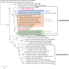
Maximum likelihood phylogenetic tree of a partial region of the RdRp gene of 65 CoVs from the four genera, including five obtained in this study (highlighted in fuchsia). The Luchacovirus clade is within the Alphacoronavirus group, each shown within labeled brackets. The CoVs most closely related to the Luchacovirus group (SADS-CoV and bat CoV HKU2) are emphasized with blue arrows. For all viruses, the virus name, host species, year, and country of collection and GeneBank accession numbers are shown at the tip of the tree. Numbers at the branches indicate bootstrap percentage values from 1,000 replicates. Branches with support <50 were collapsed. Nucleotide substitution model used: HKY + I + G.
Fig 2
Fig 2
Maximum likelihood tree of the complete nucleotide sequence of the S1 domain of the S gene of 38 coronaviruses from the four genera including the two obtained in this study (highlighted in fuchsia). In this phylogeny, the Luchacovirus clade is not within the Alphacoronavirus group, each shown within labeled brackets. Within the Luchacovirus group, the three well-supported groups (bootstrap of 100%) are shown in different colors (group 1 in blue, group 2 in orange, and group 3 in green). Four luchacoviruses, including the ones identified in this study, do not cluster within these three groups. For all viruses, the virus name, host species, year and place of collection, and GeneBank accession number are shown at the tip of the tree. Numbers at the branches indicate bootstrap percentage values from 1,000 replicates. Branches with bootstrap support of <50 were collapsed. Nucleotide substitution model used: GTR + I + G.
Fig 3
Fig 3
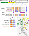
(A) Graphical representation of the S protein showing the S1 and S2 domains (top) and multiple sequence alignments (bottom) of four regions of interest within S (shown in violet circles) in which cleavage motifs are found (underlined in blue are the predicted cleavage sites and in violet are experimentally proven furin cleavage sites). The S1 domain is divided into the N-terminal domain (NTD, in green) and the C-terminal domain (CTD, in yellow). In the amino acid alignments, polar residues are in orange, acidic residues are in green, basic residues are in blue, and nonpolar residues are in gray. In the left side of the alignment, the virus name/host/year of collection/place of collection is shown. Above the alignments are the amino acid location of each region based on the S protein sequence of rodent Luchacovirus JC34 (KX964649). From left to right, there are four regions of interest: (1) Alphacoronavirus SADS-CoV has a furin cleavage site 94 amino acids upstream of its S1/S2 cleavage site that differs in only one residue when compared to four luchacoviruses and bat CoV HKU2 (shown with yellow triangles), (2) luchacoviruses have a region 37 amino acids upstream of the end of domain S1 that is highly variable and in which there are two different cleavage motifs, (3) luchacoviruses do not have an S1/S2 cleavage region in comparison to SADS-CoV and selected betacoronaviruses (SARS-CoV2 and two rodent coronaviruses), and (4) only SARS-CoV2 has an S2’ cleavage motif. (B) Amino acid sequences and the respective codons (on top) of region 1, in which SADS-CoV has a cleavage site and bat CoV HKU2, and the luchacoviruses shown differ by a single residue (458) to that of SADS-CoV (R458S, R458K, respectively), which abrogates the cleavage site. Inspection of the codon sequences shows that only a single nucleotide change (highlighted in yellow) is necessary to revert this residue change. (C) Tertiary structure of one of the monomers of the S protein of SADS-CoV showing the four regions in which we found cleavage sites in luchacoviruses or related alpha and betacoronaviruses. For this figure, we used the same colors as in A.
Fig 4
Fig 4
Graphical representation of the 3′-end of the genome of Luchacovirus, which includes the structural (S, E, M, N) and non-structural (ORFs 2, 2b, 6, 8, 9) genes. Sixteen sequences, including the one obtained in this study (on top in fuchsia), are shown. Above each genome representation is the name of each gene; below is the size of the corresponding proteins (number of amino acids). If some proteins varied in size between the viruses, all size values are shown from shortest to longest. On the left side of the figure, the name of the virus, host species, year of collection, and country of collection of all sequences included are shown. There are three genomic organizations that match the three groups (groups 1, 2, and 3 in blue, orange, and green, respectively) identified in Fig. 2. The three genomic organizations differ in the existence of ORFs between ORF1b and S (ORF2 and 2b). The sequence obtained in this study, W317/red fox/2022 (top), has a similar organization to group 2 luchacoviruses as it only has one ORF2, but it differs from other luchacoviruses because it lacks ORF8 and ORF9 is shorter. Divergent luchacoviruses P83/plateau pika/China and UMN2020/mouse/2018/USA have the same genomic organization of viruses from groups 2 and 3, respectively, but do not belong to these groups and are overall genetically different (Fig. S2).
Fig 5
Fig 5
Similarity plot of the nucleotide sequence of (A) the 3′-end of the genome of W317/red fox/2022 (top, used as reference) and (B) a partial region of the S gene of W291/bob cat/2021 (top, used as reference) compared to 15 other luchacoviruses including those in group 1 (in blue), 2 (in orange), and 3 (in green) and divergent luchacoviruses UMN2020/mouse/2018/USA (in yellow) and P83/plateau pika/China (in violet). The location of each gene or gene domain is shown above each graph. The graphs were constructed using a window of 200 bp, a step of 20 nt, and the Kimura 2 parameter distance model.

Update of

Similar articles

References

    1. Siddell SG, Walker PJ, Lefkowitz EJ, Mushegian AR, Dempsey DM, Dutilh BE, Harrach B, Harrison RL, Hendrickson RC, Junglen S, Knowles NJ, Kropinski AM, Krupovic M, Kuhn JH, Nibert M, Rubino L, Sabanadzovic S, Simmonds P, Varsani A, Zerbini FM, Davison AJ. 2019. Additional changes to taxonomy ratified in a special vote by the international committee on taxonomy of viruses (October 2018). Arch Virol 164:2417–2429. doi:10.1007/s00705-019-04306-w - DOI - PubMed
    1. Cui J, Li F, Shi Z-L. 2019. Origin and evolution of pathogenic coronaviruses. Nat Rev Microbiol 17:181–192. doi:10.1038/s41579-018-0118-9 - DOI - PMC - PubMed
    1. Li F. 2015. Receptor recognition mechanisms of coronaviruses: a decade of structural studies. J Virol 89:1954–1964. doi:10.1128/JVI.02615-14 - DOI - PMC - PubMed
    1. Belouzard S, Millet JK, Licitra BN, Whittaker GR. 2012. Mechanisms of coronavirus cell entry mediated by the viral spike protein. Viruses 4:1011–1033. doi:10.3390/v4061011 - DOI - PMC - PubMed
    1. Millet JK, Whittaker GR. 2015. Host cell proteases: critical determinants of coronavirus tropism and pathogenesis. Virus Res 202:120–134. doi:10.1016/j.virusres.2014.11.021 - DOI - PMC - PubMed

Publication types

MeSH terms

Substances