[Preprint]. 2024 Sep 4:2023.10.13.562273.
doi: 10.1101/2023.10.13.562273.

A PARP14/TARG1-Regulated RACK1 MARylation Cycle Drives Stress Granule Dynamics in Ovarian Cancer Cells

Affiliations

A PARP14/TARG1-Regulated RACK1 MARylation Cycle Drives Stress Granule Dynamics in Ovarian Cancer Cells

Sridevi Challa et al. bioRxiv. .

Abstract

Mono(ADP-ribosyl)ation (MARylation) is emerging as a critical regulator of ribosome function and translation. Herein, we demonstrate that RACK1, an integral component of the ribosome, is MARylated on three acidic residues by the mono(ADP-ribosyl) transferase (MART) PARP14 in ovarian cancer cells. MARylation of RACK1 is required for stress granule formation and promotes the colocalization of RACK1 in stress granules with G3BP1, eIF3η, and 40S ribosomal proteins. In parallel, we observed reduced translation of a subset of mRNAs, including those encoding key cancer regulators (e.g., AKT). Treatment with a PARP14 inhibitor or mutation of the sites of MARylation on RACK1 blocks these outcomes, as well as the growth of ovarian cancer cells in culture and in vivo. To re-set the system after prolonged stress and recovery, the ADP-ribosyl hydrolase TARG1 deMARylates RACK1, leading to the dissociation of the stress granules and the restoration of translation. Collectively, our results demonstrate a therapeutically targetable pathway that controls stress granule assembly and disassembly in ovarian cancer cells.


Conflict of interest statement

Conflict of Interest Statement: W.L.K. is a founder, member of the SAB, member of the BOD, and a stockholder for ARase Therapeutics, Inc. He is also coholder of U.S. Patent 9,599,606 covering the ADP-ribose detection reagents used herein, which has been licensed to and is sold by EMD Millipore.

Figures
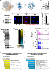
Figure 1.
Figure 1.. Site-specific MARylation of RACK1 in ovarian cancer cells.
(A) (Left) Spatial distribution of the proteins modified by MARylation in the 80S ribosome (PDB ID: 4V6X). (Middle and right) Sites of MARylation within RACK1 (Asp 144, Glu 145, and Asp 203; blue ribbon) are indicated in two expanded views, with the structure in the right rotated by 90°. (B) RACK1 is MARylated. Endogenous RACK1 was immunoprecipitated from OVCAR3 cells and subjected to immunoblotting for MAR and RACK1. (C) RACK1 is MARylated at Asp 144, Glu 145, and Asp 203. HA-tagged RACK1 was immunoprecipitated from OVCAR3 cells ectopically expressing wild-type (WT) or MARylation site mutant (Mut) RACK1 and subjected to immunoblotting for MAR and HA. (D) In situ detection of RACK1 MARylation. Proximity ligation assay (PLA) of RACK1 and MAR in OVCAR3 cells subjected to Dox-induced knockdown of endogenous and re-expression of RACK1 (WT or Mut). DNA was stained with DAPI. Scale bar is 15 μm. (E) Quantification of multiple experiments like the one shown in panel (D). Each bar represents the mean + SEM of MAR-RACK1 PLA foci from three biological replicates (Student’s t-test, **** p < 0.0001). (F) RACK1-Mut expression does not alter global protein synthesis in OVCAR3 cells. Immunoblot analysis of puromycin incorporation assays from OVCAR3 cells subjected to Dox-induced knockdown of endogenous and re-expression of RACK1. β-tubulin serves as a loading control. (G and H) Regulation of mRNA translation by RACK1 MARylation. Ribosome profiling of OVCAR3 cells subjected to Dox-induced knockdown of endogenous RACK1 followed by re-expression of exogenous RACK1 (WT or Mut). (G) Heatmap representation of mRNAs that exhibit altered translation efficiency when RACK1-Mut was expressed. (H) Gene ontology enrichment analysis of the translationally upregulated and downregulated mRNAs. (I) RACK1 MARylation regulates translation of AKT1. Example ribosome profiling and RNA-seq traces of AKT1 in OVCAR3 cells subjected to Dox-induced knockdown of endogenous RACK1 and re-expression of exogenous RACK1 (WT or Mut). A schematic of the AKT1 gene with a scale bar is shown.
Figure 2.
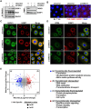
Figure 2.. Site-specific MARylation of RACK1 is required for stress granule assembly.
(A and B) Loss of RACK1 MARylation inhibits RACK1 interaction with G3BP1. (A) HA-tagged RACK1 was immunoprecipitated from OVCAR3 cells with Dox-induced knockdown of endogenous RACK1 and re-expression of exogenous RACK1. The immunoprecipitates were subjected to immunoblotting for G3BP1 and HA. (B) PLA using G3BP1 and HA antibodies. DNA was stained with DAPI. Scale bar is 15 μm. (C and D) Loss of RACK1 MARylation inhibits the recruitment of G3BP1 to ribosomes. (C) Immunoblot analysis for HA-tagged RACK1 and G3BP1 in sucrose density gradient fractions of ribosomes prepared from OVCAR3 cells subjected to Dox-induced knockdown of endogenous RACK1 and re-expression of exogenous RACK1. Each bar in the graph in (D) represents the mean + SEM of the relative abundance of G3BP1 in monosomes or polysomes (n = 3, two-way ANOVA, * p < 0.05 and ** p<0.01). (E and F) Loss of RACK1 MARylation inhibits G3BP1 localization to stress granules and its interaction with translation factors that are key components of stress granules. (E) G3BP1 was immunoprecipitated from OVCAR3 cells with Dox-induced knockdown of endogenous RACK1 and re-expression of exogenous RACK1. The immunoprecipitates were subjected to immunoblotting for eIF3η, RPS6, and G3BP1 as indicated. (F) Immunofluorescent staining assays of OVCAR3 cells with Dox-induced knockdown of endogenous RACK1 and re-expression of exogenous RACK1 subjected to 15 minutes of treatment with 250 μM sodium arsenite (NaAsO2). Staining for HA (RACK1) and G3BP1. DNA was stained with DAPI. Scale bar is 15 μm.
Figure 3.
Figure 3.. PARP14 inhibition reduces stress granule assembly.
(A) PARP14 inhibitor blocks PARP14 autoMARylation. OVCAR3 cells were treated with 10 μM PARP14 inhibitor (RBN012759) for 24 hours. PARP14 was immunoprecipitated and subjected to immunoblotting for PARP14 and MAR. (B) Inhibition of PARP14 catalytic activity blocks RACK1 MARylation. PLA using MAR and RACK1 antibodies in OVCAR3 cells treated with 10 μM PARP14 inhibitor (RBN012759) for 24 hours. DNA was stained with DAPI. Scale bar is 15 μm. (C and D) PARP14 inhibition reduces the recruitment of G3BP1 to ribosomes. (C) Immunoblot analysis of RACK1 and G3BP1 in sucrose density gradient fractions of ribosomes prepared from OVCAR3 cells treated with 10 μM PARP14 inhibitor for 24 hours. Each bar in the graph in (D) represents the mean + SEM of the relative abundance of G3BP1 in monosomes or polysomes (n = 3, two-way ANOVA, * p < 0.05 and ** p<0.01). (E and F) PARP14 inhibition reduces G3BP1 interaction with RACK1. (E) G3BP1 was immunoprecipitated from OVCAR3 cells treated with 10 μM PARP14 inhibitor for 24 hours and subjected to immunoblotting for MAR, RACK1, and G3BP1 as indicated. The band corresponding to the molecular weight of RACK1 was indicated as MARylated RACK1. Each bar in the graph in (F) represents the mean + SEM of the relative abundance of total RACK1 or MARylated RACK1 in G3BP1 immunoprecipitates (n = 3, Student’s t-test, * p < 0.05). (G and H) PARP14 inhibition reduces G3BP1 interaction with translation factors that are key components of stress granules. (G) G3BP1 was immunoprecipitated from OVCAR3 cells treated with 10 μM PARP14 inhibitor for 24 hours and subjected to immunoblotting for eIF3η, RPS6, and G3BP1 as indicated. Each bar in the graph in (H) represents the mean + SEM of the relative abundance of eIF3η and RPS6 in G3BP1 immunoprecipitates (n = 3, Student’s t-test, * p < 0.05). (I and J) PARP14 inhibition reduces G3BP1 localization to stress granules. Immunofluorescent staining assays of OVCAR3 cells treated with 10 μM PARP14 inhibitor for 24 hours and subjected to 15 minutes of treatment with 250 μM sodium arsenite (NaAsO2). Staining for RACK1 and G3BP1. DNA was stained with DAPI. Scale bar is 15 μm. Each bar in the graph in (J) represents the mean + SEM of the relative abundance of stress granules (n = 3, Student’s t-test, ** p<0.01).
Figure 4.
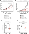
Figure 4.. Loss of RACK1 MARylation sensitizes ovarian cancer cells to stress and inhibits their growth.
(A) RACK1-Mut expressing cells are sensitive to ER stress, which inhibits their growth. Growth curves of OVCAR3 cells with Dox-induced knockdown of endogenous RACK1 and re-expression of exogenous RACK1 (WT or Mut) in the presence or absence of 3 nM thapsigargin (Thps) for the indicated times. The arrow points to the RACK-Mut growth curve beneath the RACK-WT growth curve under basal conditions. Each point represents the mean ± SEM of the growth of the cells relative to Day 0 of treatment (n = 3, two-way ANOVA, * p < 0.01). (B) PARP14 inhibition sensitizes ovarian cancer cells to ER stress and inhibits their growth. Growth curves of OVCAR3 cells in the presence or absence of 10 μM PARP14 inhibitor (PARP14i) and 3 nM thapsigargin (Thps) for the indicated times. Each point represents the mean ± SEM of the growth of the cells relative to Day 0 of treatments (n = 3, two-way ANOVA, ** p < 0.001). (C and D) Expression of RACK1-Mut or treatment with PARP14 inhibitor (PARP14i) inhibits the growth of OVCAR3 xenograft tumors derived from cells like those described in (A) and (B). The xenograft tumors were established in immunocompromised NSG mice subjected to the treatments indicated and grown until the mice reached the end-point for euthanasia as required by IACUC. (C) Tumor volume at Day 69. Each cluster in the graph shows the mean and the individual data points for n = 10 or 8 mice (WT or Mut, respectively), Student’s t-test, p = 0.0155. (D) Tumor volume at Day 19 post-treatment. Each cluster in the graph shows the mean and the individual data points for n = 5 or 6 mice (vehicle or PARP14i, respectively), Student’s t-test, p = 0.05. Different timelines in the two xenograft experiments were dictated by different growth rates of parental (D) versus Dox-treated cells (C).
Figure 5.
Figure 5.. Depletion of TARG1 enhances stress granule assembly by increasing RACK1 MARylation.
(A and B) siRNA-mediated TARG1 depletion increases RACK1 MARylation and enhances RACK1 interaction with G3BP1 in OVCAR3 cells subjected to 15 minutes of treatment with 250 μM sodium arsenite (NaAsO2). (A) RACK1 was immunoprecipitated from OVCAR3 cells with siRNA-mediated knockdown of TARG1 and subjected to immunoblotting for G3BP1, MAR, and RACK1. (B) PLA using MAR and RACK1 antibodies. DNA was stained with DAPI. Scale bar is 15 μm. (C and D) TARG1 knockdown increases the assembly of G3BP1-containing stress granules. Immunofluorescent staining assays of OVCAR3 cells with siRNA-mediated knockdown of TARG1 subjected to 15 minutes of treatment with 250 μM sodium arsenite (NaAsO2). (C) RACK1 and G3BP1, (D) RPS6 and G3BP1. DNA was stained with DAPI. Scale bar is 15 μm. (E) Changes in mRNA translation upon depletion of TARG1. Scatter plot of fold changes in ribosome profiling and RNA-seq (OVCAR3 cells subjected to siRNA-mediated TARG1 knockdown vs. WT) comparing translational control and transcriptional control. Gene ontology enrichment analysis of the mRNAs regulated at transcriptional and translational levels are shown.
Figure 6.
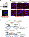
Figure 6.. Prolonged exposure to stress reduces RACK1 MARylation.
(A and B) Stress reduces RACK1 MARylation through TARG1. (A) RACK1 was immunoprecipitated from OVCAR3 cells treated with 250 μM sodium arsenite (NaAsO2) for 30 minutes or 250 nM thapsigargin (Thps) for 2 hours, and subjected to immunoblotting for MAR and RACK1. (B) PLA in OVCAR3 cells with siRNA-mediated knockdown of TARG1 using TARG1 and RACK1 (top) or MAR and RACK1 (bottom) antibodies. The cells were treated with sodium arsenite (NaAsO2) treatment for 30 minutes. DNA was stained with DAPI. Scale bar is 15 μm. (C) Knockdown of TARG1 increases the assembly of G3BP1-containing stress granules. PLA for TARG1 and RACK1 combined with immunofluorescent imaging of OVCAR3 cells expressing GFP-G3BP1 with siRNA-mediated knockdown of TARG1 and treated with 250 μM sodium arsenite (NaAsO2) for 15 minutes. DNA was stained with DAPI. Scale bar is 15 μm. (D) Schematic of the mechanisms by which PARP14 and TARG1 regulate stress granule assembly through RACK1 MARylation. Additional details are provided in the text.

Similar articles

References

    1. Adams D.R., Ron D., and Kiely P.A.. 2011. RACK1, A multifaceted scaffolding protein: Structure and function. Cell Commun Signal. 9:22. - PMC - PubMed
    1. Ahmed S., Bott D., Gomez A., Tamblyn L., Rasheed A., Cho T., MacPherson L., Sugamori K.S., Yang Y., Grant D.M., Cummins C.L., and Matthews J.. 2015. Loss of the mono-ADP-ribosyltransferase, Tiparp, increases sensitivity to dioxin-induced steatohepatitis and lethality. J Biol Chem. 290:16824–16840. - PMC - PubMed
    1. Andrews S. 2010. FASTQC. A quality control tool for high throughput sequence data.
    1. Arimoto K., Fukuda H., Imajoh-Ohmi S., Saito H., and Takekawa M.. 2008. Formation of stress granules inhibits apoptosis by suppressing stress-responsive MAPK pathways. Nature cell biology. 10:1324–1332. - PubMed
    1. Asadi M.R., Rahmanpour D., Moslehian M.S., Sabaie H., Hassani M., Ghafouri-Fard S., Taheri M., and Rezazadeh M.. 2021. Stress granules involved in formation, progression and metastasis of cancer: A scoping review. Front Cell Dev Biol. 9:745394. - PMC - PubMed

Publication types