. 2023 Oct 20;7(4):046104.
doi: 10.1063/5.0157549. eCollection 2023 Dec.

Survivin regulates intracellular stiffness and extracellular matrix production in vascular smooth muscle cells

Affiliations

Survivin regulates intracellular stiffness and extracellular matrix production in vascular smooth muscle cells

Amanda Krajnik et al. APL Bioeng. .

Abstract

Vascular dysfunction is a common cause of cardiovascular diseases characterized by the narrowing and stiffening of arteries, such as atherosclerosis, restenosis, and hypertension. Arterial narrowing results from the aberrant proliferation of vascular smooth muscle cells (VSMCs) and their increased synthesis and deposition of extracellular matrix (ECM) proteins. These, in turn, are modulated by arterial stiffness, but the mechanism for this is not fully understood. We found that survivin is an important regulator of stiffness-mediated ECM synthesis and intracellular stiffness in VSMCs. Whole-transcriptome analysis and cell culture experiments showed that survivin expression is upregulated in injured femoral arteries in mice and in human VSMCs cultured on stiff fibronectin-coated hydrogels. Suppressed expression of survivin in human VSMCs significantly decreased the stiffness-mediated expression of ECM components related to arterial stiffening, such as collagen-I, fibronectin, and lysyl oxidase. By contrast, expression of these ECM proteins was rescued by ectopic expression of survivin in human VSMCs cultured on soft hydrogels. Interestingly, atomic force microscopy analysis showed that suppressed or ectopic expression of survivin decreases or increases intracellular stiffness, respectively. Furthermore, we observed that inhibiting Rac and Rho reduces survivin expression, elucidating a mechanical pathway connecting intracellular tension, mediated by Rac and Rho, to survivin induction. Finally, we found that survivin inhibition decreases FAK phosphorylation, indicating that survivin-dependent intracellular tension feeds back to maintain signaling through FAK. These findings suggest a novel mechanism by which survivin potentially modulates arterial stiffness.


Conflict of interest statement

The authors have no conflicts to disclose.

Figures
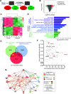
FIG. 1.
FIG. 1.
Functional network analysis and predicted network of survivin (Birc5) in VSMCs from injured mouse arteries. (a) Reductions in data magnitude by applying significance thresholds to the raw microarray data (fold change [FC] ≥ 2.0, q value ≤ 0.15). (b) Volcano plot displays the distributions of all detected genes, represented as single dots that are not statistically different (gray), significantly upregulated (red), or significantly downregulated. (c) Heat map displays the Z-scores of the 660 differentially expressed genes (DEGs). (d) Histogram presenting the significant ECM biological processes (purple), cellular components (blue), and molecular functions enriched among the 331 DEG upregulated in mouse arteries after injury. (e) Venn diagram of DEGs in mouse vascular injury model. The DEGs upregulated in response to vascular injury (331 genes; green) were compared to GO lists for extracellular matrix (red) and regulators of intracellular signaling transduction (blue). (f) Log2 fold-change and adjusted p values of the genes positively regulated after vascular injury and contained within the GO terms described above. (g) Network diagram of gene interaction pathways between Birc5 (survivin) and various ECM proteins, including collagens (Col1a1, Col1a2, and Col3a1), fibronectin (Fn1), and lysyl oxidase (Lox).
FIG. 2.
FIG. 2.
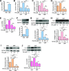
ECM synthesis in hVSMCs is reduced when survivin expression is suppressed. (a) and (f) hVSMCs were synchronized to G0 by serum starvation and plated on fibronectin-coated soft or stiff hydrogels with 10% FBS for 24 h. (b)–(e) and (g)–(j) Serum-starved hVSMCs were plated on soft or stiff hydrogels with 10% FBS with DMSO or YM155 at the indicated concentrations for 24 h. Levels of mRNA (a)–(e) and protein (f)−(j) were analyzed by RT-qPCR and immunoblotting assays, respectively. The graphs show the expression of survivin (a), (b), and (e), collagen-I (c) and (h), fibronectin (d) and (i), and Lox (e) and (j). Expression was normalized to that in hVSMCs treated with DMSO (vehicle control) on stiff hydrogels. n = 7 (a), n = 3 − 8 (b)–(e), n = 6 (f), n = 4 (g) and (j), n = 3 (h) and (i). Error bars show SEMs. *p < 0.05; **p < 0.01; ***p < 0.001; and ns, not significant by Student's t test (a) and (f) or ANOVA followed by Newman–Keuls post hoc test for multiple comparisons (b)−(e) and (g)–(j).
FIG. 3.
FIG. 3.
Survivin overexpression mimics stiffness-mediated ECM protein production in hVSMCs. hVSMCs infected with adenoviruses encoding wild-type (wt) survivin or the GFP control were plated on soft or stiff hydrogels with 10% FBS for 24 h. Total cell lysates were analyzed by immunoblotting for survivin (a), collagen and fibronectin (b), and Lox (c). Levels were normalized to those in GFP-expressing hVSMCs on soft hydrogels. n = 5 (a), n = 3–4 (b), and n = 4 (c). Error bars show SEMs. *p < 0.05; **p < 0.01; and ns, not significant by ANOVA followed by Newman–Keuls post hoc test for multiple comparisons.
FIG. 4.
FIG. 4.
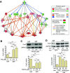
Survivin regulates VSMC stiffness. (a) Visualization of an AFM tip on VSMC surface. hVSMCs were plated on fibronectin-coated soft or stiff hydrogels with 10% FBS for 24 h. Atomic force microscopy was used to measure cellular stiffness in cells treated with YM155 (b) to reduce survivin levels and in cells infected with adenovirus encoding wild-type (wt) survivin or a GFP control for survivin overexpression (c). n = 40 − 42 cells from four experiments (b) and n = 24–35 cells from three experiments (c). Each data point represents one cell, and the means are indicated by horizontal lines. ***p < 0.001 and ns, not significant by ANOVA followed by Newman–Keuls post hoc test for multiple comparisons.
FIG. 5.
FIG. 5.
Survivin regulates stiffness-mediated Cox2 expression in hVSMCs. (a) Network diagrams of gene interaction pathways between Ptgs2 (Cox2) and genes for various ECM proteins. hVSMCs were plated on fibronectin-coated soft or stiff hydrogels with 10% FBS ± YM155 at the indicated concentrations for 24 h. Total cell lysates were analyzed by RT-qPCR (b) and immunoblotting assays (c). hVSMCs infected with adenoviruses encoding GFP or wild-type (wt) survivin were plated on hydrogels with 10% FBS for 24 h. Cell lysates were analyzed by immunoblotting (d). Expression levels were normalized to hVSMCs treated with DMSO (vehicle control) on stiff hydrogels or infected with the virus encoding GFP at an MOI of 50 and plated on soft gels. n = 5 (b), n = 3 (c) and (d). GAPDH served as a loading control. Error bars show SEMs. *p < 0.05; **p < 0.01; ***p < 0.001; and ns, not significant by ANOVA followed by Newman–Keuls post hoc test for multiple comparisons.
FIG. 6.
FIG. 6.
Survivin feeds back to regulate FAK phosphorylation. (a) Serum-starved hVSMCs were plated on fibronectin-coated soft or stiff hydrogels with 10% FBS ± YM155 at the indicated concentrations for 24 h. Total cell lysates were analyzed by immunoblotting for phospho (p)-FAK (b), total FAK (c), and cyclin D1 (d). GAPDH served as a loading control. Expression was normalized to that in hVSMCs treated with DMSO on stiff hydrogels. n = 3–6. Error bars show SEMs. *p < 0.05; **p < 0.01; ***p < 0.001; and ns, not significant by Student's t test.

Similar articles

Cited by

References

    1. Ponticos M. and Smith B. D., “ Extracellular matrix synthesis in vascular disease: Hypertension, and atherosclerosis,” J. Biomed. Res. 28(1), 25–39 (2014).10.7555/JBR.27.20130064 - DOI - PMC - PubMed
    1. Feil S. et al., “ Transdifferentiation of vascular smooth muscle cells to macrophage-like cells during atherogenesis,” Circ. Res. 115(7), 662–667 (2014).10.1161/CIRCRESAHA.115.304634 - DOI - PubMed
    1. Beamish J. A. et al., “ Molecular regulation of contractile smooth muscle cell phenotype: Implications for vascular tissue engineering,” Tissue Eng., Part B 16(5), 467–491 (2010).10.1089/ten.teb.2009.0630 - DOI - PMC - PubMed
    1. Owens G. K., Kumar M. S., and Wamhoff B. R., “ Molecular regulation of vascular smooth muscle cell differentiation in development and disease,” Physiol. Rev. 84(3), 767–801 (2004).10.1152/physrev.00041.2003 - DOI - PubMed
    1. Thyberg J. et al., “ Regulation of differentiated properties and proliferation of arterial smooth muscle cells,” Arteriosclerosis 10(6), 966–990 (1990).10.1161/01.ATV.10.6.966 - DOI - PubMed