. 2023 Oct 20;21(10):e3002331.
doi: 10.1371/journal.pbio.3002331. eCollection 2023 Oct.

Adiponectin in the mammalian host influences ticks' acquisition of the Lyme disease pathogen Borrelia

Affiliations

Adiponectin in the mammalian host influences ticks' acquisition of the Lyme disease pathogen Borrelia

Xiaotian Tang et al. PLoS Biol. .

Abstract

Arthropod-borne pathogens cause some of the most important human and animal infectious diseases. Many vectors acquire or transmit pathogens through the process of blood feeding. Here, we report adiponectin, the most abundant adipocyte-derived hormone circulating in human blood, directly or indirectly inhibits acquisition of the Lyme disease agent, Borrelia burgdorferi, by Ixodes scapularis ticks. Rather than altering tick feeding or spirochete viability, adiponectin or its associated factors induces host histamine release when the tick feeds, which leads to vascular leakage, infiltration of neutrophils and macrophages, and inflammation at the bite site. Consistent with this, adiponectin-deficient mice have diminished pro-inflammatory responses, including interleukin (IL)-12 and IL-1β, following a tick bite, compared with wild-type animals. All these factors mediated by adiponectin or associated factors influence B. burgdorferi survival at the tick bite site. These results suggest a host adipocyte-derived hormone modulates pathogen acquisition by a blood-feeding arthropod.


Conflict of interest statement

The authors have declared that no competing interests exist.

Figures

Fig 1
Fig 1. Adiponectin affects B. burgdorferi acquisition by ticks upon blood feeding.
(A) The B. burgdorferi burden in WT (n = 5) and KO mice (n = 5) after 14 days infection. The mice were needle inoculated with B. burgdorferi. (B) Pathogen-free I. scapularis nymphs were fed on B. burgdorferi-infected WT (n = 3) and KO mice (n = 3) for 48 h and then B. burgdorferi flaB levels in guts were assessed. (C) B. burgdorferi burden in the tick gut after feeding on WT and KO mice for 48 h. B. burgdorferi titers in the ticks feeding on KO mice were significantly higher compared to the ticks feeding on WT mice. (D) B. burgdorferi infected WT (n = 3) and KO mice (n = 3) were challenged with ticks and assessed for tick detachment. The percentage of ticks remaining attached on mice at a given time point was recorded. Three replicates were included, and the average values were presented. (E) The engorgement weights of nymphs feeding on WT (n = 3) and KO mice (n = 3). For all the data, each dot represents 1 biological replicate. Statistical significance was assessed using a nonparametric Mann–Whitney test (**p < 0.01; ns, p > 0.05). Data underlying this figure can be found in S1 Data. We would like to acknowledge that figures were created using BioRender (https://www.biorender.com/) with permission. KO, knock out; WT, wild-type.
Fig 2
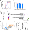
Fig 2. Host adiponectin significantly increases tick HBP expression.
(A) No interaction of adiponectin with B. burgdorferi was identified, as analyzed by flow cytometry. PGLYRP1 was used as positive control. The background of Alexa Fluor 488-His antibody alone with B. burgdorferi is shown in gray. (B) Mouse adiponectin has no effect on B. burgdorferi viability as determined by BacTiter-Glo assay. (C) RNA-seq of ticks feeding on WT (n = 3) and KO mice (n = 3). The ticks that fed on the same mouse were pooled for RNA extraction. (D) GO enrichment analysis of transcriptome data from the ticks feeding on WT (n = 3) and KO mice (n = 3). The second level GO terms were shown in the plot and enrichment analysis was performed using the functional annotation tool DAVID. (E) Volcano plot of differentially expressed genes between the ticks feeding on WT (n = 3) and KO mice (n = 3). The Top 3 genes were highlighted by orange color. The gene names can be found in S1 Table. (F) qPCR validation of HBP gene expression in SGs and MG of the ticks feeding on WT (n = 3) and KO mice (n = 3). The ticks feeding on different mice were collected for analysis. For all the data, each dot represents one biological replicate. Statistical significance was assessed using a nonparametric Mann–Whitney test (*p < 0.05; ns, p > 0.05). Data underlying this figure can be found in S1 Data. We would like to acknowledge that figures were created using BioRender (https://www.biorender.com/) with permission. GO, Gene Ontology; HBP, histamine-binding protein; KO, knock out; MG, midgut; PGLYRP1, peptidoglycan recognition protein 1; qPCR, quantitative real-time PCR; SG, salivary gland; WT, wild-type.
Fig 3
Fig 3. Adiponectin-deficient mice exhibit less inflammation at the tick bite site.
(A) Histamine concentration in WT (n = 5) and KO naïve mice (n = 5), or after tick bite (n = 10). (B) Injection of Evans blue during tick feeding. The white circles indicated WT (n = 6) mice have more vascular leakage than KO mice (n = 6) at the tick bite site. Quantification of Evans blue leakage at the tick bite site of WT (n = 6) and KO mice (n = 6). (C) The CD11b+Ly6G+/CD45+ PMN population and the percentage of PMNs in the total CD45+ leukocyte cell population in WT (n = 4) and KO mice (n = 4) after tick bite. The CD11b+CD11c/CD45+Ly6G MAC population and the percentage of MACs in the total CD45+ leukocyte cell population in WT (n = 4) and KO mice (n = 4) after tick bite. (D) Semiquantitative histopathologic scoring of tick bite/inflammation sites show there is no significant difference in the severity of injury (Injury) but there is an overall increase in the degree of inflammation (Inflammation) in WT mice (n = 5) compared to adiponectin KO mice (n = 4). Representative HE-stained sections of WT and adiponectin KO tick bite lesions (arrows). Scale bars = 50 μm, * = subcutis, and ** = ear cartilage. For all the data, statistical significance was assessed using a nonparametric Mann–Whitney test (*p < 0.05; **p < 0.01; ns, p > 0.05). Data underlying this figure can be found in S1 Data. HE, hematoxylin and eosin; KO, knock out; MAC, macrophage; PMN, polymorphonuclear neutrophil; WT, wild-type.
Fig 4
Fig 4. Adiponectin-deficient mice showed an attenuated inflammation signature at the tick bite site.
(A) Serum cytokines and chemokines production in WT (n = 5) and KO mice (n = 5) after tick feeding. WT mice have higher production of IL-12p40 and IL-13. (B) Gene expression of cytokines and chemokines at the tick bite site of WT (n = 6) and KO mice (n = 6) after tick feeding. WT mice have higher gene expression of IL-1β, IL-12, and TLR2. For all the data, each dot represents 1 biological replicate. Statistical significance was assessed using a nonparametric Mann–Whitney test (*p < 0.05). (C) Cluster dendrogram and heatmap of transcriptome data of WT (n = 3) and KO murine skin (n = 3) during tick bite. (D) Volcano plot of differentially expressed genes. The significant differentially expressed genes were highlighted in blue and orange. (E) Immune signaling pathways identified by KEGG pathway enrichment analysis, which was performed using functional annotation tool DAVID. MAPK signaling pathway is the most enriched pathway. (F) Schematic diagram of the mechanism that adiponectin or associated factors induce inflammation during tick bite, inhibiting acquisition of the Lyme disease agent. Data underlying this figure can be found in S1 Data. We would like to acknowledge that figures were created using BioRender (https://www.biorender.com/) with permission. KEGG, Kyoto Encyclopedia of Genes and Genomes; KO, knock out; WT, wild-type.

Similar articles

References

    1. Graça-Souza AV, Maya-Monteiro C, Paiva-Silva GO, Braz GR, Paes MC, Sorgine MH, et al.. Adaptations against heme toxicity in blood-feeding arthropods. Insect Biochem Mol Biol. 2006;36(4):322–335. doi: 10.1016/j.ibmb.2006.01.009 - DOI - PubMed
    1. Pakpour N, Akman-Anderson L, Vodovotz Y, Luckhart S. The effects of ingested mammalian blood factors on vector arthropod immunity and physiology. Microbes Infect. 2013;15(3):243–254. doi: 10.1016/j.micinf.2013.01.003 - DOI - PMC - PubMed
    1. Zhu Y, Tong L, Nie K, Wiwatanaratanabutr I, Sun P, Li Q, et al.. Host serum iron modulates dengue virus acquisition by mosquitoes. Nat Microbiol. 2019;4(12):2405–2415. doi: 10.1038/s41564-019-0555-x - DOI - PubMed
    1. Smith AA, Navasa N, Yang X, Wilder CN, Buyuktanir O, Marques A, et al.. Cross-species interferon signaling boosts microbicidal activity within the tick vector. Cell Host Microbe. 2016;20(1):91–98. doi: 10.1016/j.chom.2016.06.001 - DOI - PMC - PubMed
    1. Rana VS, Kitsou C, Dutta S, Ronzetti MH, Zhang M, Bernard Q, et al.. Dome1–JAK–STAT signaling between parasite and host integrates vector immunity and development. Science. 2023;379(6628):eabl3837. doi: 10.1126/science.abl3837 - DOI - PMC - PubMed