Review
. 2023 Jul 20;6(10):1310-1322.
doi: 10.1021/acsptsci.3c00079. eCollection 2023 Oct 13.

The Many Faces of Histidine Triad Nucleotide Binding Protein 1 (HINT1)

Affiliations
Review

The Many Faces of Histidine Triad Nucleotide Binding Protein 1 (HINT1)

Maxwell Dillenburg et al. ACS Pharmacol Transl Sci. .

Abstract

The histidine triad nucleotide binding protein 1 (HINT1) is a nucleoside phosphoramidase that has garnered interest due to its widespread expression and participation in a broad range of biological processes. Herein, we discuss the role of HINT1 as a regulator of several CNS functions, tumor suppressor, and mast cell activator via its interactions with multiple G-protein-coupled receptors and transcription factors. Importantly, altered HINT1 expression and mutation are connected to the progression of multiple disease states, including several neuropsychiatric disorders, peripheral neuropathy, and tumorigenesis. Additionally, due to its involvement in the activation of several clinically used phosphoramidate prodrugs, tremendous efforts have been made to better understand the interactions behind nucleoside binding and phosphoramidate hydrolysis by HINT1. We detail the substrate specificity and catalytic mechanism of HINT1 hydrolysis, while highlighting the structural biology behind these efforts. The aim of this review is to summarize the multitude of biological and pharmacological functions in which HINT1 participates while addressing the areas of need for future research.


Conflict of interest statement

The authors declare no competing financial interest.

Figures
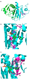
Figure 1
Figure 1
Crystal structures of AMP bound HINT1 dimer (Protein Data Bank (PDB) ID 3TW2). (A) Overall cartoon structure of homodimeric human HINT1 bound to AMP, with each monomer depicted in a different color. (B) AMP in green bound to the HINT1 nucleoside binding site. The side chains of the histidine triad (H110, H112, and H114) and contributing His51 are labeled and highlighted in magenta. (C) AMP in green bound to the nucleobase binding pocket. The hydrophobic residues (I18, F19, I22, F41, and I44) forming the hydrophobic nucleobase binding pocket are labeled in magenta. Key H-bonds from D43 to the 2′- and 3′-OH are measured and labeled in black, with the side chain of D43 highlighted in magenta.
Figure 2
Figure 2
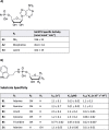
Structures of HINT1 interacting molecules. Series (A) Modifications to the leaving group for HINT1 substrates. Series (B) Modifications to the nucleobase with the corresponding tryptamine leaving group for HINT1 substrates. Series (C) HINT1 slow substrate used for isolating HINT1 catalytic intermediates. Series (D) Modifications to the ribose sugar for HINT1 substrates. Series (E) HINT1 inhibitors.
Figure 3
Figure 3
Kinetics data for two series of HINT1 substrates (compound structures detailed in Figure 2). (A) Specific activity of human HINT1 for a series of adenosine phosphoramidate substrates. (B) Substrate specificity of human HINT1 following changes to the substrate nucleobase and ribose.
Figure 4
Figure 4
Kinetic mechanism of HINT1 acyl-AMP HINT1 hydrolysis. (A) Kinetic equation for hHINT1 hydrolysis of tryptamine adenosine phosphoramidate B1. (B) Scheme for HINT1 hydrolysis of B1. His112 first attacks the substrate phosphorus, releasing tryptamine. Water then attacks the substrate/HINT1 complex, releasing HINT1 and the AMP product.
Figure 5
Figure 5
Isolated transition states of HINT1 hydrolysis of C1. (A) Overlay of the ES* complex (teal, PDB ID 5IPC) with a known AMP product complex (gray, 3TW2). H-bonds between C1 and HINT1 are shown in black. Residues of interest are labeled in black. (B) Overlay of ES* (teal, PDB ID 5IPC) and the nucleotidylated HINT1 complex E* (green, PDB ID 5IPD). His112 is bound to the phosphorus center and the electron density for the tryptamine leaving group is no longer observed. (C) Overlay of the product of C1 phosphoramidate hydrolysis (magenta, PDB ID 5IPE) and AMP (gray, PDB ID 3TW2).
Figure 6
Figure 6
Clinically approved ProTides. (A) Tenofovir alafenamide (Vemlidy), (B) sofosbuvir (Sovaldi), and (C) remdesivir (Veklury).
Figure 7
Figure 7
Protide activation mechanism. Steps of ProTide activation: (A) ester hydrolysis by carboxyesterase 1 or other carboxyesterase-type enzyme, (B) intramolecular cyclization via nucleophilic displacement of phenol by carboxylate, (C) nonenzymatic chemical hydrolysis by water, and (D) phosphoramidate hydrolysis via HINT1 or another phosphoramidase-type enzyme.
Figure 8
Figure 8
Proposed schematic pathway showing the role of HINT1 in downregulation of MOR signaling via NMDAR. In the resting state, HINT1 is associated with the C-terminus of the MOR and the regulator of G-protein signaling, RGSZ2., Following activation of MOR by opioid therapy, G-protein signaling triggers activation of the nNOS pathway and subsequent generation of nitric oxide (NO) leading to the release of free zinc ions. Additionally, RGSZ2 binds to the Gα subunit, freeing ΗΙΝΤ1 to bind to C-terminus of the NR1 subunit of NMDAR. PKCγ is then recruited to the cell membrane to bind HINT1 in a zinc dependent manner. RGSZ2 activation of PLCβ results in the release of DAG and subsequent activation of PKCγ, which can then activate NMDAR via phosphorylation of the NR1 subunit. The resulting calcium influx following NMDAR activation positively regulates the CaMKII signaling pathway, leading to phosphorylation of the mu opioid receptor and the development of opioid tolerance.

Similar articles

Cited by

References

    1. Krakowiak A.; Fryc I. Histidine triad protein superfamily-biological function and enzymatic activity. Postepy Biochem. 2012, 58 (3), 302–313. - PubMed
    2. (in Polish).

    1. Séraphin B. The HIT protein family: a new family of proteins present in prokaryotes, yeast and mammals. DNA Seq 1992, 3 (3), 177–179. 10.3109/10425179209034013. - DOI - PubMed
    1. Brenner C. Hint, Fhit, and GalT: function, structure, evolution, and mechanism of three branches of the histidine triad superfamily of nucleotide hydrolases and transferases. Biochemistry 2002, 41 (29), 9003–9014. 10.1021/bi025942q. - DOI - PMC - PubMed
    1. Rajasekaran R.; Felser A.; Nuoffer J. M.; Dufour J. F.; St-Pierre M. V. The histidine triad nucleotide-binding protein 2 (HINT-2) positively regulates hepatocellular energy metabolism. FASEB J. 2018, 32 (9), 5143–5161. 10.1096/fj.201701429R. - DOI - PubMed
    1. Chou T. F.; Cheng J.; Tikh I. B.; Wagner C. R. Evidence that human histidine triad nucleotide binding protein 3 (Hint3) is a distinct branch of the histidine triad (HIT) superfamily. J. Mol. Biol. 2007, 373 (4), 978–989. 10.1016/j.jmb.2007.08.023. - DOI - PubMed

LinkOut - more resources