

Influence of hypoxia on retinal progenitor and ganglion cells in human induced pluripotent stem cell-derived retinal organoids
- PMID: 37854379
- PMCID: PMC10559029
- DOI: 10.18240/ijo.2023.10.03
Influence of hypoxia on retinal progenitor and ganglion cells in human induced pluripotent stem cell-derived retinal organoids
Abstract
Aim: To observe the effect of low oxygen concentration on the neural retina in human induced pluripotent stem cell (hiPSC)-derived retinal organoids (ROs).
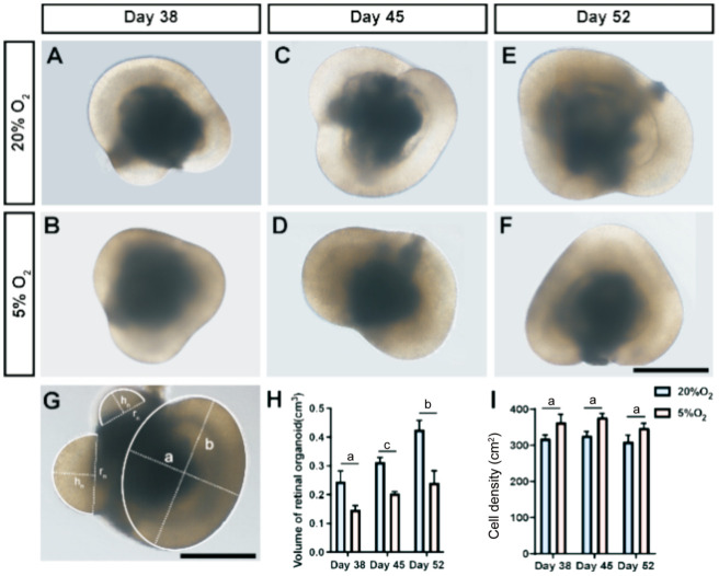
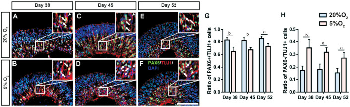
Methods: The hiPSC and a three-dimensional culture method were used for the experiments. Generated embryoid bodies (EBs) were randomly and equally divided into hypoxic and normoxic groups. Photographs of the EBs were taken on days 38, 45, and 52, and the corresponding volume of EBs was calculated. Simultaneously, samples were collected at these three timepoints, followed by fixation, sectioning, and immunofluorescence.
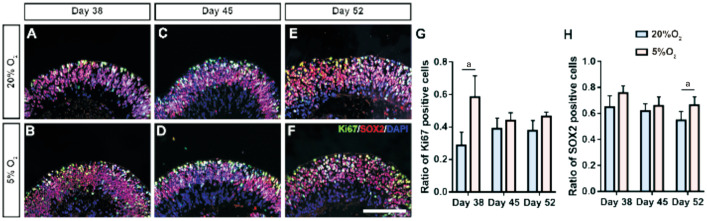
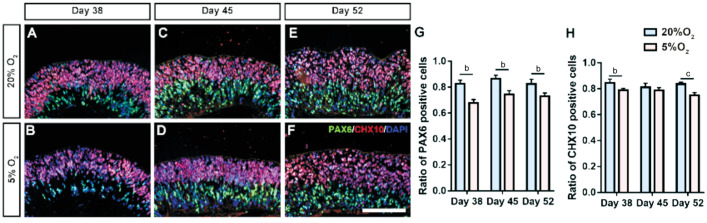
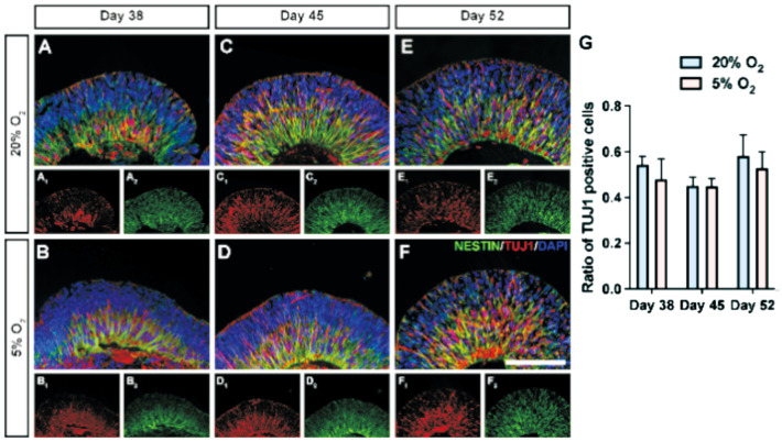
Results: The proportion of Ki67-positive proliferating cells increased steadily on day 38; this proliferation-promoting effect tended to increase tissue density rather than tissue volume. On days 45 and 52, the two groups had relatively similar ratios of Ki67-positive cells. Further immunofluorescence analysis showed that the ratio of SOX2-positive cells significantly increased within the neural retina on day 52 (P<0.05). In contrast, the percentage of PAX6- and CHX10-positive cells significantly decreased following hypoxia treatment at all three timepoints (P<0.01), except for CHX10 at day 45 (P>0.05). Moreover, the proportion of PAX6-/TUJ1+ cells within the neural retinas increased considerably (P<0.01, <0.05, <0.05 respectively).
Conclusion: Low oxygen promotes stemness and proliferation of neural retinas, suggesting that hypoxic conditions can enlarge the retinal progenitor cell pool in hiPSC-derived ROs.
Keywords: hypoxia; retinal ganglion cells; retinal organoid; retinal progenitor cells.
International Journal of Ophthalmology Press.
Figures
Similar articles
-
[Hypoxia promotes differentiation of human induced pluripotent stem cells into embryoid bodies in vitro].Nan Fang Yi Ke Da Xue Xue Bao. 2022 Jun 20;42(6):929-936. doi: 10.12122/j.issn.1673-4254.2022.06.18. Nan Fang Yi Ke Da Xue Xue Bao. 2022. PMID: 35790445 Free PMC article. Chinese.
-
Notch signaling pathway regulates proliferation and differentiation of immortalized Müller cells under hypoxic conditions in vitro.Neuroscience. 2012 Jul 12;214:171-80. doi: 10.1016/j.neuroscience.2012.04.025. Epub 2012 Apr 21. Neuroscience. 2012. PMID: 22525134
-
Hypoxia enhances the generation of retinal progenitor cells from human induced pluripotent and embryonic stem cells.Stem Cells Dev. 2012 May 20;21(8):1344-55. doi: 10.1089/scd.2011.0225. Epub 2011 Oct 27. Stem Cells Dev. 2012. PMID: 21875341
-
HUMAN CELLULAR MODELS FOR RETINAL DISEASE: From Induced Pluripotent Stem Cells to Organoids.Retina. 2022 Oct 1;42(10):1829-1835. doi: 10.1097/IAE.0000000000003571. Retina. 2022. PMID: 35858274 Free PMC article. Review.
-
Deciphering retinal diseases through the generation of three dimensional stem cell-derived organoids: Concise Review.Stem Cells. 2019 Dec;37(12):1496-1504. doi: 10.1002/stem.3089. Epub 2019 Oct 31. Stem Cells. 2019. PMID: 31617949 Free PMC article. Review.
References
-
- Watson AL, Palmer ME, Jauniaux E, Burton GJ. Variations in expression of copper/zinc superoxide dismutase in villous trophoblast of the human placenta with gestational age. Placenta. 1997;18(4):295–299. - PubMed
-
- Grimm C, Willmann G. Hypoxia in the eye: a two-sided coin. High Alt Med Biol. 2012;13(3):169–175. - PubMed
-
- Hawkins KE, Sharp TV, McKay TR. The role of hypoxia in stem cell potency and differentiation. Regen Med. 2013;8(6):771–782. - PubMed
LinkOut - more resources
Full Text Sources