. 2023 Oct 13;80(11):326.
doi: 10.1007/s00018-023-04977-4.

The phosphatidylserine receptor TIM1 promotes infection of enveloped hepatitis E virus

Affiliations

The phosphatidylserine receptor TIM1 promotes infection of enveloped hepatitis E virus

Laura Corneillie et al. Cell Mol Life Sci. .

Abstract

The hepatitis E virus (HEV) is an underestimated RNA virus of which the viral life cycle and pathogenicity remain partially understood and for which specific antivirals are lacking. The virus exists in two forms: nonenveloped HEV that is shed in feces and transmits between hosts; and membrane-associated, quasi-enveloped HEV that circulates in the blood. It is suggested that both forms employ different mechanisms for cellular entry and internalization but little is known about the exact mechanisms. Interestingly, the membrane of enveloped HEV is enriched with phosphatidylserine, a natural ligand for the T-cell immunoglobulin and mucin domain-containing protein 1 (TIM1) during apoptosis and involved in 'apoptotic mimicry', a process by which viruses hijack the apoptosis pathway to promote infection. We here investigated the role of TIM1 in the entry process of HEV. We determined that HEV infection with particles derived from culture supernatant, which are cloaked by host-derived membranes (eHEV), was significantly impaired after knockout of TIM1, whereas infection with intracellular HEV particles (iHEV) was unaffected. eHEV infection was restored upon TIM1 expression; and enhanced after ectopic TIM1 expression. The significance of TIM1 during entry was further confirmed by viral binding assay, and point mutations of the PS-binding pocket diminished eHEV infection. In addition, Annexin V, a PS-binding molecule also significantly reduced infection. Taken together, our findings support a role for TIM1 in eHEV-mediated cell entry, facilitated by the PS present on the viral membrane, a strategy HEV may use to promote viral spread throughout the infected body.

Keywords: Apoptotic mimicry; Cell surface receptor; HAVCR1; Hepatitis E virus; T cell immunoglobulin and mucin domain-containing protein 1; Viral entry; Viral hepatitis; Virus-host interaction.


Conflict of interest statement

The authors declare no conflict of interest.

Figures
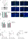
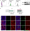
Fig. 1
Fig. 1
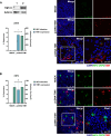
Knockout or blocking of TIM1 decreases eHEV infection in Huh-7.5 cells. A Upper left panel shows an overlay of TIM1 cell surface expression in Huh-7.5WT (green) and Huh-7.5TIM1−KO (purple) cells. Expression evaluated by flow cytometry using a TIM1 recombinant rabbit monoclonal antibody. Middle left panel shows confirmation of TIM1 KO by western blot, 1 = Huh-7.5WT 2 = Huh-7.5TIM1−KO. Lower left panel shows infection in Huh-7.5WT and Huh-7.5TIM1−KO exposed to different preparations of HEV inoculum: extracellular HEV (eHEV), intracellular HEV (iHEV) and extracellular HEV treated with deoxycholate/trypsin (tHEV). Infection was determined 3 days post infection by indirect immunostaining against the HEV ORF2 protein. Representative confocal images of the different HEV preparations are on the right with DAPI staining in blue and HEV ORF2 in green. B Quantification of HEV RNA by qRT-PCR in supernatants of Huh-7.5WT and Huh-7.5TIM1−KO at two different time-points after challenge with eHEV on the left or iHEV on the right. C Huh-7.5WT were preincubated with different concentrations (1 µg/mL, 3 µg/mL or 10 µg/mL) of a polyclonal anti-TIM1 antibody or normal IgG as control for 30 min. Preincubated cells were challenged with eHEV (left panel) or iHEV (middle panel) and infection was determined 3 days later by HEV ORF2 immunostaining. Right panel shows cell viability as measured by MTT assay and normalized to untreated control. The data are represented as mean ± SEM of at least two independent experiments. Each experiment consisted of at least 3 technical replicates. Significance was calculated using a two-tailed t test (A&B) or two-way ANOVA (C). ****p < 0.0001, ***p < 0.001, **p < 0.01, *p < 0.05. ns non-significant
Fig. 2
Fig. 2
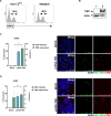
Rescue of TIM1 enhances HEV infection. A Huh-7.5TIM1−KO cells were transfected with an empty plasmid (MOCK) or pcDNA3.1-TIM1 expression vector. Expression was verified via western blot, 1 = Huh-7.5TIM1−KO/MOCK and 2 = Huh-7.5TIM1−KO/pcDNA-TIM1. eHEV infection was performed 24 h after transfection and analyzed 3 days later. eHEV infection levels determined by HEV ORF2 indirect immunofluorescence staining (right axis, green). TIM1 expression on the same day is depicted on the left y axis (blue) and was measured by TIM1 immunofluorescence staining. On the right side, representative confocal microscopy images of MOCK and TIM1 transfected Huh-7.5TIM1−KO cells are depicted. Cells were fixed and permeabilized, followed by immunofluorescence staining against TIM1 (red) and HEV ORF2 (green), DAPI nuclei in blue. Lowest panel shows a merged image where a region of interest was acquired by 40× objective lens instead of 20× objective (white square) to demonstrate overlap between the cells expressing TIM1 and being infected with HEV. B Huh-7.5TIM1−KO cells were transfected with an empty plasmid (MOCK) or pcDNA3.1-TIM1 expression vector and infected with iHEV. Experiment and graph same as for the upper panel. On the right side, representative confocal microscopy images are depicted, the same as described for the upper panel. Data are represented as means ± SEM from two independent experiments. Each experiment included at least 3 technical replicates. Significance was calculated using a two-tailed t test. *p < 0.05 ns nonsignificant
Fig. 3
Fig. 3
Ectopic expression of TIM1 facilitates eHEV infection in HEK293T cells. A HEK293T cells support HEV replication after transfection with HEV RNA, comparison made with electroporated Huh-7.5 cells. Percentages indicate expression of HEV, as determined by flow cytometry after intracellular staining using an HEV ORF2 monoclonal antibody, followed by labeling with an Alexa Fluor 488-conjugated secondary antibody. B HEK293T were transiently transfected with an empty plasmid (MOCK) or pcDNA3.1-TIM1 expression vector and expression was verified via western blot. 1 = HEK293T/MOCK 2 = HEK293T/pcDNA-TIM1. C HEK293T were transfected with an empty plasmid (MOCK) or pcDNA3.1-TIM1 expression vector one day before eHEV infection. left panel shows results with eHEV infection percentage on the right y axis 3 days after, as measured by HEV ORF2 immunofluorescence staining. TIM1 expression on the same day is depicted on the left y-axis and was measured by TIM1 immunofluorescence staining. Right panel are representative confocal images of eHEV infected HEK293T/MOCK (parental) compared to the HEK293T/pcDNA-TIM1, DAPI staining in blue, HEV ORF2 in green and TIM1 in red. D HEK293T cells transfected the same way as described and infected with iHEV. iHEV percentage on the right y axis on day 3 and TIM1 expression on the same day depicted on the left y axis. Right panel shows representative confocal images of iHEV infected HEK293T/MOCK compared to HEK293T/pcDNA-TIM1. Data represented as means ± SEM from two independent experiments. Each experiment consisted of at least 3 technical replicates. Significance was calculated using a t test. **p < 0.01 ns nonsignificant
Fig. 4
Fig. 4
TIM1 facilitates eHEV entry. A Adherence of eHEV and iHEV to Huh-7.5WT and Huh-7.5TIM1−KO at 4 °C for 1 h. After washing, RNA was extracted and determined by qRT-PCR (left panel). Right panel shows viral binding of eHEV and iHEV to Huh-7.5WT and Huh-7.5TIM1−KO, followed by a shift to 37 °C for 2 h. Cells were washed, trypsinized and total RNA was extracted and measured by qRT-PCR. B Adherence of eHEV and iHEV to HEK293Tparental and HEK293TTIM1−WT at 4 °C for 1 h. After washing, RNA was extracted and determined by qRT-PCR (left panel). Right panel shows viral binding of eHEV and iHEV to Huh-7.5WT and Huh-7.5TIM1−KO, followed by a shift to 37 °C for 2 h. Cells were washed, trypsinized and total RNA was extracted and measured by qRT-PCR. Data represented as means ± SEM from two independent experiments. Each experiment consisted of at least two technical replicates. Significance was calculated using a t test. **p < 0.01 *p < 0.05 ns nonsignificant
Fig. 5
Fig. 5
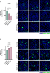
Annexin V inhibits eHEV infection. A eHEV containing viral supernatant was preincubated with different amounts of ANX5 (1 µL, 10 µL) followed by addition to Huh-7.5 cells. Infection normalized to infection without ANX5. On the right, representative confocal microscopy images of the different conditions are depicted. B iHEV containing viral lysate was preincubated with different amounts of ANX5 (1 µL, 10 µL) followed by addition to Huh-7.5 cells. Infection normalized to infection without ANX5. On the right, representative confocal microscopy images of the different conditions are depicted. Data represented as means ± SEM from two independent experiments. Each experiment consisted of at least three technical replicates. Significance was calculated using a two-way ANOVA. *p < 0.05 ns nonsignificant
Fig. 6
Fig. 6
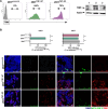
The PS-binding cavity is important for viral binding to TIM1. A TIM1 cell surface expression of parental HEK293T (purple) compared to TIM1-WT transduced HEK293T (green) and TIM1-ND mutant transduced HEK293T (grey). Expression was evaluated by flow cytometry using a TIM1 recombinant monoclonal antibody (left panel). Right panel shows expression verified by western blot. 1 = HEK293Tparental 2 = HEK293TTIM1−WT 3 = HEK293TTIM1−ND. B Stable transduced HEK293TTIM1−WT and HEK293TTIM1−ND mutant as well as parental HEK293T cells were challenged with eHEV (left panels) or iHEV (right panels) and analyzed by HEV ORF2 immunofluorescence staining three days later. Lower panels are representative confocal images of the different conditions, DAPI staining in blue, HEV ORF2 in green and TIM1 in red. Data represented as means ± SEM from two independent experiments. Each experiment consisted of at least three technical replicates. Significance was calculated using a two-way ANOVA. ****p < 0.0001 ns nonsignificant
Fig. 7
Fig. 7
The PS-binding cavity is important for viral binding to TIM1. A TIM1 cell surface expression of Huh-7.5TIM1−KO (purple) compared to Huh-7.5TIM1−WT (green) and Huh-7.5TIM1−ND (grey). Expression was evaluated by flow cytometry using a TIM1 recombinant monoclonal antibody (left panel). Right panel shows expression verified by western blot. 1 = Huh-7.5TIM1−WT 2 = Huh-7.5TIM1−ND 3 = Huh-7.5TIM1−KO. B Huh-7.5TIM1−KO, Huh-7.5TIM1−WT, and Huh-7.5TIM1−ND were challenged with eHEV (left panels) or iHEV (right panels) and analyzed by HEV ORF2 immunofluorescence staining three days later. Lower panels are representative confocal images of the different conditions, DAPI staining in blue, HEV ORF2 in green and TIM1 in red. Data represented as means ± SEM from two independent experiments. Each experiment consisted of at least three technical replicates. Significance was calculated using a two-way ANOVA. ****p < 0.0001 ns nonsignificant

Similar articles

Cited by

References

    1. Lhomme S, et al. Clinical manifestations, pathogenesis and treatment of hepatitis E virus infections. J Clin Med. 2020 doi: 10.3390/jcm9020331. - DOI - PMC - PubMed
    1. Webb GW, Dalton HR. Hepatitis E: an expanding epidemic with a range of complications. Clin Microbiol Infect. 2020;26(7):828–832. doi: 10.1016/j.cmi.2020.03.039. - DOI - PubMed
    1. Sayed IM, et al. Is hepatitis E virus an emerging problem in industrialized countries? Hepatology (Baltimore, MD) 2015;62(6):1883–1892. doi: 10.1002/hep.27990. - DOI - PubMed
    1. Navaneethan U, Al-Mohajer M, Shata MT. Hepatitis E and pregnancy: understanding the pathogenesis. Liver Int. 2008;28(9):1190–1199. doi: 10.1111/j.1478-3231.2008.01840.x. - DOI - PMC - PubMed
    1. Kamar N, et al. Hepatitis E virus infection. Nat Rev Dis Primers. 2017;3:17086. doi: 10.1038/nrdp.2017.86. - DOI - PubMed

MeSH terms

Substances