

Extracellular vesicle biopotentiated hydrogels for diabetic wound healing: The art of living nanomaterials combined with soft scaffolds
- PMID: 37810755
- PMCID: PMC10550777
- DOI: 10.1016/j.mtbio.2023.100810
Extracellular vesicle biopotentiated hydrogels for diabetic wound healing: The art of living nanomaterials combined with soft scaffolds
Abstract
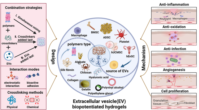
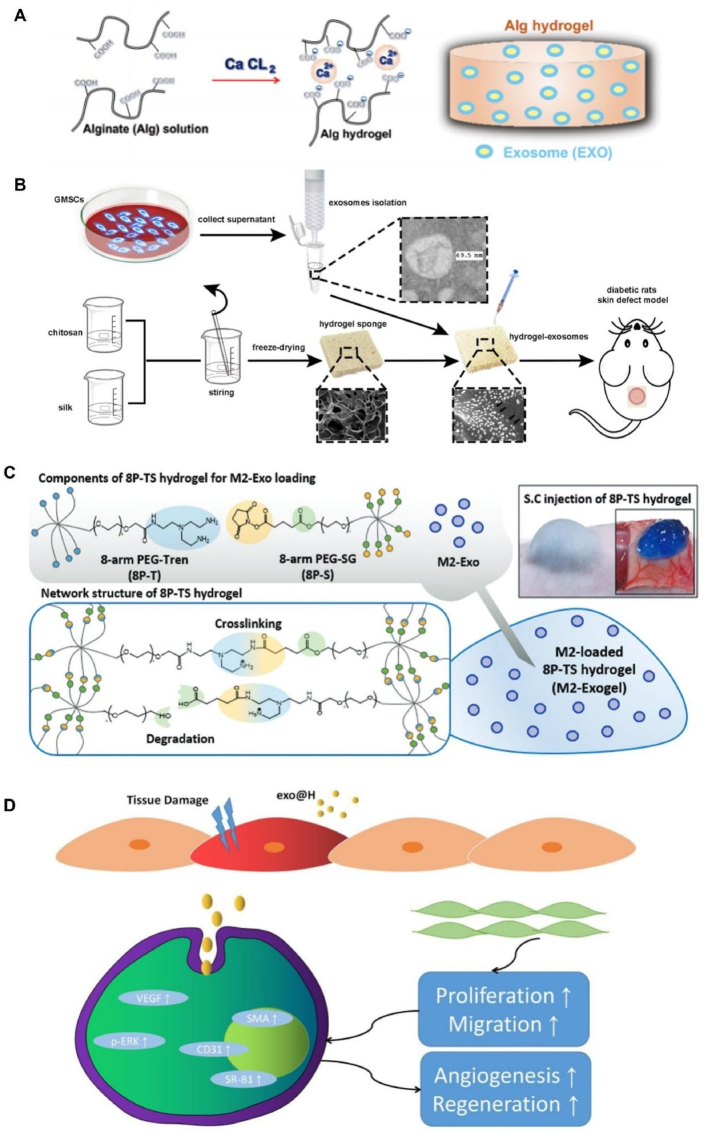
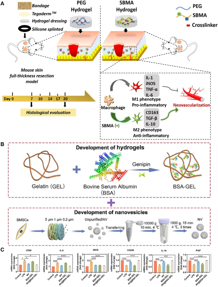
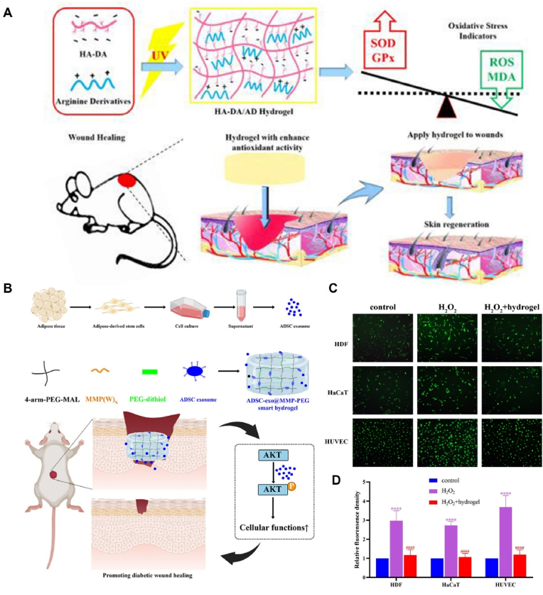
Diabetic wounds (DWs) pose a major challenge for the public health system owing to their high incidence, complex pathogenesis, and long recovery time; thus, there is an urgent need to develop innovative therapies to accelerate the healing process of diabetic wounds. As natural nanovesicles, extracellular vesicles (EVs) are rich in sources with low immunogenicity and abundant nutritive molecules and exert potent therapeutic effects on diabetic wound healing. To avoid the rapid removal of EVs, a suitable delivery system is required for their controlled release. Owing to the advantages of high porosity, good biocompatibility, and adjustable physical and chemical properties of hydrogels, EV biopotentiated hydrogels can aid in achieving precise and favorable therapy against diabetic wounds. This review highlights the different design strategies, therapeutic effects, and mechanisms of EV biopotentiated hydrogels. We also discussed the future challenges and opportunities of using EV biopotentiated hydrogels for diabetic wound healing.
Keywords: Design strategy; Diabetic wounds; Extracellular vesicle; Hydrogel; Mechanism.
© 2023 The Authors. Published by Elsevier Ltd.
Conflict of interest statement
The authors declare that they have no known competing financial interests or personal relationships that could have appeared to influence the work reported in this paper.
Figures

Similar articles
-
Hydrogel Loaded with Extracellular Vesicles: An Emerging Strategy for Wound Healing.Pharmaceuticals (Basel). 2024 Jul 10;17(7):923. doi: 10.3390/ph17070923. Pharmaceuticals (Basel). 2024. PMID: 39065772 Free PMC article. Review.
-
Adipose-Derived Mesenchymal Stem Cell-Derived Exosomes Biopotentiated Extracellular Matrix Hydrogels Accelerate Diabetic Wound Healing and Skin Regeneration.Adv Sci (Weinh). 2023 Oct;10(30):e2304023. doi: 10.1002/advs.202304023. Epub 2023 Sep 15. Adv Sci (Weinh). 2023. PMID: 37712174 Free PMC article.
-
VH298-loaded extracellular vesicles released from gelatin methacryloyl hydrogel facilitate diabetic wound healing by HIF-1α-mediated enhancement of angiogenesis.Acta Biomater. 2022 Jul 15;147:342-355. doi: 10.1016/j.actbio.2022.05.018. Epub 2022 May 16. Acta Biomater. 2022. PMID: 35580827
-
Extracellular Vesicle-Based Hydrogels for Wound Healing Applications.Int J Mol Sci. 2023 Feb 18;24(4):4104. doi: 10.3390/ijms24044104. Int J Mol Sci. 2023. PMID: 36835516 Free PMC article. Review.
-
GelMA Hydrogel Loaded with Extracellular Vesicles Derived from Umbilical Cord Mesenchymal Stem Cells for Promoting Cutaneous Diabetic Wound Healing.ACS Omega. 2023 Mar 8;8(11):10030-10039. doi: 10.1021/acsomega.2c07291. eCollection 2023 Mar 21. ACS Omega. 2023. PMID: 36969465 Free PMC article.
Cited by
-
Exosome-based cell therapy for diabetic foot ulcers: Present and prospect.Heliyon. 2024 Oct 11;10(20):e39251. doi: 10.1016/j.heliyon.2024.e39251. eCollection 2024 Oct 30. Heliyon. 2024. PMID: 39498056 Free PMC article. Review.
-
Engineered mesenchymal stem cell-derived extracellular vesicles: kill tumors and protect organs.Theranostics. 2024 Sep 23;14(16):6202-6217. doi: 10.7150/thno.99618. eCollection 2024. Theranostics. 2024. PMID: 39431009 Free PMC article. Review.
-
Multifunctional type lll recombinant human collagen incorporated sodium alginate hydrogel with sustained release of extra cellular vehicles for wound healing multimodal therapy in diabetic mice.Regen Ther. 2024 Apr 16;27:329-341. doi: 10.1016/j.reth.2024.03.010. eCollection 2024 Dec. Regen Ther. 2024. PMID: 38873636 Free PMC article.
-
Biomimetic Scaffolds-A Novel Approach to Three Dimensional Cell Culture Techniques for Potential Implementation in Tissue Engineering.Nanomaterials (Basel). 2024 Mar 16;14(6):531. doi: 10.3390/nano14060531. Nanomaterials (Basel). 2024. PMID: 38535679 Free PMC article. Review.
-
Multifunctional hydrogel-based engineered extracellular vesicles delivery for complicated wound healing.Theranostics. 2024 Jul 8;14(11):4198-4217. doi: 10.7150/thno.97317. eCollection 2024. Theranostics. 2024. PMID: 39113809 Free PMC article. Review.
References
-
- Harreiter J., Roden M. [Diabetes mellitus-Definition, classification, diagnosis, screening and prevention (Update 2019)] Wien Klin. Wochenschr. 2019;131(Suppl 1):6–15. - PubMed
-
- Hicks C.W., Canner J.K., Sherman R.L., Black J.H., 3rd, Lum Y.W., Abularrage C.J. Evaluation of revascularization benefit quartiles using the Wound, Ischemia, and foot Infection classification system for diabetic patients with chronic limb-threatening ischemia. J. Vasc. Surg. 2021;74(4):1232–1239 e3. - PMC - PubMed
-
- Mponponsuo K., Sibbald R.G., Somayaji R. A comprehensive review of the pathogenesis, diagnosis, and management of diabetic foot infections. Adv. Skin Wound Care. 2021;34(11):574–581. - PubMed
Publication types
LinkOut - more resources
Full Text Sources
Research Materials
