. 2023 Oct 6;118(1):43.
doi: 10.1007/s00395-023-01013-1.

Outcomes of hypothalamic oxytocin neuron-driven cardioprotection after acute myocardial infarction

Affiliations

Outcomes of hypothalamic oxytocin neuron-driven cardioprotection after acute myocardial infarction

Kathryn J Schunke et al. Basic Res Cardiol. .

Abstract

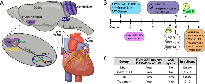
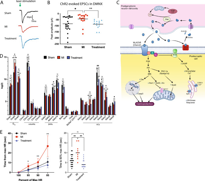
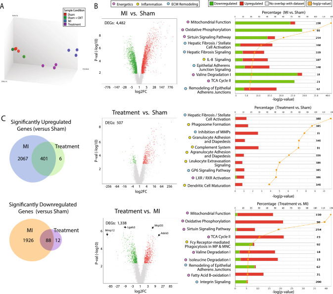
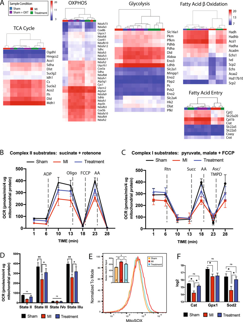
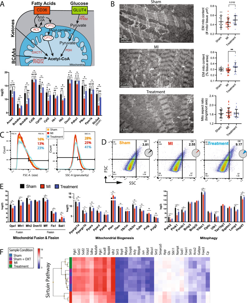
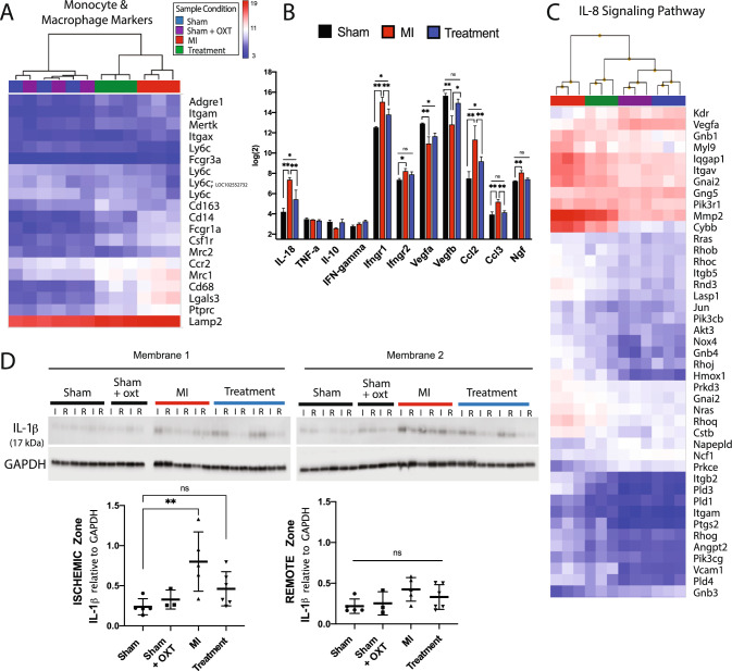
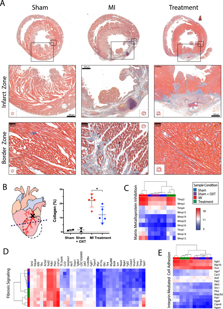
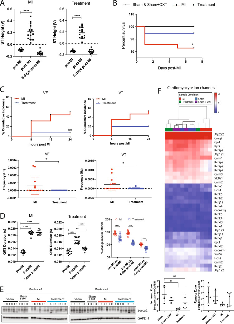
Altered autonomic balance is a hallmark of numerous cardiovascular diseases, including myocardial infarction (MI). Although device-based vagal stimulation is cardioprotective during chronic disease, a non-invasive approach to selectively stimulate the cardiac parasympathetic system immediately after an infarction does not exist and is desperately needed. Cardiac vagal neurons (CVNs) in the brainstem receive powerful excitation from a population of neurons in the paraventricular nucleus (PVN) of the hypothalamus that co-release oxytocin (OXT) and glutamate to excite CVNs. We tested if chemogenetic activation of PVN-OXT neurons following MI would be cardioprotective. The PVN of neonatal rats was transfected with vectors to selectively express DREADDs within OXT neurons. At 6 weeks of age, an MI was induced and DREADDs were activated with clozapine-N-oxide. Seven days following MI, patch-clamp electrophysiology confirmed the augmented excitatory neurotransmission from PVN-OXT neurons to downstream nuclei critical for parasympathetic activity with treatment (43.7 ± 10 vs 86.9 ± 9 pA; MI vs. treatment), resulting in stark improvements in survival (85% vs. 95%; MI vs. treatment), inflammation, fibrosis assessed by trichrome blue staining, mitochondrial function assessed by Seahorse assays, and reduced incidence of arrhythmias (50% vs. 10% cumulative incidence of ventricular fibrillation; MI vs. treatment). Myocardial transcriptomic analysis provided molecular insight into potential cardioprotective mechanisms, which revealed the preservation of beneficial signaling pathways, including muscarinic receptor activation, in treated animals. These comprehensive results demonstrate that the PVN-OXT network could be a promising therapeutic target to quickly activate beneficial parasympathetic-mediated cellular pathways within the heart during the early stages of infarction.

Keywords: Arrhythmia; Cardioprotection; Infarction; Mitochondria; Oxytocin; Parasympathetic nervous system.


Conflict of interest statement

On behalf of all authors, the corresponding author states that there is no conflict of interest.

Figures

Fig. 1
Fig. 1
A In vivo activation of brainstem parasympathetic neurons. Selective expression of excitatory hM3D(Gq) DREADDs within PVN-OXT neurons, and subsequent activation via CNO, increases the firing rate of PVN-OXT neurons, which co-release OXT and GLUT at synapses on parasympathetic DMNX neurons of the medulla. Elevated release of synaptic OXT and GLUT increases excitatory neurotransmission to DMNX parasympathetic neurons, elevating their firing rate to increase downstream activation of post-ganglionic parasympathetic ganglia neurons that release acetylcholine at their target tissue. Ultimately, the increased release of acetylcholine within the myocardium elevates the level of myocyte muscarinic pathway activation via cholinergic muscarinic (Chrm2/M2) receptors. B Protocol timeline from animal birth to sacrifice at 7 weeks of age with subsequent ex vivo assessments. Three viruses encoding DREADDs, ChR2, and OXT-Cre were injected into the PVN of all rats at 1 week of age, followed by surgical implantation of an ECG transmitter at 3 weeks of age. Baseline ECG and HRR data were collected at 5 weeks, followed by either sham MI or MI surgery. Immediately following surgery, and daily for 7 days, animals were injected with either saline or CNO. At 7 weeks of age (or 1-week post-MI), animals were sacrificed, and brains and hearts were collected for ex vivo assessments. C The four animal groups with the assigned interventions are shown in the table. Abbreviations: PVN, paraventricular nucleus of the hypothalamus; DMNX, dorsal motor nucleus of the vagus; DREADDs, designer receptor exclusively activated by designer drugs; CNO, clozapine-N-oxide; OXT, oxytocin; GLUT, glutamate; CVN, cardiac vagal neuron; AAV, adeno-associated virus; ChR2, channelrhodopsin; EPSC, excitatory post-synaptic current; EM, electron microscopy
Fig. 2
Fig. 2
Daily activation of PVN-OXT neurons after MI maintained excitatory neurotransmission to parasympathetic neurons in the DMNX and rapid HRR. A Typical voltage clamp recordings of evoked EPSCs upon photoexcitation of ChR2-expressing PVN-OXT neurons show diminished EPSC amplitude for MI animals. B Distribution of evoked EPSC amplitudes recorded 7 days after MI for each group illustrates maintenance of EPSC in Treatment and loss of EPSC in MI animals. EPSC amplitude was significantly lower in MI animals (− 44 ± 10 pA) compared to Sham (− 84 ± 12 pA) and Treatment animals (− 87 ± 9 pA) (Kruskal–Wallis test with post hoc Dunn’s test, mean ± SEM, p = 0.0045; *p = 0.02 and **p = 0.007). Sham: n = 24 cells, 24 brainstem slices, 11 animals; MI: n = 17 cells, 17 brainstem slices, 6 animals; Treatment: n = 14 cells, 14 brainstem slices, 5 animals. (C) A cholinergic synapse showing ACh production and release and the pathways that are activated by the primary Gi/o coupling of the muscarinic ACh type 2 receptors (m2AChRs) of the post-synaptic cell. Preserved gene expression of myocyte m2AChRs and elevated release of ACh from cardiac cholinergic axon varicosities could activate cellular cardioprotective pathways that would reduce sarcoplasmic reticulum (SR) stress and mitochondrial ROS, inhibit activation of the mitochondrial permeability transition pore (MPTP), and reduce nuclear production of inflammatory cytokines. Small blue arrows indicate increased or decreased abundance/activity. D Gene expression profiles of proteins that are integral for myocyte muscarinic signaling (n = 3 per group; student’s t test; mean ± SD; *p < 0.05). E HRR time 5 days after MI as a percentage of HR at peak running effort (the maximum HR). Recovery time to 95%, 90%, and 85% of maximum HR was significantly longer for MI animals. HRR time was not significantly different between Treatment and Sham animals (Sham n = 24; MI, n = 9; Treatment, n = 13; two-way ANOVA; mean ± SD, *p < 0.05, **p < 0.01). Abbreviations: PVN, paraventricular nucleus; OXT, oxytocin; CVN, cardiac vagal neuron; ChR2, channelrhodopsin; EPSC, excitatory post-synaptic current; HR, heart rate; HRR, heart rate recovery
Fig. 3
Fig. 3
Transcriptome analysis of LV myocardium DEGs. A PCA of each group (Sham, n = 3; Sham + OXT, n = 3; MI, n = 3; Treatment, n = 3). B Volcano plots of DEGs between groups (left; 1.7 < FC < − 1.7, FDR < 0.08), and top ten corresponding differentially regulated canonical pathways represented by the DEGs (right). The stacked bar chart depicts the percentage (upper x-axis) of pathway genes up-, down-, or not differentially expressed (bar color), with the total number of pathway genes shown on the right of each bar, and the -log significance of the differential pathway regulation (orange line; lower x-axis). Only transcripts with FDR < 0.08 were entered into the analysis; all pathways depicted exhibit p < 0.05. C Venn diagrams of differentially upregulated or downregulated genes compared to Sham. Abbreviations: FC, fold change; FDR, false discovery rate; MI, myocardial infarction; MMPs, matrix metalloproteases; MP, macrophage; MNC, monocyte
Fig. 4
Fig. 4
Mitochondrial respiration was preserved in hearts of Treatment animals. A Transcriptome expression heat maps and hierarchical clustering of key genes involved in mitochondrial respiration: TCA cycle, OXPHOS, glycolysis, fatty acid beta-oxidation, and fatty acid entry. B Seahorse analysis of substrate-stimulated respiration of isolated mitochondria with succinate or (C) uncoupled respiration with pyruvate and malate. D Quantitation of oxygen consumption rate (OCR) for succinate-stimulated respiration. For all respiration assays: Sham, n = 3; MI and Treatment, n = 4 each; one-way ANOVA; mean ± SD; *p < 0.05. E Flow cytometric analysis of isolated mitochondrial superoxide (mitoSOX; Sham and MI, n = 4; Treatment, n = 5; one-way ANOVA; mean ± SD; *p < 0.05), and F microarray expression of mitochondrial antioxidant enzymes (n = 3 per group; one-way ANOVA; mean ± SD; *p < 0.05)
Fig. 5
Fig. 5
LV mitochondria respiration and morphology were preserved in Treatment animals. A Schematic of myocyte substrate utilization and substrate-specific key processes that drive ATP production (top), with expression of genes involved in each component for each group (bottom, n = 3 per group; one-way ANOVA; mean ± SD; *p < 0.05). B Representative electron micrographs of mitochondrial ultrastructure with derived measurements of mitochondrial content and aspect ratio (n = 4 animals per group; one-way ANOVA; **p < 0.01). C Representative flow cytometric mitochondrial forward scatter (FSC, left panel) and side scatter (SSC, right panel) depicting size and granularity, respectively. Counts are 50,000 ± 200 mitochondria per group. (Sham and MI, n = 4; Treatment, n = 5). D Representative forward vs. side mitochondrial scatter indicating increased size and complexity (Q6 – top right quadrant) in Treatment animals. E Expression of genes involved in mitochondrial fusion and fission (left), biogenesis (middle), and mitophagy (right) (n = 3 per group; one-way ANOVA; mean ± SD; *p < 0.05, **p < 0.01). F Microarray heat map expression and hierarchical clustering of the sirtuin pathway involved in regulation of mitochondrial dynamics (n = 3 per group)
Fig. 6
Fig. 6
Cardiac inflammation was reduced 7 days after MI in Treatment animals. Microarray expression and hierarchical clustering of A cardiac monocyte and macrophage markers, B cytokines (n = 3 per group; one-way ANOVA; mean ± SD; *p < 0.05, **p < 0.01) and C the IL-8 signaling pathway. D Western blot of IL-1β expression in ischemic (I) and remote (R) areas of the infarct (Sham, n = 5; Sham + OXT, n = 3; MI, n = 5; Treatment, n = 6; one-way ANOVA; mean ± SD, **p < 0.01)
Fig. 7
Fig. 7
Myocardial remodeling and infarct size were reduced in Treatment animals 7 days after MI. A Representative Masson’s trichrome images of myocardial sections from Sham, MI, and Treatment animals are shown in each column. Large and small bounding boxes on images of the full section (top row) indicate the bounding area of the high-resolution images for the infarct zone (middle row) and border zone (bottom row). Blue denotes the presence of collagen. B Schematic of the heart (left) illustrates the cross-section of histological assessment (dashed line). The dashed circle represents the location of the ischemic zone (area at risk) from which tissue was harvested for microarray analysis and Western blotting. Percent collagen content (right) within a region of the infarct zone was measured using the trichrome images (Sham, n = 3; Sham + OXT, n = 2; MI, n = 5; Treatment, n = 5; one-way ANOVA; mean ± SD; *p < 0.05). Hierarchical clustering and heat map expression of genes involved in C inhibition of matrix metalloproteins, D fibrosis signaling, and E integrin-mediated cell adhesion are provided (n = 3 per group)
Fig. 8
Fig. 8
Arrhythmia incidence and mortality were reduced in Treatment animals. A Average 24 h ST segment elevation immediately following MI and 5 days post-MI is similar between MI and Treatment animals, indicating that both groups experienced a similar degree of ischemic damage. Pre-MI: MI, n = 12; Treatment, n = 13. Post-MI: MI, n = 14; Treatment, n = 13. Five days post-MI: MI, n = 14; Treatment, n = 15. One-way ANOVA; ****p < 0.0001. B Post-MI survival is significantly improved in Treatment animals compared to MI animals (Sham, n = 25; MI, n = 35; Treatment, n = 38; Kaplan–Meier; *p = 0.046). C In the 24 h immediately following MI, Treatment animals had lower incidence of arrhythmias (MI, n = 14; Treatment, n = 12; unpaired t test; **p < 0.01, *p < 0.05) VF ventricular fibrillation, VT ventricular tachycardia. D QRS duration significantly increased after MI in both groups but was significantly lower 5 days post-MI in Treatment animals. Multiple comparisons of percent increases in QRS duration from pre-MI values reveal that QRS widening was also significantly less for Treatment animals compared to MI animals (same animal numbers as panel A, one-way ANOVA, **p < 0.01, ****p < 0.0001). E Western blots for Serca2a (Atp2a2) expression of LV tissue (I: ischemic) and RV tissue (R: remote) 7 days after MI are shown (left). Serca2a was reduced in the I zone for MI animals but not Treatment animals (right, Sham, n = 5; Sham + OXT, n = 3; MI, n = 5; Treatment, n = 6; one-way ANOVA; mean ± SD, **p < 0.01). F Expression and hierarchical clustering of genes that are key contributors to the cardiac action potential. MI animals cluster alone yet Treatment animals cluster with Sham and Sham + OXT animals, indicating preserved expression of key genes in Treatment animals (n = 3 per group)

Similar articles

Cited by

References

    1. Abe M, Rastelli DD, Gomez AC, Cingolani E, Lee Y, Soni PR, Fishbein MC, Lehman TJA, Shimada K, Crother TR, Chen S, Noval Rivas M, Arditi M. IL-1-dependent electrophysiological changes and cardiac neural remodeling in a mouse model of Kawasaki disease vasculitis. Clin Exp Immunol. 2020;199:303–313. doi: 10.1111/cei.13401. - DOI - PMC - PubMed
    1. Agarwal R, Mokelke E, Ruble SB, Stolen CM. Vagal nerve stimulation evoked heart rate changes and protection from cardiac remodeling. J Cardiovasc Transl Res. 2016;9:67–76. doi: 10.1007/s12265-015-9668-7. - DOI - PubMed
    1. Allard MF, Schonekess BO, Henning SL, English DR, Lopaschuk GD. Contribution of oxidative metabolism and glycolysis to ATP production in hypertrophied hearts. Am J Physiol - Hear Circ Physiol. 1994;267:H742–H750. doi: 10.1152/ajpheart.1994.267.2.h742. - DOI - PubMed
    1. Androne AS, Hryniewicz K, Goldsmith R, Arwady A, Katz SD. Acetylcholinesterase inhibition with pyridostigmine improves heart rate recovery after maximal exercise in patients with chronic heart failure. Heart. 2003;89:854–858. doi: 10.1136/heart.89.8.854. - DOI - PMC - PubMed
    1. Basalay MV, Mastitskaya S, Mrochek A, Ackland GL, Del Arroyo AG, Sanchez J, Sjoquist P-O, Pernow J, Gourine AV, Gourine A. Glucagon-like peptide-1 (GLP-1) mediates cardioprotection by remote ischaemic conditioning. Cardiovasc Res. 2016;112:669–676. doi: 10.1093/cvr/cvw216. - DOI - PMC - PubMed

Publication types