. 2023 Oct 6;325(6):H1360-H1372.
doi: 10.1152/ajpheart.00279.2023. Online ahead of print.

Endothelial KIR2 channel dysfunction in aged cerebral parenchymal arterioles

Affiliations

Endothelial KIR2 channel dysfunction in aged cerebral parenchymal arterioles

Felipe D Polk et al. Am J Physiol Heart Circ Physiol. .

Abstract

Aging is associated with cognitive decline via incompletely understood mechanisms. Cerebral microvascular dysfunction occurs in aging, particularly impaired endothelium-mediated dilation. Parenchymal arterioles are bottlenecks of the cerebral microcirculation, and dysfunction causes a mismatch in nutrient demand and delivery, leaving neurons at risk. Extracellular nucleotides elicit parenchymal arteriole dilation by activating endothelial purinergic receptors (P2Y), leading to opening of K+ channels, including inwardly-rectifying K+ channels (KIR2). These channels amplify hyperpolarizing signals, resulting in dilation. However, it remains unknown if endothelial P2Y and KIR2 signaling are altered in brain parenchymal arterioles during aging. We hypothesized that aging impairs endothelial P2Y and KIR2 function in parenchymal arterioles. We observed reduced dilation to the purinergic agonist 2-methyl-S-ADP (1 µM) in arterioles from Aged (>24-month-old) mice when compared to Young (4-6 months of age) despite similar hyperpolarization in endothelial cells tubes. No differences were observed in vasodilation or endothelial cell hyperpolarization to activation of small- and intermediate-conductance Ca2+-activated K+ channels (KCa2.3 / KCa3.1) by NS309. Hyperpolarization to 15 mM [K+]E was smaller in Aged than Young mice, despite a paradoxical increased dilation in Aged arterioles to 15 mM [K+]E that was unchanged by endothelium removal. KIR2 Inhibition attenuated vasodilatory responses to 15 mM [K+]E and 1 µM 2-me-S-ADP in both Young and Aged arterioles. Further, we observed a significant increase in myogenic tone in Aged parenchymal arterioles, which was not enhanced by endothelium removal. We conclude that aging impairs endothelial KIR2 channel function in the cerebral microcirculation with possible compensation by smooth muscle cells.

Keywords: aging; endothelium-dependent hyperpolarization; inwardly-rectifying K+ channel; parenchymal arterioles; purinergic receptors.


Conflict of interest statement

No conflicts of interest, financial or otherwise, are declared by the authors.

Figures

Figure 1.
Figure 1.
Increased spontaneous myogenic tone of parenchymal arterioles isolated from aged mice. A: representative traces of the lumen diameter of young (black, top trace) and aged (blue, bottom trace) pressurized parenchymal arterioles after equilibration at 40 mmHg. Note the larger decrease in lumen diameter of parenchymal arterioles isolated from aged mice. B: summary graph showing a significant increase in resting myogenic tone of parenchymal arterioles isolated from aged mice than those isolated from young mice. *P < 0.05, unpaired one-tailed Student’s t test; n = 10–12 different mice/group, n = 3 mice. Data are means ± SE. C: representative lumen diameter traces of young (black, top trace) and aged (blue, bottom trace) pressurized endothelium-denuded parenchymal arterioles after equilibration at 40 mmHg. D: summary graph showing no change in spontaneous myogenic tone after removal of the endothelium between young and aged mice.
Figure 2.
Figure 2.
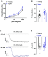
Reduced dilation of aged parenchymal arterioles to purinergic receptor activation despite no differences in membrane potential (Vm). A: summary line graph showing that dilation to the pan-P2Y agonist 2-methyl-thio-ADP (2-me-S-ADP) was similar between aged and young arterioles. Increased dilatory response to 2-me-S-ADP in young, pressurized arterioles at highest tested concentration (1 µM). B: summary bar graph of 1 µM 2-me-S-ADP dilation highlighting the blunted vasodilatory response in aged compared with young arterioles. *P < 0.05, unpaired one-tailed Student’s t test, n = 8 or 9 different mice/group. C: representative traces of sharp electrode electrophysiology recordings of endothelial cells tubes Vm after exposure to the pan-P2Y agonist 2-me-S-ADP. D: summary graph showing similar endothelial cell hyperpolarization in endothelial cell tubes isolated from young and aged mice. All data are means ± SE; n = 5–7 mice/group.
Figure 3.
Figure 3.
Parenchymal arteriole endothelial cell responses to small- and intermediate-conductance Ca2+-activated K+ channels (KCa2.3/KCa3.1) are not altered by aging. A: summary line graph showing that dilation to increasing concentrations of NS309 was similar between aged and young arterioles. B: summary bar graph of the dilation to 1 µM NS309 highlighting that vasodilatory responses of parenchymal arterioles were similar between young and aged mice. n = 5 different mice/group. C: representative traces of sharp electrode electrophysiology recordings of endothelial cell tubes membrane potential (Vm) from young (black, top trace) and aged (blue, bottom trace) mice after exposure to the KCa2.3/KCa3.1 activator NS309 (1 µM). D: summary graph showing that endothelial cell hyperpolarization was similar between endothelial cell tubes isolated from young and aged mice. All data are means ± SE; n = 13 or 14 mice/group.
Figure 4.
Figure 4.
Blunted inwardly rectifying K+ channels (Kir2)-induced hyperpolarization in aged endothelial cell tubes. A: representative traces of sharp electrode electrophysiology recordings of endothelial cells tubes membrane potential (Vm) after exposure to 15 mM extracellular K+ concentration ([K+]e). Note the smaller hyperpolarization in endothelial cell tubes isolated from aged parenchymal arterioles (blue, bottom trace) compared with those from young mice (black, top trace). B: summary graph showing blunted hyperpolarization to Kir2 activation in endothelial cell tubes from aged parenchymal arterioles. *P < 0.05, unpaired one-tailed Student’s t test; n = 13 mice/group. All data are means ± SE.
Figure 5.
Figure 5.
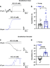
Preserved inwardly rectifying K+ channels (Kir2)-induced dilation of aged parenchymal arterioles via compensation by smooth muscle Kir2. A: representative traces of lumen diameter of young (black, top trace) and aged (blue, bottom trace) pressurized parenchymal arterioles exposed to 15 mM extracellular K+ concentration ([K+]e). Note the larger dilation observed in parenchymal arterioles isolated from aged mice. B: summary graph showing Kir2-dependent dilation of parenchymal arterioles was increased in aged parenchymal arterioles when compared with those isolated from young mice. *P < 0.05, one-tailed Student’s t test; n = 10 or 11 different mice/group. C: representative traces of lumen diameter of young (black, upper trace) and aged (blue, lower trace) pressurized parenchymal arterioles after removal of the endothelium. Preserved dilation in parenchymal arterioles isolated from aged mice was observed after 15 mM [K+]e exposure. D: summary graph showing that removal of the endothelium by an intraluminal air bolus elicited a robust contraction in young parenchymal arterioles in response to 15 mM [K+]e, while vasodilatory effects were maintained in aged arterioles. ****P < 0.001, one-tailed Student’s t test; n = 10 different mice/group, except for endothelium denuded arterioles, where n = 3 mice. All data are means ± SE.
Figure 6.
Figure 6.
Inwardly rectifying K+ channels (Kir2) blockade attenuates 15 mM extracellular K+ concentration ([K+]e)-induced dilation and response to P2Y receptor activation. A: representative trace of the lumen diameter of young (black, top trace) and aged (blue, bottom trace) pressurized parenchymal arterioles exposed to 15 mM [K+] in the presence of BaCl2 (100 μM). B: summary graph of Kir2 blockade with BaCl2 highlighting contraction in young and lack of response in aged parenchymal arterioles to 15 mM [K+]e; n = 6 different mice/group. *P < 0.05, one-tailed Student's t test. C: representative traces of lumen diameter of young (black, top trace) and aged (blue, bottom trace) pressurized parenchymal arterioles exposed 2-methyl-S-ADP (2-me-S-ADP; 1 μM) in the presence of BaCl2. D: summary graph showing attenuation of vasodilatory response to 2-me-S-ADP in the presence of BaCl2 in both young and aged arterioles. *P < 0.05, one-tailed Student’s t test; n = 4 or 5 different mice/group. All data are means ± SE.
Figure 7.
Figure 7.
Graphical summary highlighting endothelial inwardly rectifying K+ channels (Kir2) dysfunction with possible compensation by smooth muscle Kir2 in aged parenchymal arterioles. In young arterioles, activation of endothelial P2Y receptors leads to the opening of small- and intermediate-conductance Ca2+-activated K+ channels (KCa2.3/KCa3.1) causing an increase in extracellular K+ concentration ([K+]e). Endothelial Kir2 channels are activated by localized increases in [K+]e, resulting in the boosting of a hyperpolarizing signal that is then transmitted to smooth muscle cells. In aged parenchymal arterioles, activation of P2Y receptors does not induce vasodilation despite functional KCa2.3/KCa3.1 channels. This impairment is likely a consequence of the loss of endothelial Kir2 function observed in our study. Although Kir2 activity seems to be upregulated in smooth muscle, it is not sufficient to recover purinergic-induced dilation.

Comment in

  • To err, KIR2 that is, on the side of vasodilation in aging.
    Bernardino de Paula R, Wenceslau CF, McCarthy CG. Bernardino de Paula R, et al. Am J Physiol Heart Circ Physiol. 2023 Dec 1;325(6):H1415-H1417. doi: 10.1152/ajpheart.00670.2023. Epub 2023 Oct 20. Am J Physiol Heart Circ Physiol. 2023. PMID: 37861649 No abstract available.

Similar articles

Cited by

References

    1. 2023 Alzheimer's disease facts and figures. Alzheimers Dement 19: 1598–1695, 2023. doi:10.1002/alz.13016. - DOI - PubMed
    1. Knopman DS, Roberts R. Vascular risk factors: imaging and neuropathologic correlates. J Alzheimers Dis 20: 699–709, 2010. doi:10.3233/JAD-2010-091555. - DOI - PMC - PubMed
    1. Smith CD, Andersen AH, Kryscio RJ, Schmitt FA, Kindy MS, Blonder LX, Avison MJ. Altered brain activation in cognitively intact individuals at high risk for Alzheimer’s disease. Neurology 53: 1391–1396, 1999. doi:10.1212/wnl.53.7.1391. - DOI - PubMed
    1. Bookheimer SY, Strojwas MH, Cohen MS, Saunders AM, Pericak-Vance MA, Mazziotta JC, Small GW. Patterns of brain activation in people at risk for Alzheimer’s disease. N Engl J Med 343: 450–456, 2000. doi:10.1056/NEJM200008173430701. - DOI - PMC - PubMed
    1. Nishimura N, Schaffer CB, Friedman B, Lyden PD, Kleinfeld D. Penetrating arterioles are a bottleneck in the perfusion of neocortex. Proc Natl Acad Sci USA 104: 365–370, 2007. doi:10.1073/pnas.0609551104. - DOI - PMC - PubMed

LinkOut - more resources