. 2023 Sep 30;14(1):6111.
doi: 10.1038/s41467-023-41705-9.

A CK2 and SUMO-dependent, PML NB-involved regulatory mechanism controlling BLM ubiquitination and G-quadruplex resolution

Affiliations

A CK2 and SUMO-dependent, PML NB-involved regulatory mechanism controlling BLM ubiquitination and G-quadruplex resolution

Shichang Liu et al. Nat Commun. .

Erratum in

Abstract

The Boom syndrome helicase (BLM) unwinds a variety of DNA structures such as Guanine (G)-quadruplex. Here we reveal a role of RNF111/Arkadia and its paralog ARKL1, as well as Promyelocytic Leukemia Nuclear Bodies (PML NBs), in the regulation of ubiquitination and control of BLM protein levels. RNF111 exhibits a non-canonical SUMO targeted E3 ligase (STUBL) activity targeting BLM ubiquitination in PML NBs. ARKL1 promotes RNF111 localization to PML NBs through SUMO-interacting motif (SIM) interaction with SUMOylated RNF111, which is regulated by casein kinase 2 (CK2) phosphorylation of ARKL1 at a serine residue near the ARKL1 SIM domain. Upregulated BLM in ARKL1 or RNF111-deficient cells leads to a decrease of G-quadruplex levels in the nucleus. These results demonstrate that a CK2- and RNF111-ARKL1-dependent regulation of BLM in PML NBs plays a critical role in controlling BLM protein levels for the regulation of G-quadruplex.


Conflict of interest statement

The authors declare no competing interests.

Figures
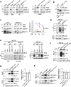
Fig. 1
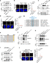
Fig. 1. RNF111 ubiquitinates BLM and regulates BLM protein level.
a Loss of RNF111 leads to increased BLM protein levels. RNF111 bands are indicated by arrows. Cells transfected with control (Ctrl) or RNF111 siRNAs or two independent clones of RNF111 knockout (KO) cells (KO-15 and KO-17) are shown. b Expression of RNF111 but not empty vector (Vec) reduces elevated BLM levels in RNF111 KO U2OS cells. “*” non-specific band. c Increased BLM protein stability in RNF111 knockdown cells. U2OS cells transfected with indicated siRNAs were treated with cycloheximide (CHX) (100 μg/ml). “*” non-specific band. The relative BLM level at each time point normalized by GAPDH and relative to that at 0 h is quantified and shown with mean ± SEM (n = 3 independent experiments). Two way Anova was used for statistics. Band intensity was measured using Image J. d RNF111 knockdown leads to decreased BLM ubiquitination. U2OS cells transfected with siRNAs were treated with MG132 (10 μM, 4 h). Immunoprecipitation (IP) was performed under denature condition. e BLM ubiquitination is dependent on RNF111 catalytic activity. The IPs were performed under denature condition using lysates from WT or RNF111 KO U2OS cells expressing indicated constructs and treated with MG132. f RNF111 catalytic activity is required for its self-ubiquitination. The IPs were performed under denature condition using lysates from 293 T cells expressing indicated constructs and treated with MG132. g The SIM mutant of RNF111 interacts with BLM. 293T cells expressing indicated constructs were used. The relative BLM level in the HA immunoprecipitates (normalized by immunoprecipitated HA-RNF111) is quantified (n = 2 independent experiments). h The SIMs and SUMOylation mutant of BLM interacts with RNF111. 293 T cells expressing indicated constructs were used. The relative RNF111 level present in the GFP immunoprecipitates (normalized by immunoprecipitated GFP-BLM) is quantified (n = 2 independent experiments). Source data are provided as a Source Data file.
Fig. 2
Fig. 2. ARKL1 promotes BLM ubiquitination and regulates BLM protein level.
a A schematic of RNF111 and its N-terminal paralog ARKL1. b Loss of ARKL1 leads to an increase of BLM protein level in various cell lines. c Increased BLM protein stability in ARKL1 knockdown cells. U2OS cells transfected with indicated siRNAs were treated with cycloheximide (CHX) (100 μg/ml). The relative BLM level at each time point (normalized by GAPDH and relative to that at 0 h) is quantified and shown with mean ± SEM (n = 3 independent experiments). Two way Anova was used for statistics. Band intensity was measured using Image J. d Loss of ARKL1 leads to decreased BLM ubiquitination. The IPs were performed under denature condition using lysates from WT or ARKL1 KO Hela cells treated with MG132 (10 μM, 4 h). e Loss of ARKL1 leads to increased RNF111 protein stability. The relative RNF111 level (indicated by arrow) at each time point after CHX treatment (normalized by GAPDH and relative to the beginning of treatment) is quantified and shown with mean ± SEM (n = 3 independent experiments). Two way Anova was used for statistics. f Loss of ARKL1 leads to reduced RNF111 self-ubiquitination. The IPs were performed under denature condition using lysates from WT or ARKL1 KO Hela cells treated with MG132. g ARKL1 interacts with both BLM and RNF111. HA IPs were performed using lysates from U2OS cells expressing vector or HA-ARKL1. Source data are provided as a Source Data file.
Fig. 3
Fig. 3. ARKL1 interacts with RNF111 in a SIM-SUMO dependent manner.
a RNF111 knockdown disrupts the interaction of ARKL1 and BLM. HA IP was performed with lysates of U2OS cells expressing vector, HA-ARKL1 and treated with indicated siRNAs. b Loss of RNF111 abolishes the interaction of ARKL1 and BLM in proximity ligation assay (PLA). Quantifications of WT (n = 170), RNF111 KO (n = 189), ARKL1 KO (n = 115) U2OS cells from two biological replicates are shown. c The SIMs of ARKL1 are required for ARKL1 interaction with both RNF111 and BLM. Lysates from 293 T cells expressing indicated constructs were used. d The co-localization/interaction of RNF111 and ARKL1 is dependent on the SIMs of ARKL1. PLA using antibodies against HA and RNF111 are qunatified for ARKL1 KO U2OS cells complemented with HA-ARKL1WT (n = 28), HA-ARKL1SIM* (n = 20) or vector (n = 17) from two biological replicates. e A schematic of RNF111 deletion mutants generated. f RNF111Δ214–246 deletion mutant exhibits decreased SUMOylation. HA IPs were performed under denature condition using lysates of 293 T cells expressing indicated constructs. g Identification of RNF111 SUMOylation sites at K237 and K238. Sequences flanking the SUMOylation sites from various species are aligned. h RNF111K237/238R mutant exhibits reduced SUMOylation. Flag IP was performed under denature condition using lysates of 293T cells expressing indicated constructs. i The interaction of RNF111 and ARKL1 requires RNF111 K237/238. 293T cells co-expressing GFP-ARKL1 and HA-Flag-tagged RNF111WT or RNF111K237/238R mutant were used. j The co-localization/interaction of RNF111 and ARKL1 requires RNF111 K237/238. PLA using antibodies to HA and ARKL1 are quantified for RNF111 KO U2OS cells complemented with vector (n = 19), HA-RNF111WT (n = 18), and HA-RNF111 K237/238R (n = 28). For b, d, j quantification is shown with mean ± 95% confidence interval (CI), One-way ANOVA with Sidak’s correction was used for statistics, scale bar, 10 μm. Source data are provided as a Source Data file.
Fig. 4
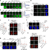
Fig. 4. ARKL1 promotes RNF111 localization to PML NBs.
a, b FRAP of ARKL1-GFP and GFP-RNF111CS in PML NBs. Mcherry-PML and ARKL1-GFP or GFP-RNF111CS are co-expressed in U2OS cells. Photobleaching was performed on indicated PML-NB (marked by mcherry-PML in the “merge” image). Representative images of live-cell imaging at indicated times are shown. Relative GFP intensity in the bleached PML NBs before and after bleach was quantified. Exponential fit and recovery half time (T1/2) is shown with mean ± SD (n = 23 events). c Percentage of mobile fraction after photo bleach was quantified with mean ± SD (n = 23 events). d RNF111-ARKL1 PLA was performed in indicated cells expressing GFP-PML. Arrows point to PLA foci that overlap with PML NBs. e Decreased RNF111 localization to PML NBs in ARKL1-deficient cells. Immunofluorescence (IF) was quantified for RNF111 KO cells complemented with HA-RNF111 and treated with either siCtrl (n = 98) or siARKL1 (n = 100) from two biological replicates. f RNF111 co-localization/interaction with PML is dependent on ARKL1. RNF111-PML PLA was quantified for WT (n = 71), ARKL1 KO (n = 62) and RNF111 KO (n = 77) cells from two biological replicates. g ARKL1 SIMs are required for RNF111 recruitment to PML NBs. RNF111-PML PLA was quantified for WT U2OS cells expressing vector (n = 164) or ARKL1 KO expressing vector (n = 133), ARKL1WT (n = 171), ARKL1ΔS (n = 200) from two biological replicates. Expression of the indicated proteins is shown in Supplementary Fig. 4g. h RNF111 K237/K238R mutation disrupts RNF111 localization to PML NBs. IF was quantified for RNF111 KO cells complemented with RNF111WT (n = 53) or RNF111K237/238R (n = 60) from two biological replicates. Quantification is shown with mean ± 95% CI for eh. Statistics: two-tailed unpaired t-test for c, e, h, one-way Anova with Sidak’s correction for f, g. Scale bar, 10 μm for a, b, dh. Source data are provided as a Source Data file.
Fig. 5
Fig. 5. RNF111-mediated ubiquitination of BLM is associated with PML NBs.
a Increased BLM levels in PML NBs in RNF111 and ARKL1 KO cells. BLM intensity in each PML NB in immunofluorescence (IF) was quantified with arbitrary (arb.) units with mean ± 95% CI for WT (n = 1532), ARKL1 KO (n = 1617), RNF111 KO (n = 952) U2OS cells from two biological replicates. One-way ANOVA with Sidak’s correction was used for statistics. b Detection of RNF111 and BLM interaction in PML NBs by PLA in WT or RNF111 KO U2OS cells expressing GFP-PML. RNF111-BLM PLA foci that colocalize with PML NBs are indicated by arrows. c RNF111 SIM domains are required for BLM ubiquitination. The IPs were performed under denature condition using lysates from WT or RNF111 KO U2OS cells expressing indicated constructs and treated with MG132. CHK1 was used as a loading control. d RNF111SIM* mutant fails to reduce the elevated BLM levels in RNF111 KO cells. e RNF111 SIM domains are required for self-ubiquitination. 293T cells expressing indicated constructs were used. f BLM ubiquitination is dependent on its SIMs and SUMOylation at K317/331. GFP IP was performed under denature condition using lysates of 293T cells expressing GFP or GFP-tagged BLM constructs. g ARKL1 SIMs are required for BLM ubiquitination. WT or ARKL1 KO Hela cells expressing indicated constructs were used. In the Input panel, “*” non-specific band; left arrow, endogenous ARKL1; right arrow, Flag-tagged ARKL1. h Expression of WT but not SIM mutant of ARKL1 reduced the elevated BLM levels in ARKL1 KO cells. i RNF111 SUMOylation sites K237/K238 are required for BLM ubiquitination. WT or RNF111 KO U2OS cells expressing indicated constructs were used. Scale bar, 10 μm for a, b. Source data are provided as a Source Data file.
Fig. 6
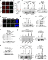
Fig. 6. CK2 phosphorylates ARKL1 promoting ARKL1-RNF111 interaction and BLM ubiquitination.
a CK2 inhibitor TBB treatment abolishes HA-RNF111 localization to PML NBs. Immunofluorescence (IF) in RNF111 KO U2OS cells expressing HA-RNF111 was quantified for DMSO-treated (n = 101) or TBB (50 μM, 4 h)-treated (n = 100) cells from two biological replicates. b PLA in RNF111 KO cells expressing HA-RNF111 was quantified for DMSO-treated (n = 43) or TBB-treated (n = 37) cells. c TBB treatment reduces BLM ubiquitination. d TBB treatment leads to increased BLM levels in PML NBs. IF was quantified with arbitrary (arb.) units for DMSO-treated (n = 1354) or TBB-treated (n = 903) cells. e The serine-rich region of ARKL1 is required for promoting RNF111-PML colocalization. PLA was quantified for Hela WT expressing vector (n = 152) or ARKL1 KO expressing vector (n = 178), ARKL1WT (n = 180), or ARKL1ΔS (n = 130) from two biological replicates. Expression of the indicated proteins is shown in Supplementary Fig. 6c. f ARKL1WT but not ARKL1ΔS or ARKL1SIM* mutant reduces the elevated BLM levels in PML NBs in ARKL1 KO cells. Quantifications are shown for U2OS WT expressing vector (n = 1302) or ARKL1 KO cells expressing vector (n = 2109), ARKL1WT (n = 1467), ARKL1ΔS (n = 2307), ARKL1SIM (n = 3015). Expression of the indicated proteins is shown in Supplementary Fig. 6d. g TBB treatment leads to decreased CK2 phosphorylation of ARKL1. The IPs were performed under denature condition. h ARKL1ΔS mutant cannot be phosphorylated by CK2. Hela cells expressing indicated constructs were used. i Identification of CK2 phosphorylation site on ARKL1. 293T cells expressing vector, HA-tagged ARKL1 WT, S328A or 4 A (S327/328/330/385 A) mutant were used. j TBB treatment reduces the interaction of ARKL1-Flag with RNF111. k ARKL1ΔS and ARKL1S328A mutants exhibit decreased interaction with RNF111. Quantifications are shown with mean ± 95% CI for a, b, df. Statistics: two-tailed unpaired t-test for ad, one-way ANOVA with Sidak’s correction for e, f. Scale bar, 10 μm for a, b, df. Source data are provided as a Source Data file.
Fig. 7
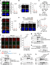
Fig. 7. RNF111-ARKL1 and CK2 regulate G-quadruplex in the nucleus through modulating BLM levels.
a Decreased G4 levels in RNF111 KO nucleus can be rescued by BLM knockdown. Data from two biological replicates are quantified with arbitrary (arb.) units for U2OS WT treated with siCtrl (n = 68), RNF111 KO treated with siCtrl (n = 101) or siBLM (n = 57). Western blots are shown in Supplementary Fig. 7b. b Decreased G4 levels in RNF111 KO cells are rescued by treatment with BLM helicase inhibitor ML216 (12.5 μM, 72 h). Data from two biological replicates are quantified for WT cells treated with DMSO (n = 66), RNF111 KO treated with DMSO (n = 95) or ML216 (n = 94). c Decreased G4 intensity in RNF111 KO cells is rescued by expression of RNF111WT (n = 56), but not empty vector (n = 68), RNF111CS (n = 61), or RNF111SIM* (n = 62) from two biological replicates. d Decreased G4 intensity in ARKL1 KO U2OS nucleus can be rescued by BLM knockdown. Western blots are shown in Supplementary Fig. 7d. Data of two biological replicates are quantified for WT cells treated with siCtrl (n = 85), ARKL1 KO cells treated with siCtrl (n = 98) or SiBLM (n = 68). e CK2 inhibitor TBB treatment leads to decreased G4 intensity in the nucleus. Data of two biological replicates are quantified for U2OS cells treated with DMSO (n = 87) or TBB (50 μM, 4 h) (n = 86). f A proposed model for ARKL1-RNF111-dependent ubiquitination of BLM in PML NBs limiting BLM protein level for the regulation of G4 in the nucleus. Quantifications are shown with mean ± 95% CI. Statistics: one-way ANOVA with Sidak’s correction for ad and two-tailed unpaired t-test for e. Scale bar, 10 μm for ae. Source data are provided as a Source Data file.

Similar articles

Cited by

References

    1. Vindigni A, Hickson ID. RecQ helicases: multiple structures for multiple functions? HFSP J. 2009;3:153–164. doi: 10.2976/1.3079540. - DOI - PMC - PubMed
    1. Bohm S, Bernstein KA. The role of post-translational modifications in fine-tuning BLM helicase function during DNA repair. DNA Repair (Amst) 2014;22:123–132. doi: 10.1016/j.dnarep.2014.07.007. - DOI - PMC - PubMed
    1. Mendoza O, Bourdoncle A, Boule JB, Brosh RM, Jr, Mergny JL. G-quadruplexes and helicases. Nucleic Acids Res. 2016;44:1989–2006. doi: 10.1093/nar/gkw079. - DOI - PMC - PubMed
    1. Huber MD, Lee DC, Maizels N. G4 DNA unwinding by BLM and Sgs1p: substrate specificity and substrate-specific inhibition. Nucleic Acids Res. 2002;30:3954–3961. doi: 10.1093/nar/gkf530. - DOI - PMC - PubMed
    1. Sun H, Karow JK, Hickson ID, Maizels N. The Bloom’s syndrome helicase unwinds G4 DNA. J. Biol. Chem. 1998;273:27587–27592. doi: 10.1074/jbc.273.42.27587. - DOI - PubMed

Publication types

MeSH terms