Review
. 2024 Jan 30;36(2):227-245.
doi: 10.1093/plcell/koad254.

Biomolecular condensates in plant RNA silencing: insights into formation, function, and stress responses

Affiliations
Review

Biomolecular condensates in plant RNA silencing: insights into formation, function, and stress responses

Qi Li et al. Plant Cell. .

Abstract

Biomolecular condensates are dynamic structures formed through diverse mechanisms, including liquid-liquid phase separation. These condensates have emerged as crucial regulators of cellular processes in eukaryotic cells, enabling the compartmentalization of specific biological reactions while allowing for dynamic exchange of molecules with the surrounding environment. RNA silencing, a conserved gene regulatory mechanism mediated by small RNAs (sRNAs), plays pivotal roles in various biological processes. Multiple types of biomolecular condensate, including dicing bodies, processing bodies, small interfering RNA bodies, and Cajal bodies, have been identified as key players in RNA silencing pathways. These biomolecular condensates provide spatial compartmentation for the biogenesis, loading, action, and turnover of small RNAs. Moreover, they actively respond to stresses, such as viral infections, and modulate RNA silencing activities during stress responses. This review summarizes recent advances in understanding of dicing bodies and other biomolecular condensates involved in RNA silencing. We explore their formation, roles in RNA silencing, and contributions to antiviral resistance responses. This comprehensive overview provides insights into the functional significance of biomolecular condensates in RNA silencing and expands our understanding of their roles in gene expression and stress responses in plants.


Conflict of interest statement

Conflict of interest statement. The authors declare no competing interests, and we have included this information for all co-authors.

Figures
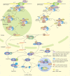
Figure 1.
Figure 1.
The interplay of biomolecular condensates with miRNA expression and processing. Initially, pri-miRNAs are transcribed by RNA Pol II and subsequently processed into miRNAs. D-body formation relies on core components such as DCL1, HYL1, SE, and RNA helicases RH6/8/12. Notably, both SE and RH6/8/12 contain IDR regions. SE undergoes LLPS and incorporates DCL1, HYL1, and pri/pre-miRNAs, thereby facilitating the formation of D-bodies. RH6/8/12 interacts with SE and promotes its phase separation, facilitating D-body formation. Additionally, various other proteins involved in regulating pri-miRNA transcription, stability, and maturation interact with D-body components and may also localize in and influence D-body formation. Following processing, miRNA duplexes are methylated by HEN1 and then either sorted into AGO1 or transported out of the nucleus for cytoplasmic loading into AGO1. In the cytoplasm, miRNA deceases target gene expression by mRNA cleavage and/or translation inhibition. The translation inhibition processes of miRNAs take places on ER. The cleavage products are partially degraded, possibly by XRN4 within P bodies. Additionally, SUO and VARICOSE may facilitate the translation inhibition activity of miRNAs within P bodies. The turnover of unmethylated miRNAs is likely fulfilled by ATRM2 in P bodies and/or related biomolecular condensates. Mature miRNAs are predominantly degraded through SDN1-3, URT1, and HESO1-mediated degradation within P bodies and/or related biomolecular condensates. Furthermore, XRN4 participates in the degradation of miRNA* in P bodies.
Figure 2.
Figure 2.
Prediction of the disordered segments of plant AGO1 by IUPred2A. Protein residues with score above 0.5 is identified as disordered. The prediction was performed at web site version of IUPred2A using default parameters. The address is http://iupred2a.elte.hu.
Figure 3.
Figure 3.
siRNA body and ta-siRNA biogenesis. phasiRNA-generating (PHAS) loci and ta-siRNA-generating (TAS) loci are transcribed by RNA Pol II. After miRNA-induced silencing complex biosynthesis, miRNAs that direct ta-siRNA biosynthesis are sorted in AGOs. They can fulfill transcript cleavage either in a “one-hit” manner with the AGO1 RISC complex or in a “two-hit” manner with the AGO7 RISC complex. The PrLD domain-containing protein SGS3, which interacts with RDR6, plays a crucial role in driving the formation of siRNA bodies through phase separation. Within siRNA bodies, the cleavage products of AGO1 and AGO7 are stabilized by SGS3 and converted into dsRNAs by RDR6. These dsRNAs are subsequently by DCL2/4 to produce ta-siRNAs and other phased-secondary siRNAs.
Figure 4.
Figure 4.
Cajal body and heterochromatin siRNA production. The generation of 24-nt heterochromatin siRNA initiates with Pol IV/Pol II transcripts generated from transposable elements and repetitive DNA elements. These transcripts are converted into dsRNAs by RDR2 within Cajal bodies. Subsequently, DCL3 cleaves the dsRNAs, leading to the production of 24-nt hc-siRNAs. The hc-siRNAs are loaded into AGO4 complex within Cajal bodies and then transported to target sites for DNA methylation. Alternatively, the hc-siRNAs can also be exported into the cytoplasm for AGO4 assembly, and transported back into the nucleus to perform their functions. By base pairing with the ssRNA transcribed by Pol V, the AGO4-complex is recruited to the methylation site. It interacts with DRM2, RDM1, and NRPD1b/NRPE1, a Pol IV/Pol V subunit containing the PrLD domain. This complex confers RNA-dependent DNA methylation.

Similar articles

References

    1. Alberti S, Dormann D. Liquid-liquid phase separation in disease. Annu Rev Genet. 2019:53(1):171–194. 10.1146/annurev-genet-112618-043527 - DOI - PubMed
    1. Alberti S, Gladfelter A, Mittag T. Considerations and challenges in studying liquid-liquid phase separation and biomolecular condensates. Cell. 2019:176(3):419–434. 10.1016/j.cell.2018.12.035 - DOI - PMC - PubMed
    1. Alberti S, Hyman AA. Biomolecular condensates at the nexus of cellular stress, protein aggregation disease and ageing. Nat Rev Mol Cell Biol. 2021:22(3):196–213. 10.1038/s41580-020-00326-6 - DOI - PubMed
    1. Atkinson N, Mao Y, Chan KX, McCormick AJ. Condensation of rubisco into a proto-pyrenoid in higher plant chloroplasts. Nat Commun. 2020:11(1):6303. 10.1038/s41467-020-20132-0 - DOI - PMC - PubMed
    1. Baeg K, Iwakawa HO, Tomari Y. The poly(A) tail blocks RDR6 from converting self mRNAs into substrates for gene silencing. Nat Plants. 2017:3(4):17036. 10.1038/nplants.2017.36 - DOI - PubMed

LinkOut - more resources