

Navigating the kidney organoid: insights into assessment and enhancement of nephron function
- PMID: 37767571
- PMCID: PMC10878724
- DOI: 10.1152/ajprenal.00166.2023
Navigating the kidney organoid: insights into assessment and enhancement of nephron function
Abstract
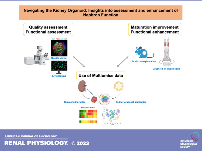
Kidney organoids are three-dimensional structures generated from pluripotent stem cells (PSCs) that are capable of recapitulating the major structures of mammalian kidneys. As this technology is expected to be a promising tool for studying renal biology, drug discovery, and regenerative medicine, the functional capacity of kidney organoids has emerged as a critical question in the field. Kidney organoids produced using several protocols harbor key structures of native kidneys. Here, we review the current state, recent advances, and future challenges in the functional characterization of kidney organoids, strategies to accelerate and enhance kidney organoid functions, and access to PSC resources to advance organoid research. The strategies to construct physiologically relevant kidney organoids include the use of organ-on-a-chip technologies that integrate fluid circulation and improve organoid maturation. These approaches result in increased expression of the major tubular transporters and elements of mechanosensory signaling pathways suggestive of improved functionality. Nevertheless, continuous efforts remain crucial to create kidney tissue that more faithfully replicates physiological conditions for future applications in kidney regeneration medicine and their ethical use in patient care.NEW & NOTEWORTHY Kidney organoids are three-dimensional structures derived from stem cells, mimicking the major components of mammalian kidneys. Although they show great promise, their functional capacity has become a critical question. This review explores the advancements and challenges in evaluating and enhancing kidney organoid function, including the use of organ-on-chip technologies, multiomics data, and in vivo transplantation. Integrating these approaches to further enhance their physiological relevance will continue to advance disease modeling and regenerative medicine applications.
Keywords: development; function; kidney; organoid; physiology.
Conflict of interest statement
R.M. is an inventor on a patent related to this work filed by the President and Fellows of Harvard College and Mass General Brigham (PCT/US2018/036677). R.M. holds a stock option in Trestle Biotherapeutics. None of the other authors has any conflicts of interest, financial or otherwise, to disclose.
Figures
Similar articles
-
Depressing time: Waiting, melancholia, and the psychoanalytic practice of care.In: Kirtsoglou E, Simpson B, editors. The Time of Anthropology: Studies of Contemporary Chronopolitics. Abingdon: Routledge; 2020. Chapter 5. In: Kirtsoglou E, Simpson B, editors. The Time of Anthropology: Studies of Contemporary Chronopolitics. Abingdon: Routledge; 2020. Chapter 5. PMID: 36137063 Free Books & Documents. Review.
-
Unlocking data: Decision-maker perspectives on cross-sectoral data sharing and linkage as part of a whole-systems approach to public health policy and practice.Public Health Res (Southampt). 2024 Nov 20:1-30. doi: 10.3310/KYTW2173. Online ahead of print. Public Health Res (Southampt). 2024. PMID: 39582242
-
Trends and challenges in organoid modeling and expansion with pluripotent stem cells and somatic tissue.PeerJ. 2024 Nov 27;12:e18422. doi: 10.7717/peerj.18422. eCollection 2024. PeerJ. 2024. PMID: 39619184 Free PMC article. Review.
-
Using Experience Sampling Methodology to Capture Disclosure Opportunities for Autistic Adults.Autism Adulthood. 2023 Dec 1;5(4):389-400. doi: 10.1089/aut.2022.0090. Epub 2023 Dec 12. Autism Adulthood. 2023. PMID: 38116059 Free PMC article.
-
Trends in Surgical and Nonsurgical Aesthetic Procedures: A 14-Year Analysis of the International Society of Aesthetic Plastic Surgery-ISAPS.Aesthetic Plast Surg. 2024 Oct;48(20):4217-4227. doi: 10.1007/s00266-024-04260-2. Epub 2024 Aug 5. Aesthetic Plast Surg. 2024. PMID: 39103642 Review.
Cited by
-
Efficient proximal tubule-on-chip model from hiPSC-derived kidney organoids for functional analysis of renal transporters.iScience. 2024 Aug 19;27(9):110760. doi: 10.1016/j.isci.2024.110760. eCollection 2024 Sep 20. iScience. 2024. PMID: 39286490 Free PMC article.
-
Postnatal renal tubule development: roles of tubular flow and flux.Curr Opin Nephrol Hypertens. 2024 Sep 1;33(5):518-525. doi: 10.1097/MNH.0000000000001007. Epub 2024 Jun 24. Curr Opin Nephrol Hypertens. 2024. PMID: 38913022 Review.
-
Advancements in therapeutic development: kidney organoids and organs on a chip.Kidney Int. 2024 Apr;105(4):702-708. doi: 10.1016/j.kint.2023.11.035. Epub 2024 Jan 29. Kidney Int. 2024. PMID: 38296026 Review.
-
3D Hydrogel Encapsulation Regulates Nephrogenesis in Kidney Organoids.Adv Mater. 2024 Apr;36(14):e2308325. doi: 10.1002/adma.202308325. Epub 2024 Jan 10. Adv Mater. 2024. PMID: 38180232
-
Advancing preclinical drug evaluation through automated 3D imaging for high-throughput screening with kidney organoids.Biofabrication. 2024 Apr 8;16(3):10.1088/1758-5090/ad38df. doi: 10.1088/1758-5090/ad38df. Biofabrication. 2024. PMID: 38547531
References
Publication types
MeSH terms
Grants and funding
- U24 DK135157/DK/NIDDK NIH HHS/United States
- P50 DK133943/DK/NIDDK NIH HHS/United States
- U54 DK137329/DK/NIDDK NIH HHS/United States
- U01 EB028899/EB/NIBIB NIH HHS/United States
- UG3 DK114933/DK/NIDDK NIH HHS/United States
- R01 DK129285/DK/NIDDK NIH HHS/United States
- R21 DK129909/DK/NIDDK NIH HHS/United States
- U01 DK114933/DK/NIDDK NIH HHS/United States
- UC2 DK126023/DK/NIDDK NIH HHS/United States
- R01 DK038470/DK/NIDDK NIH HHS/United States
- U54 DK134301/DK/NIDDK NIH HHS/United States
- U01 DK127587/DK/NIDDK NIH HHS/United States
- DP2 DK133821/DK/NIDDK NIH HHS/United States
- UH3 DK114933/DK/NIDDK NIH HHS/United States
LinkOut - more resources
Full Text Sources
Research Materials