Review
. 2023 Aug 31;15(9):1847.
doi: 10.3390/v15091847.

Feline Infectious Peritonitis: European Advisory Board on Cat Diseases Guidelines

Affiliations
Review

Feline Infectious Peritonitis: European Advisory Board on Cat Diseases Guidelines

Séverine Tasker et al. Viruses. .

Abstract

Feline coronavirus (FCoV) is a ubiquitous RNA virus of cats, which is transmitted faeco-orally. In these guidelines, the European Advisory Board on Cat Diseases (ABCD) presents a comprehensive review of feline infectious peritonitis (FIP). FCoV is primarily an enteric virus and most infections do not cause clinical signs, or result in only enteritis, but a small proportion of FCoV-infected cats develop FIP. The pathology in FIP comprises a perivascular phlebitis that can affect any organ. Cats under two years old are most frequently affected by FIP. Most cats present with fever, anorexia, and weight loss; many have effusions, and some have ocular and/or neurological signs. Making a diagnosis is complex and ABCD FIP Diagnostic Approach Tools are available to aid veterinarians. Sampling an effusion, when present, for cytology, biochemistry, and FCoV RNA or FCoV antigen detection is very useful diagnostically. In the absence of an effusion, fine-needle aspirates from affected organs for cytology and FCoV RNA or FCoV antigen detection are helpful. Definitive diagnosis usually requires histopathology with FCoV antigen detection. Antiviral treatments now enable recovery in many cases from this previously fatal disease; nucleoside analogues (e.g., oral GS-441524) are very effective, although they are not available in all countries.

Keywords: FCoV; FIP; antiviral; diagnosis; feline coronavirus; mutation; treatment.


Conflict of interest statement

The authors are members of the European Advisory Board on Cat Diseases (ABCD), a scientifically independent board of experts in feline medicine. Some individual authors have been consultants for, or have received research funding and honoraria from, animal health companies and other organisations, but all authors declare no conflicts of interest for the writing of these independent guidelines. The ABCD sponsors had no role in the writing, nor in the decision to publish this review.

Figures

Figure 1
Figure 1
Schematic diagrams of type I FCoV, not drawn to scale. (a) Schematic FCoV genome. FCoV is a positive-sense single-stranded RNA virus. The FCoV genome of 27–32 kilobases encodes a replicase polyprotein, four structural proteins (spike [S], membrane [M], nucleocapsid [N] and envelope [E]) and non-structural accessory proteins 3a, 3b and 3c and 7a and 7b. UTR indicates an untranslated region. Image Emi Barker, Langford Vets, University of Bristol, UK [54]. (b) Schematic FCoV spike protein (based on [55,56,57]) sequence showing the division into the S1 and S2 subunits representing the receptor-binding and fusion domains, respectively, with N- and C-terminals shown. The S1/S2 and S2′ sites represent cleavage sites (in red), and the fusion peptide domain is also shaded in red. The positions of the M1058* and S1060* amino acid residues (blue lines) are shown because these correspond to the FCoV nucleotide sequences in specific spike gene mutations that are evaluated in some commercially available molecular assays. * Convention is to label amino acid substitutions by initials surrounding the numbered amino acid residue location (e.g., M1058L indicates that methionine is replaced by leucine at position 1058; similarly, S1060A indicates that serine is replaced by alanine at position 1060). Image Séverine Tasker, University of Bristol, UK.
Figure 2
Figure 2
Schematic diagram of FCoV structure showing single-stranded (ss) RNA and the structural proteins: spike, envelope, membrane and nucleocapsid proteins. The spike protein is the part of the virus particle that interacts with the host-cell receptor. The spikes on the surface present a coronal (i.e., crown-like) appearance under electron microscopy [60]. Image Emi Barker, Langford Vets, University of Bristol, UK [54].
Figure 3
Figure 3
Origin of feline coronavirus (FCoV) type II; Image Peter Rottier, University of Utrecht, The Netherlands. Schematic diagram showing how type II FCoV arises from recombination of FCoV type I (shown in white) with CCoV (shown in brown).
Figure 4
Figure 4
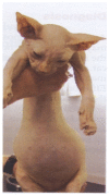
Ascites in a young Sphinx cat presenting with FIP. Image Hannah Dewerchin, Ghent University, Belgium [46].
Figure 5
Figure 5
Icterus can occur in cases with FIP, particularly in cats with effusive FIP. Image Séverine Tasker, Bristol Veterinary School, University of Bristol, UK.
Figure 6
Figure 6
Ataxia can occur in cats with neurological FIP. Image Séverine Tasker, Bristol Veterinary School, University of Bristol, UK.
Figure 7
Figure 7
Ataxia (wide-based stance) and obtundation in a cat with neurological FIP. Image Allan May, University of Glasgow, UK through Diane Addie, www.catvirus.com.
Figure 8
Figure 8
FIP-associated anterior uveitis can manifest variably such as with the presence of hyphaema. Image Maria Bonino and Erica Carter.
Figure 9
Figure 9
FIP-associated anterior uveitis can manifest variably such as with the presence of hyphaema. Image Albert Lloret, Universitat Autònoma Barcelona, Spain [46].
Figure 10
Figure 10
FIP-associated anterior uveitis can manifest variably such as with the presence of inflammatory keratic precipitates. Image Eric Déan, Vet-Oeil Ophthalmology Clinic, France [46].
Figure 11
Figure 11
European Advisory Board on Cat Diseases (ABCD) Feline Infectious Peritonitis (FIP) Diagnostic Approach Tools: diagnostic approach I, showing evidence that can contribute to being highly suspicious of a diagnosis of FIP. This tool is available online [211], with revisions made to the online version as required. Many features of the cat’s signalment, history and clinical examination can contribute to a suspicion of FIP. Effusion analysis is always extremely helpful, so looking for evidence of an effusion and then sampling should be prioritised whenever possible. Certain haematological features can also contribute to the suspicion of FIP as a diagnosis.
Figure 12
Figure 12
European Advisory Board on Cat Diseases (ABCD) Feline Infectious Peritonitis (FIP) Diagnostic Approach Tools: diagnostic approach IIa, showing diagnostic testing evidence that can confirm FIP as a diagnosis following being highly suspicious of FIP in cats with an effusion (1) and cats that neither have effusions nor specific clinical signs (2). This tool is available online [211], with revisions made to the online version as required.
Figure 13
Figure 13
European Advisory Board on Cat Diseases (ABCD) Feline Infectious Peritonitis (FIP) Diagnostic Approach Tools: diagnostic approach IIb, showing diagnostic testing evidence that can confirm FIP as a diagnosis following being highly suspicious of FIP in cats with neurological signs (3) and cats with aqueous humour cytology consistent with FIP (4). This tool is available online [211], with revisions made to the online version as required. In this figure, the confirmation of a diagnosis of FIP requires the collection of cerebrospinal fluid (CSF) or aqueous humour. However, it is generally easier to sample effusions, if present, or accessible abnormal organs or tissues (e.g., mesenteric lymph node, identified by imaging) by fine-needle aspiration, if present, as indicated in Figure 12.
Figure 14
Figure 14
European Advisory Board on Cat Diseases (ABCD) Feline Infectious Peritonitis (FIP) Diagnostic Approach Tools: differential diagnoses. This tool is available online [211], with revisions made to the online version as required.
Figure 15
Figure 15
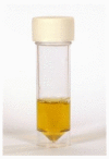
Abdominal effusion sample collected from a cat with FIP showing typical clear straw-yellow-coloured fluid. Image Séverine Tasker, Bristol Veterinary School, University of Bristol, UK.
Figure 16
Figure 16
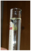
This image shows a positive Rivalta’s test; a drop of abdominal effusion has been placed onto the surface of a mixture of 8 mL of distilled water and one drop of 98% acetic acid (or white vinegar) in a test tube, and it has retained its shape with a connection to the solution surface. This is not very specific for FIP but can be performed in-house; a positive test increases the likelihood of FIP, while a negative test makes FIP very unlikely. Image Diane Addie, www.catvirus.com.
Figure 17
Figure 17
Ultrasonogram of a young cat with FIP showing pericardial and pleural effusion; ultrasonography can be used to guide sampling of the effusion. Green arrow = pericardial fluid. Yellow arrow = cardiac ventricle. White arrow = pleural effusion. Image Séverine Tasker, Bristol Veterinary School, University of Bristol, UK.
Figure 18
Figure 18
Ultrasonogram of a young cat with FIP showing abdominal effusion; ultrasonography can be used to guide sampling of the effusion. Yellow arrow = small intestinal loop in transverse section. White arrows = peritoneal effusion—note the effusion is echogenic, suggesting cellularity. Green arrow = mesenteric fat. Image Séverine Tasker, Bristol Veterinary School, University of Bristol, UK.
Figure 19
Figure 19
Lateral thoracic radiograph showing the presence of a pleural effusion; Yellow arrow = pleural fissure lines. White arrow = border effacement of the cardiac silhouette. Green arrow = the pleural effusion has displaced air-filled lung dorsally and the lungs are reduced in size. Image Andrew Parry, Willows Veterinary Centre, Solihull, UK.
Figure 20
Figure 20
Ultrasonogram of a cat with FIP and renomegaly with a loss of normal renal architecture; ultrasonography might be useful to guide fine-needle aspirate or tissue core-biopsy sampling of organs by targeting abnormal tissue. The kidney is enlarged (50 mm, normal size range is 33–44 mm). White arrow = loss of corticomedullary distinction with heterogeneously echogenic renal parenchyma. Green arrow = pericapsular hypoechoic material is sometimes seen in cases with FIP. However, this is also encountered with other diseases (e.g., lymphoma). Image Séverine Tasker, Bristol Veterinary School, University of Bristol, UK.
Figure 21
Figure 21
CT of the head of a cat with neurological signs due to FIP post-contrast—reconstructed to show a sagittal midline view. White arrow = there is evidence of generalised ventriculomegaly, suggesting alteration/obstruction to CSF flow. Green arrow = homogeneous contrast enhancement of the lining of the ventricles (ependyma) is sometimes seen in patients with FIP. Yellow arrow = the increased ventricular size in this patient has led to an increase in volume of the contents of the calvarium. This patient has coning of the cerebellum—the cerebellar vermis is beginning to pass through the foramen magnum—a life threatening finding. Image Séverine Tasker, Bristol Veterinary School, University of Bristol, UK.
Figure 22
Figure 22
T2W transverse MRI of a cat with neurological signs due to FIP. The brain also appears swollen with a lack of visible sulci. Yellow arrow = the gyri in this patient are enlarged, with narrowing of the sulci. This will be due to parenchymal inflammation. Green arrow = the edge of the left lateral ventricle is identified and appears dilated. White arrow = the T2W isointense (to grey matter) structure is an enlarged right choroid plexus. Image Séverine Tasker, Bristol Veterinary School, University of Bristol, UK.
Figure 23
Figure 23
(a) Histopathology: Hematoxylin and eosin stain and (b) positive FCoV antigen immunostaining in a cat with FIP: liver, fibrinous perihepatitis with embedded FCoV-infected macrophages (shorter arrows) and focal granulomatous infiltrate (longer arrows) with FCoV-positive macrophages. Image Anja Kipar, University of Zurich, Switzerland.
Figure 24
Figure 24
(a) Histopathology: Hematoxylin and eosin stain and (b) positive FCoV antigen immunostaining in a cat with FIP: Mesentery with focal granulomatous infiltrate with embedded small veins (arrows) and abundant FCoV-positive macrophages. Image Anja Kipar, University of Zurich, Switzerland.
Figure 25
Figure 25
(a) Histopathology: Hematoxylin and eosin stain and (b) positive FCoV antigen immunostaining in a cat with FIP: Mesenteric lymph node with focal granulomatous infiltrate with extensive central necrosis (N) and abundant FCoV-infected macrophages (arrows) in the surrounded infiltrate. Image Anja Kipar, University of Zurich, Switzerland.
Figure 26
Figure 26
Positive FCoV antigen immunostaining in a cat with FIP: Kidney, stellate vein in subcapsular cortex with granulomatous (peri)phlebitis. Focally, the granulomatous infiltration has destroyed the vascular basement membrane (left arrow), protrudes into the lumen of the vein (wall-bound thrombus; right arrow) and is present in surrounding tissue, containing abundant FCoV-infected macrophages (cells stained in brown). Shorter arrows outline the remnants of the basement membrane. Image Anja Kipar, University of Zurich, Switzerland.
Figure 27
Figure 27
Gross appearance of fibrinous plaque-like inflammation present on the surface of the spleen in a case of FIP with an abdominal effusion that underwent post-mortem examination. Image Séverine Tasker and the Pathology Department, Bristol Veterinary School, University of Bristol, UK.
Figure 28
Figure 28
(a,b) Gross appearance of the kidneys from two cats with FIP, showing renomegaly with pyogranulomas visible on the renal surface on post-mortem examination. (b) shows how pyogranulomas can be centred on blood vessels. These lesions could be mistaken for tumours on gross post-mortem examination, which is why histopathology, and ideally, immunohistochemistry is necessary. Images Pathology Department, Bristol Veterinary School, University of Bristol, UK.
Figure 29
Figure 29
Immunocytochemistry showing the presence of FCoV antigen in macrophages in an effusion of a cat with FIP. Overview at 40× magnification, cytospin, scattered positive macrophages clearly visible at low power (e.g., black arrowheads) before cells further identifiable. Image Alex Malbon and Anja Kipar, University of Zurich, Switzerland.
Figure 30
Figure 30
Immunocytochemistry showing the presence of FCoV antigen in macrophages in an effusion of a cat with FIP. 400× magnification, cytospin, highly cellular—macrophages (black arrowhead), neutrophils (white arrowhead) and fibrin (white asterisk). Scattered positive macrophages (black arrows). Image Alex Malbon and Anja Kipar, University of Zurich, Switzerland.

Similar articles

Cited by

References

    1. Pedersen N.C., Boyle J.F., Floyd K., Fudge A., Barker J. An enteric coronavirus infection of cats and its relationship to feline infectious peritonitis. Am. J. Vet. Res. 1981;42:368–377. - PubMed
    1. Hayashi T., Watabe Y., Nakayama H., Fujiwara K. Enteritis due to feline infectious peritonitis virus. Jpn. J. Vet. Res. 1982;44:97–106. doi: 10.1292/jvms1939.44.97. - DOI - PubMed
    1. Addie D.D., Jarrett O. A study of naturally occurring feline coronavirus infections in kittens. Vet. Rec. 1992;130:133–137. doi: 10.1136/vr.130.7.133. - DOI - PubMed
    1. Kipar A., Kremendahl J., Addie D.D., Leukert W., Grant C.K., Reinacher M. Fatal enteritis associated with coronavirus infection in cats. J. Comp. Pathol. 1998;119:1–14. doi: 10.1016/S0021-9975(98)80067-4. - DOI - PMC - PubMed
    1. Addie D.D., Jarrett O. Use of a reverse-transcriptase polymerase chain reaction for monitoring the shedding of feline coronavirus by healthy cats. Vet. Rec. 2001;148:649–653. doi: 10.1136/vr.148.21.649. - DOI - PubMed

Publication types

Grants and funding

ABCD Europe gratefully acknowledges the support of Boehringer Ingelheim (the founding sponsor of the ABCD), Virbac, MSD; and IDEXX.

LinkOut - more resources