Review
. 2023 Sep 11;12(18):2253.
doi: 10.3390/cells12182253.

Therapeutic Oligonucleotides: An Outlook on Chemical Strategies to Improve Endosomal Trafficking

Affiliations
Review

Therapeutic Oligonucleotides: An Outlook on Chemical Strategies to Improve Endosomal Trafficking

Priyanka Mangla et al. Cells. .

Abstract

The potential of oligonucleotide therapeutics is undeniable as more than 15 drugs have been approved to treat various diseases in the liver, central nervous system (CNS), and muscles. However, achieving effective delivery of oligonucleotide therapeutics to specific tissues still remains a major challenge, limiting their widespread use. Chemical modifications play a crucial role to overcome biological barriers to enable efficient oligonucleotide delivery to the tissues/cells of interest. They provide oligonucleotide metabolic stability and confer favourable pharmacokinetic/pharmacodynamic properties. This review focuses on the various chemical approaches implicated in mitigating the delivery problem of oligonucleotides and their limitations. It highlights the importance of linkers in designing oligonucleotide conjugates and discusses their potential role in escaping the endosomal barrier, a bottleneck in the development of oligonucleotide therapeutics.

Keywords: antisense oligonucleotides (ASOs); cleavable linkers; endosomal escape; endosomolytic agents; linker chemistry; non-cleavable linkers; oligonucleotide conjugates; oligonucleotide delivery; oligonucleotide therapeutics; pH-sensitive linkers; siRNA.


Conflict of interest statement

The authors declare no conflict of interest.

Figures

Figure 1
Figure 1
Biological barriers preventing therapeutic activity of oligonucleotides. Systemically administered oligonucleotides encounter nuclease degradation, RES phagocyte uptake, protein interaction, and renal excretion before reaching the specific tissue/cell of interest. On reaching the tissue/cell, intracellular uptake occurs via endocytosis. Most oligonucleotides either get trapped in endosomes and further transferred to lysosomes where they encounter enzymatic degradation or are recycled back into extracellular matrix via exocytosis. Only 1–2% of administered oligonucleotides escape the endosomes to reach the target in cytosol or nucleus. Created with BioRender.com, AO25FLAM37.
Figure 2
Figure 2
Summary of trafficking and mode of action of ASOs and conjugated siRNAs. (1) Oligonucleotides are first internalized into the endosomal pathway by gymnotic uptake or by receptor-mediated uptake, (2) in early endosomes, dissociation with the targeting moiety or ligand is initiated, and (3) maturation of the early endosome leads to late endosomes where oligonucleotides are found to escape in the cytosol. Late endosomes can either mature to (4) lysosomes, where most of the material is degraded by the low-pH and degradation enzymes, or (5) to Multivesicular bodies where they are (6) exported to the external environment. In the cytoplasm, (A.I) siRNAs are loaded into RISC, where (A.II) the passenger strand is degraded and the guide strand selected. After target recognition by the guide strand (A.III), the mRNA transcript is cleaved and degraded. For ASOs, they can either (B) degrade the target mRNA through the RNAse H1 enzyme or (C) block the translation machinery of the transcript. In the nucleus, ASOs can interfere with (D) the splicing machinery and generate a modified mature mRNA or (B) directly downregulate the target through RNAse H1. Created with BioRender.com, VP25FLPEXQ.
Figure 3
Figure 3
Examples of chemical modifications used to improve the therapeutic activity of oligonucleotides.
Figure 4
Figure 4
Schematic representation of mentioned delivery strategies applied to oligonucleotides. (A): (A1) Receptor agonist conjugates as exemplified with the GLP1R peptide agonist-ASO conjugate, (A2) GalNAc conjugate as found in liver targeting therapeutics, and (A3) lipid conjugates exemplified with the 2′-O-hexadecyl modified siRNA; (B): (B1) antibody–oligonucleotide conjugate, and (B2) divalent siRNAs scaffold; (C): (C1) LNPs as for instance used in patirisan, (C2) dendrimer–oligonucleotide complexes like PANAM, (C3) exosome delivery, (C4) DNA-nanoconstruct like the tetrahedron DNA origami, functionalized with an ASO, and (C5) spherical nucleic acid, made with a gold nanoparticle and packed ASOs. Figure created with BioRender.com, FU25OEFFUQ.
Figure 5
Figure 5
Proposed mechanisms of endosomal escape. (A) Proton-sponge effect: buffering polymers or small molecules induce increased inflow of protons, counterions, and water molecules into endosomes, resulting in high osmotic pressure and hence endosomal rupture; (B) membrane fusion: ionizable lipids containing fusogenic lipids fuse with endosomal bilayer and hence releases the oligonucleotides into cytosol; (C) membrane destabilization: polymers containing pH-sensitive scaffolds interact with anionic endosomal membrane and induce membrane destabilization; (D) pore formation: some endosomolytic peptides form pores in endosomal membrane, which allows escaping of oligonucleotides into cytosol. It can be of two types such as barrel-stave pore formation and toroidal pore formation. (E) Vesicle budding and collapse: some endosomolytic peptides induce budding and collapse of CPP-containing vesicles from endosomal membrane. Created with BioRender.com, JF25FK1RLE.
Figure 6
Figure 6
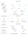
Chemical structures of selected endosomal buffering polymers that are commonly used in gene delivery studies. Poly(L-lysine) and polyethylenimine (PEI) are among the earliest used buffering polymers. Several other polymeric vectors, such as polyamidoamine (PAMAM) dendrimers, degradable poly(β-amino esters) (PBAEs), poly[(2-dimethylamino)ethyl methacrylate] (pDMAEMA), and various carbohydrate-based polymers (chitosan and β-cyclodextrin-containing polycations), have also been explored to improve safety and efficacy.
Figure 7
Figure 7
Few examples of chemical structures of OECs used in oligonucleotide delivery.
Figure 8
Figure 8
Chemical structures of some examples of lipids used in LNPs. Dioleylphosphatidylamine (DOPE), phosphatidylglycerol (PG), phosphatidylserine (PS), phosphatidylcholine (PC), and cholesteryl hemisuccinate (CHEMS).
Figure 9
Figure 9
(A) Ligand conjugation sites in oligonucleotides. (B) Preferred ligand site conjugation in ASO and siRNA. Created with BioRender.com, LN25GKFR3K.
Figure 10
Figure 10
Commonly used linkages for ligand-oligonucleotide conjugation. Note: Generalized linker structures are shown in this figure, which may be different from original linker structures.
Figure 11
Figure 11
Oligonucleotide conjugates containing amide linkages. (A) GalNAc-siRNA conjugate; (B) cholesterol-siRNA conjugate; (C) fatty acid-siRNA conjugate; and (D) antibody-ssDNA conjugate.
Figure 12
Figure 12
Oligonucleotides containing triazole linkages formed using SPAAC reaction. (A) mAb-siRNA conjugates containing DBCO as strained alkyne; (B) Ang II peptide-ASO conjugate containing BCN as strained alkyne.
Figure 13
Figure 13
Oligonucleotide conjugates containing maleimide linkages. (A) Reaction of sulfhydryl-containing anti-CD71 Fab fragment with maleimide modified siRNA. (B) Conjugation of ThiomabTM antibodies with siRNA using non-cleavable SMCC linker.
Figure 14
Figure 14
(A) Conjugation of ThiomabTM antibodies with siRNA using cleavable SPDB linker; (B) conjugation of oligonucleotides with disulfide units enhances cellular permeability through disulfide exchange reactions with the thiol group on the cellular surface.
Figure 15
Figure 15
Trafficking of oligonucleotide conjugates containing pH-sensitive linker via endocytosis. The conjugates are internalized into the endosomal pathway by gymnotic uptake or by receptor-mediated uptake. pH-sensitive linkers are relatively stable in extracellular matrix at pH 7.4 and labile in endosomal compartments at pH 5.5–6.5. They can selectively release the oligonucleotide in endosomes and hence, can potentially enhance the endosomal escape of oligonucleotides. Created with BioRender.com, JQ25FK563G.
Figure 16
Figure 16
General chemical structure of examples of pH-sensitive linkers used in ADCs.
Figure 17
Figure 17
Oligonucleotide conjugates containing pH–sensitive hydrazone linkages. (A) Solid–phase synthesis of 5′–liphophilic conjugates of oligonucleotides via hydrazone bond formation. (B) Hydralink techniques for antibody–oligonucleotide conjugates preparation.
Figure 18
Figure 18
Design of FRET probes incorporating acetal-based pH-sensitive linkers.
Figure 19
Figure 19
(A) Improvement in hydrolytic stability of aryl hydrazones by introducing a boronic group in the ortho-position. (B) Dual-responsive bioconjugates, containing two dynamic covalent linkages (hydrazone and catechol–boronate), for cytosolic delivery of impermeable peptides.
Figure 20
Figure 20
(A) No hydrolytic cleavage of amide bond present in succinic amide at pH 5.5. (B) Hydrolysis of maleic acid derivative (e.g., doxorubicin prodrug). Hydrolysis at neutral pH competes with formation of stable imide, while acidic conditions promote drug release. (C) Replacement of double bond in maleic acid derivative promotes amine scaffold release under acidic conditions.
Figure 21
Figure 21
(A) Structures of tunable pH-sensitive phosphoramidate-based pH-sensitive linkers; (B) proposed mechanism for the hydrolysis of 2-carboxbenzyl phosphoramidates.
Figure 22
Figure 22
Solid-phase synthesis of oligonucleotide conjugates containing pH–sensitive phosphoramidate linkage.

Similar articles

Cited by

References

    1. Roberts T.C., Langer R., Wood M.J.A. Advances in oligonucleotide drug delivery. Nat. Rev. Drug Discov. 2020;19:673–694. doi: 10.1038/s41573-020-0075-7. - DOI - PMC - PubMed
    1. Kulkarni J.A., Witzigmann D., Thomson S.B., Chen S., Leavitt B.R., Cullis P.R., van der Meel R. The current landscape of nucleic acid therapeutics. Nat. Nanotechnol. 2021;16:630–643. doi: 10.1038/s41565-021-00898-0. - DOI - PubMed
    1. Juliano R., Bauman J., Kang H., Ming X. Biological barriers to therapy with antisense and siRNA oligonucleotides. Mol. Pharm. 2009;6:686–695. doi: 10.1021/mp900093r. - DOI - PMC - PubMed
    1. Geary R.S., Norris D., Yu R., Bennett C.F. Pharmacokinetics, biodistribution and cell uptake of antisense oligonucleotides. Adv. Drug Deliv. Rev. 2015;87:46–51. doi: 10.1016/j.addr.2015.01.008. - DOI - PubMed
    1. Kim Y. Drug Discovery Perspectives of Antisense Oligonucleotides. Biomol. Ther. 2023;31:241–252. doi: 10.4062/biomolther.2023.001. - DOI - PMC - PubMed

Publication types

Substances

Grants and funding

This review received no external funding.

LinkOut - more resources