. 2023 Sep 8;9(9):912.
doi: 10.3390/jof9090912.

Paracoccidioides brasiliensis Induces α3 Integrin Lysosomal Degradation in Lung Epithelial Cells

Affiliations

Paracoccidioides brasiliensis Induces α3 Integrin Lysosomal Degradation in Lung Epithelial Cells

Bruna Rocha Almeida et al. J Fungi (Basel). .

Abstract

Studies on the pathogen-host interaction are crucial for the understanding of the mechanisms involved in the establishment, maintenance, and spread of infection. In recent years, our research group has observed that the P. brasiliensis species interact with integrin family receptors and increase the expression of α3 integrin in lung epithelial cells within 5 h of infection. Interestingly, α3 integrin levels were reduced by approximately 99% after 24 h of infection with P. brasiliensis compared to non-infected cells. In this work, we show that, during infection with this fungus, α3 integrin is increased in the late endosomes of A549 lung epithelial cells. We also observed that the inhibitor of the lysosomal activity bafilomycin A1 was able to inhibit the decrease in α3 integrin levels. In addition, the silencing of the charged multivesicular body protein 3 (CHMP3) inhibited the reduction in α3 integrin levels induced by P. brasiliensis in A549 cells. Thus, together, these results indicate that this fungus induces the degradation of α3 integrin in A549 lung epithelial cells by hijacking the host cell endolysosomal pathway.

Keywords: Paracoccidioides brasiliensis; epithelial cell; lysosomal degradation; vesicular traffic; α3 integrin.


Conflict of interest statement

The authors declare no conflict of interest.

Figures
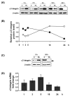
Figure 1
Figure 1
α3 integrin levels in lung epithelial cells during infection with P. brasiliensis. Human lung epithelial A549 cells were incubated for different periods with P. brasiliensis yeasts (Pb) (MOI 1:1). Control (C) was performed in the absence of yeasts. (A) Protein extracts were submitted to SDS-PAGE, and α3 integrin protein levels were analyzed using Western blot. β-actin was used as a loading control. (B) Values represent the intensity of the integrin band divided by the corresponding intensity of the β-actin band shown in panel (A) (■ Represents infected cells; ○ Represents uninfected cells). (C) α3 integrin levels were analyzed using Western blot after 5 h and 24 h infection of A549 cells with P. brasiliensis. Arrow points to bands with lower molecular weight than intact α3 integrin (150 kDa). Similar results were obtained in three independent experiments. (D) α3 integrin mRNA levels were analyzed via RT-qPCR. GAPDH was used as housekeeping gene. Values represent relative fold change (2−ΔΔCt) in target gene transcription levels compared to control sample (without fungi). Mean of three experiments ± standard deviation.
Figure 2
Figure 2
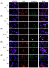
Colocalization analysis of α3 integrin with the early endosome marker Rab5 in A549 epithelial cells infected with P. brasiliensis. A549 epithelial cells were infected with P. brasiliensis yeasts (MOI 1:1) for (A) 5 h, (B) 8 h, (C) 12 h, (D) 16 h, or (E) 24 h. Control (C) was performed in the absence of yeasts. Indirect immunofluorescence was performed using anti-α3 integrin antibodies (green) and anti-Rab5 antibodies (red). In blue, the nucleus of A549 cells was stained with DAPI and the fungus cell wall, with calcofluor White. Arrows indicate P. brasiliensis yeasts. Colocalization sites of Rab5 and α3 integrin were identified using the ImageJ software and are indicated by the arrowheads. Bar = 20 µm. This result is representative of three independent experiments.
Figure 3
Figure 3
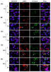
Colocalization analysis of α3 integrin with the late endosome/lysosome marker LAMP-1 in A549 epithelial cells infected with P. brasiliensis. A549 epithelial cells were infected with P. brasiliensis yeasts (MOI 1:1) for (A) 5 h, (B) 8 h, (C) 12 h, (D) 16 h, or (E) 24 h. Control (C) was performed in the absence of yeasts. Indirect immunofluorescence was performed using anti-α3 integrin antibodies (green) and anti-LAMP-1 antibodies (red). In blue, the nucleus of A549 cells was stained with DAPI and the fungus cell wall, with calcofluor White. Arrows indicate P. brasiliensis yeasts. Colocalization sites of LAMP-1 and α3 integrin were identified using the ImageJ software and are indicated by the arrowheads. Bar = 20 µm. This result is representative of three independent experiments.
Figure 4
Figure 4
α3 integrin levels in endosome-enriched fractions of A549 epithelial cells infected with P. brasiliensis. A549 cells were incubated in the absence (C) or presence of P. brasiliensis yeasts (Pb) for 16 h (MOI 1:1). Then, the epithelial cells were collected, lysed, and submitted to sucrose density gradient/ultracentrifugation. Aliquots of endosome-enriched fractions 1, 2, 5, and 6 were submitted side by side to the same SDS-PAGE. Next, Western blot was performed using antibodies anti-α3 integrin, -EEA1, -Rab5, -Rab7, and -LAMP-1. LE: Late endosome markers; EE: Early endosome markers. This result is representative of three independent experiments.
Figure 5
Figure 5
Effect of CHMP3 silencing on the α3 integrin levels during infection of A549 epithelial cells with P. brasiliensis. A549 cells were transfected with siRNA directed to CHMP3 (CHMP3) or negative control (NC) siRNA. Next, A549 cells were infected (+) or not (−) with P. brasiliensis yeasts (Pb) (MOI 1:1) for 24 h. (A) Protein extract aliquots were submitted to SDS-PAGE and α3 integrin levels were analyzed using Western blot. β-actin was used as loading protein control. (B) Values represent the intensity of the integrin band divided by the intensity of the corresponding β-actin band shown in (A). (C) CHMP3 silencing was analyzed by RT-qPCR. β-actin was used as housekeeping gene. Values represent relative fold change (2−ΔΔCt) in the target gene transcription levels compared to control group (without siRNA and Pb). Mean of triplicates ± standard deviation. *, p < 0.01 when compared to NC. Similar results were obtained in two independent experiments.
Figure 6
Figure 6
α3 integrin levels during infection with P. brasiliensis in the presence of bafilomycin A1. A549 cells were pre-incubated for 1 h with 20 nM of bafilomycin A1 (Baf+) and subsequently with P. brasiliensis yeasts (Pb) (MOI 1:1) for 24 h. Control (C) was performed in the absence of yeasts (−). (A) Protein extracts were submitted to SDS-PAGE and α3 integrin levels were analyzed using Western blot. β-actin was used for sample normalization. (B) Values represent the intensity of the α3 integrin band divided by the intensity of the corresponding β-actin band. Similar results were obtained in three independent experiments.

Similar articles

References

    1. Hahn R.C., Hagen F., Mendes R.P., Burger E., Nery A.F., Siqueira N.P., Guevara A., Rodrigues A.M., de Camargo Z.P. Paracoccidioidomycosis: Current Status and Future Trends. Clin. Microbiol. Rev. 2022;35:e0023321. doi: 10.1128/cmr.00233-21. - DOI - PMC - PubMed
    1. Shikanai-Yasuda M.A., Mendes R.P., Colombo A.L., de Queiroz Telles F., Kono A., Paniago A.M.M., Nathan A., do Valle A.C.F., Bagagli E., Benard G., et al. Brazilian guidelines for the clinical management of paracoccidioidomycosis. Epidemiol. Serv. Saude. 2018;27:e0500001. doi: 10.1590/0037-8682-0230-2017. - DOI - PubMed
    1. do Valle A.C.F., De Macedo P.M., Almeida-Paes R., Romão A.R., Lazéra M.D.S., Wanke B. Paracoccidioidomycosis after Highway Construction, Rio de Janeiro, Brazil. Emerg. Infect. Dis. 2017;23:1917–1919. doi: 10.3201/eid2311.170934. - DOI - PMC - PubMed
    1. Barros B.C.S.C., Almeida B.R., Barros D.T.L., Toledo M.S., Suzuki E. Respiratory Epithelial Cells: More Than Just a Physical Barrier to Fungal Infections. J. Fungi. 2022;8:548. doi: 10.3390/jof8060548. - DOI - PMC - PubMed
    1. Branchett W.J., Lloyd C.M. Regulatory cytokine function in the respiratory tract. Mucosal Immunol. 2019;12:589–600. doi: 10.1038/s41385-019-0158-0. - DOI - PMC - PubMed

LinkOut - more resources