Review
. 2022 Oct 26:3:948477.
doi: 10.3389/ffunb.2022.948477. eCollection 2022.

Vacuolar proteases and autophagy in phytopathogenic fungi: A review

Affiliations
Review

Vacuolar proteases and autophagy in phytopathogenic fungi: A review

Margarita Juárez-Montiel et al. Front Fungal Biol. .

Abstract

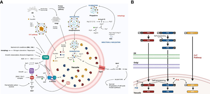
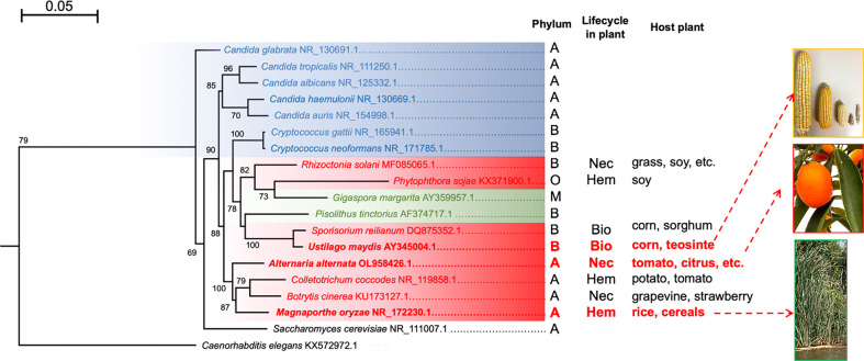
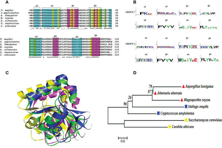
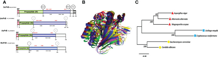
Autophagy (macroautophagy) is a survival and virulence mechanism of different eukaryotic pathogens. Autophagosomes sequester cytosolic material and organelles, then fuse with or enter into the vacuole or lysosome (the lytic compartment of most fungal/plant cells and many animal cells, respectively). Subsequent degradation of cargoes delivered to the vacuole via autophagy and endocytosis maintains cellular homeostasis and survival in conditions of stress, cellular differentiation, and development. PrA and PrB are vacuolar aspartyl and serine endoproteases, respectively, that participate in the autophagy of fungi and contribute to the pathogenicity of phytopathogens. Whereas the levels of vacuolar proteases are regulated by the expression of the genes encoding them (e.g., PEP4 for PrA and PRB1 for PrB), their activity is governed by endogenous inhibitors. The aim of the current contribution is to review the main characteristics, regulation, and role of vacuolar soluble endoproteases and Atg proteins in the process of autophagy and the pathogenesis of three fungal phytopathogens: Ustilago maydis, Magnaporthe oryzae, and Alternaria alternata. Aspartyl and serine proteases are known to participate in autophagy in these fungi by degrading autophagic bodies. However, the gene responsible for encoding the vacuolar serine protease of U. maydis has yet to be identified. Based on in silico analysis, this U. maydis gene is proposed to be orthologous to the Saccharomyces cerevisiae genes PRB1 and PBI2, known to encode the principal protease involved in the degradation of autophagic bodies and its inhibitor, respectively. In fungi that interact with plants, whether phytopathogenic or mycorrhizal, autophagy is a conserved cellular degradation process regulated through the TOR, PKA, and SNF1 pathways by ATG proteins and vacuolar proteases. Autophagy plays a preponderant role in the recycling of cell components as well as in the fungus-plant interaction.

Keywords: ATG8 and TOR; autophagic body degradation; autophagy; phytopathogenic fungus; vacuolar proteases PrA and PrB.


Conflict of interest statement

The authors declare that the research was conducted in the absence of any commercial or financial relationships that could be construed as a potential conflict of interest.

Figures

Figure 1
Figure 1
The role of autophagy, including the regulation of vacuolar proteases, in the yeast S. cerevisiae. (A) The TOR and RAS/cAMP kinases negatively regulate autophagy when the yeast is growing in nutrient rich conditions (glucose, NH+ 4, and Gln), thus avoiding G0 arrest. Both kinases inactivate Atg1, Atg13, and Atg8 proteins, which are involved in the first step of development of autophagosomes (Cebollero and Reggiori, 2009) and in the inactivation of transcription factor Gln3. The latter activates NCR genes and the Snf1 kinase. Under conditions of nutrient scarcity, Snf1 kinase is located in the vicinity of the vacuole and is released from the inhibition exerted by TOR and PKA, allowing it to inactivate TOR and PKA kinases. This inactivation takes place in part due to the targeting of Ras2 to the vacuole for proteolysis, mediated by the complex cell cycle regulator Whi2-phosphatase Psr1 (Leadsham et al., 2009). In addition to degrading specific proteins (e.g., Ras2), proteases and lipases break down autophagic bodies and their cargos, releasing amino acids and other biosynthetic units. These are either reused directly or induce Glu and Asp synthesis to replenish the amino acid sinks and therefore assure cell survival (Liu et al., 2021). In response to carbon availability, PKA and TOR regulate assembly and disassembly of V-ATPase through their effector Sch9 (Wilms et al., 2017). Vacuolar pH is regulated to maintain intracellular pH homeostasis and allow for the auto maturation carried out by protease PrA (Kane, 2006). (B) PrA matures to some extent in the endoplasmic reticulum (ER) before arriving to the vacuole and completing the process through automaturation. Subsequently, it stimulates the maturation of other vacuolar proteases, which are also synthesized as zymogens. Figure created by BioRender.com (accessed in April 2022).
Figure 2
Figure 2
Maximum likelihood phylogenetic tree constructed with the sequences of the ITS regions of different fungi. Human pathogenic fungi are portrayed by blue branches, phytopathogenic fungi by red branches, and mycorrhizal fungi by green branches. S. cerevisiae and C. elegans are illustrated with black branches because the former is rarely isolated as a pathogen (its interest being purely biotechnological) and the latter served as an outgroup. The tree was generated with the MEGA11 program based on sequence alignment by using the MUSCLE algorithm, the substitution model HKY+G calculated by JModelTest, and 1,000 bootstrap replicates, as explained in the supplementary material. It was edited with the FigTree program. The phyla: A = Ascomycota, B = Basidiomycota, M = Mucoromycota, and O = Oomycota. The types of phytopathogen: Nec =necrotrophic, Bio = biotrophic, and Hem = hemibiotrophic. The host plant for each species is shown. The tree was generated with the MEGA11 program based on sequence alignment by using the MUSCLE algorithm, the substitution model HKY+G calculated by JModelTest, and 1,000 bootstrap replicates, as indicated in the supplementary material. It was edited with the FigTree program.
Figure 3
Figure 3
The role of autophagy and vacuolar proteases PrA and PrB in the pathosystem of U. maydis, M. oryzae, and A alternata. (A) In the course of an infection by U. maydis, nitrogen scarcity promotes filamentation of haploid sporidia mediated by the bE and bW genes (in an independent and nonredundant manner) and the Ump2 transporter, probably preparing the cells for mating (Wallen et al., 2021). After the mating of two compatible basidiospores, Rbf1 and bE/bW induce cell cycle arrest and dikaryotic filaments extend apically over the host tissue. Vacuolated areas separated from empty sections by septa are generated during filament extension. Subsequently, non-melanized appressoria develop and penetrate the plant cells by means of CWDE’s (Nadal et al., 2010). During this early stage, autophagy-related genes and vacuolar proteases PRB1 and PEP4 are overexpressed. Then the cell cycle restarts, fungal effectors are produced, and plant tissue is colonized. At the time of tumor formation, carbon, nitrogen and oligopeptide transporters, as well as autophagy-related proteins and proteases, all of them are overexpressed. Furthermore, the transcription factors nit2 and snf1 are upregulated (Lanver et al., 2018). In this late stage, there is a metabolic change in plant tissue that favors the growth of reproductive over vegetative tissue. Thus, carbon transporters Hxt1 and Suc1 are indispensable for fungal virulence (Schmitz et al., 2018). During the biotrophic phase, U. maydis faces conditions of nutrient stress and deploys many strategies to establish an effective infection system. (B) When three-celled conidia of M. oryzae arrive to the plant surface, the germ tube emerges, mitosis takes place, and the nuclei travel. As the appressoria mature, the conidia undergo autophagic programmed cell death to sustain appressorium function. The formation of appressoria is positively and negatively regulated by the Ras/cAMP and TOR pathways, respectively (Marroquin-Guzman et al., 2017), which in turn are regulated by the Whis2-Psr1 complex. The latter maintains the appropriate levels of cAMP and perhaps targets the Ras22 protein to the vacuole (Shi et al., 2021). (C) When the multicellular conidia of A alternata arrive to the plant tissue, they germinate and enter the plant cells through stomates or breaches by using an appressorium-like structure and CWDE’s. The plant cell membrane is immediately disrupted by ACT, which causes plant cell necrosis. Then the fungus proliferates and conidia are formed and released. The generation of H2O2 by the plant cell as a defense mechanism gives rise to pexophagy. The good functioning of peroxisomes is vital for the production of ACT toxin (Wu et al., 2021). Additionally, different aspects of autophagy are important for the pathogenicity of A alternata. The Δatg8 strain is unable of either to form aerial hyphae and provoke necrotic lesions, similarly to the Δprb and a pep4-silenced strain. Interestingly PrB participating in the synthesis of secreted proteases (Fu et al., 2020). Nutrient scarcity leads to the expression of the entire autophagic machinery as well as the induction of Snf1, an essential protein for carbon utilization, vegetative growth, conidiation, and cell wall functions (Tang et al., 2020). Schemes were drawn whit BioRender.com (accessed in April 2022) and Corel Drawn v. 19.
Figure 4
Figure 4
Atg8 proteins are conserved in pathogenic and nonpathogenic fungi. (A) Multiple alignments were performed with Clustal Omega. In the alignment, the amino acids associated with alpha-helical (cyan) or beta-strand (fuchsia) secondary structures are highlighted with boxes. Likewise, the important amino acid residues for the two deep hydrophobic pockets HP1 and HP2 function are highlighted in yellow and green, respectively. An asterisk (*) denotes a completely conserved residue; a colon (:) represents the conservation of the properties of the residual side chain; a dot (.) indicates residues with side chains of weakly similar properties; and a hyphen (-) designates a gap. (B) Consensus logos were generated with WebLogo. Group 1 (S. cerevisiae, C albicans, A fumigatus, M. oryzae, and A alternata) and group 2 (C. amylolentus and U. maydis). (C) 3D model of the Atg8 proteins. The overlap of S. cerevisiae (green) with the fungal phytopathogens is represented by utilizing the best model of the Atg8 of each fungus: U. maydis (blue), M. oryzae (fuchsia), and A alternata (yellow). The model was generated by homology modeler by using the Modeller 9.23 program, as explained in the supplementary material. (D) Phylogenetic analysis of the Atg8 of different organisms, carried out in the MEGA6 program with the maximum likelihood method, the WAG+G model, and 100 bootstrap replicates. The phylogenetic tree is drawn to scale, with the length of the branches depicting the corresponding evolutionary distances. Fungi that are grouped together in the same clade are portrayed with red, blue, and yellow symbols.
Figure 5
Figure 5
Serine vacuolar endoproteases PrBs are conserved in pathogenic and nonpathogenic fungi. (A) The characteristic domains of vacuolar proteases of the S8 family are illustrated, including the DHS catalytic triad of serine proteases, the probable maturation sites, and the I9 inhibitor domain of the propeptide (predicted in the Expasy server). Proteases such as ScPrB are apparently synthesized as zymogens, which are inactive until propeptides are removed in the vacuole. The cysteines in the protein are also shown. The circles in the S. cerevisiae protease designate the regions where autocatalysis and PrA processing are conducted. In the rest of the proteases, the circles denote regions of theoretical maturation. The alignment of sequences was performed with Clustal X and the drawing with BioRender.com (accessed in April 2022). (B) Tertiary structure of fungal PrBs. The overlap is represented by utilizing the best model of PrB of S. cerevisiae (green) and of the three fungal phytopathogens herein studied: U. maydis (blue), M. oryzae (fuchsia), and A alternata (yellow). (C) Phylogenetic analysis of the PrB of different organisms. Carried out in the MEGA6 program with the maximum likelihood method, the WAG+G model, and 100 bootstrap replicates. The phylogenetic tree is drawn to scale, with the length of the branches depicting the corresponding evolutionary distances. Fungi that are grouped together in the same clade are portrayed with red, blue, and yellow symbols.
Figure 6
Figure 6
Intermolecular interactions of inhibitory peptide Um10059 (Um2129) (cyan) with PrBSc (A), PrBUm (B), and PrBAa (C). Dotted lines indicate the type of interactions. For the purpose of clarity, the interactions between the amino acid residues of the PrB catalytic triad (Asp, His, and Ser) and the inhibitory Um10059 are shown. The interactions with M. oryzae were not shown since none of the residues of the catalytic triad interact with the inhibitor of U. maydis.

Similar articles

References

    1. Abeliovich H., Klionsky D. J. (2001). Autophagy in yeast: Mechanistic insights and physiological function. microbiol. Mol. Biol. Rev. 65 (3), 463–479. doi: 10.1128/MMBR.65.3.463-479.2001 - DOI - PMC - PubMed
    1. Aman Y., Schmauck-Medina T., Hansen M., Morimoto R. I., Simon A. K., Bjedov I., et al. . (2021). Autophagy in healthy aging and disease. Nat. Aging 1 (8), 634–650. doi: 10.1038/s43587-021-00098-4 - DOI - PMC - PubMed
    1. Azzopardi M., Farrugia G., Balzan R. (2017). Cell-cycle involvement in autophagy and apoptosis in yeast. Mech. Ageing Dev. 161, 211–224. doi: 10.1016/j.mad.2016.07.006 - DOI - PubMed
    1. Baba M., Takeshige K., Baba N., Ohsumi Y. (1994). Ultrastructural analysis of the autophagic process in yeast: Detection of autophagosomes and their characterization. J. Cell Biol. 124 (6), 903–913. doi: 10.1083/jcb.124.6.903 - DOI - PMC - PubMed
    1. Böhmer M., Colby T., Böhmer C., Bräutigam A., Schmidt J., Bölker M. (2007). Proteomic analysis of dimorphic transition in the phytopathogenic fungus ustilago maydis. Proteomics 7 (5), 675–685. doi: 10.1002/pmic.200600900 - DOI - PubMed