. 2024 Jan 1;75(1):45-59.
doi: 10.1093/jxb/erad364.

Characterization of intracellular membrane structures derived from a massive expansion of endoplasmic reticulum (ER) membrane due to synthetic ER-membrane-resident polyproteins

Affiliations

Characterization of intracellular membrane structures derived from a massive expansion of endoplasmic reticulum (ER) membrane due to synthetic ER-membrane-resident polyproteins

Andras Sandor et al. J Exp Bot. .

Abstract

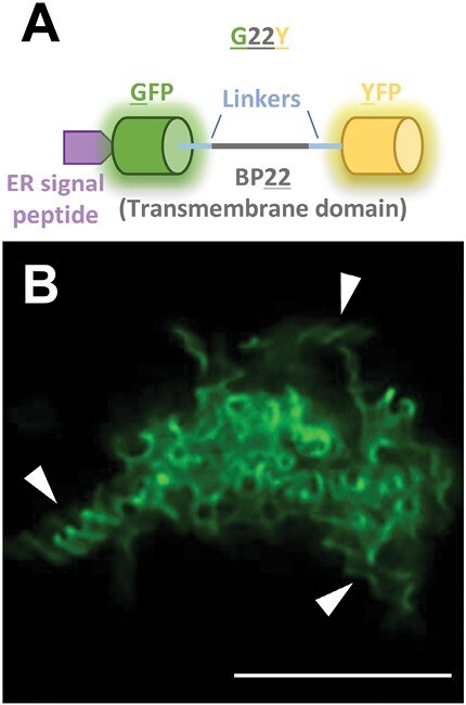
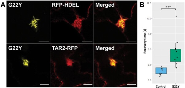
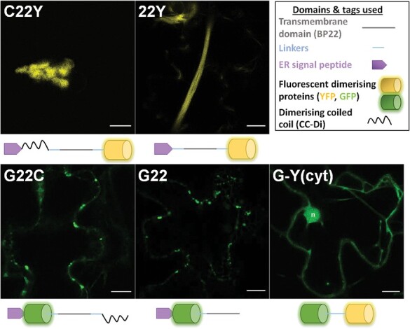
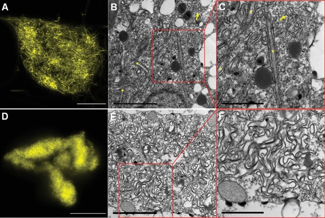
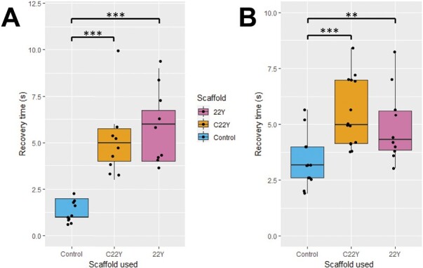
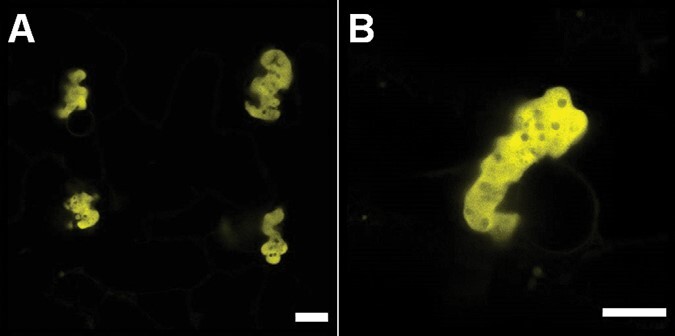
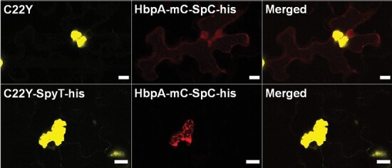
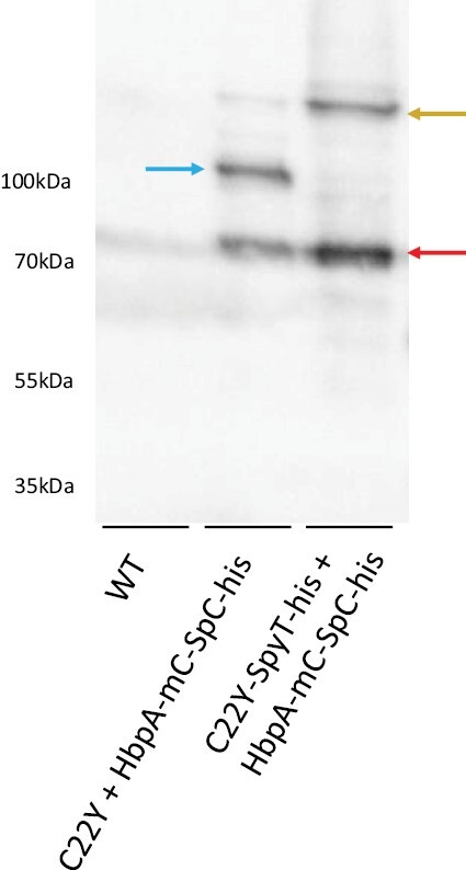
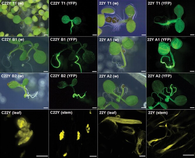
The endoplasmic reticulum (ER) is a dynamic organelle that is amenable to major restructuring. Introduction of recombinant ER-membrane-resident proteins that form homo oligomers is a known method of inducing ER proliferation: interaction of the proteins with each other alters the local structure of the ER network, leading to the formation large aggregations of expanded ER, sometimes leading to the formation of organized smooth endoplasmic reticulum (OSER). However, these membrane structures formed by ER proliferation are poorly characterized and this hampers their potential development for plant synthetic biology. Here, we characterize a range of ER-derived membranous compartments in tobacco and show how the nature of the polyproteins introduced into the ER membrane affect the morphology of the final compartment. We show that a cytosol-facing oligomerization domain is an essential component for compartment formation. Using fluorescence recovery after photobleaching, we demonstrate that although the compartment retains a connection to the ER, a diffusional barrier exists to both the ER and the cytosol associated with the compartment. Using quantitative image analysis, we also show that the presence of the compartment does not disrupt the rest of the ER network. Moreover, we demonstrate that it is possible to recruit a heterologous, bacterial enzyme to the compartment, and for the enzyme to accumulate to high levels. Finally, transgenic Arabidopsis constitutively expressing the compartment-forming polyproteins grew and developed normally under standard conditions.

Keywords: Compartment; OSER; endoplasmic reticulum; membrane; proliferation; synthetic biology.


Conflict of interest statement

No conflict of interest declared.

Figures

Fig. 1.
Fig. 1.
Remodelling the ER using the synthetic oligomerizing polyprotein G22Y. (A) Schematic of the G22Y polyprotein, composed of two dimerizing fluorescent proteins (GFP and YFP, facing the ER lumen and the cytosol, respectively) flanking a transmembrane domain (BP22) and a N-terminal ER targeting signal peptide. (B) Fluorescence confocal microscopy images of the structures formed in mature N. tabacum leaves 7 d after agroinfiltration. Arrowheads point to some notable helical organizations in the structure. The image was captured using high-resolution confocal Airyscan imaging. Scale bar=10 µm.
Fig. 2.
Fig. 2.
Co-expression of G22Y with fluorescent ER markers shows the connection of the compartment to the ER membrane and lumen. (A) G22Y (yellow) was co-expressed with the fluorescent ER-luminal marker protein RFP-HDEL or ER-membrane marker TAR2-RFP (both in red). The signals overlap, confirming that the G22Y compartment derives its membrane and lumen from the ER. The fluorescence confocal microscopy images were captured 7 d after agroinfiltrating mature N. tabacum leaves. Scale bars=10 µm. (B) The recovery rate of RFP-HDEL after FRAP of the G22Y compartment is slower when compared with the peripheral ER (Control). Asterisks depict significance levels: ***P<0.001 (Student’s t-test), n=9.
Fig. 3.
Fig. 3.
Visualizing alternative G22Y-derived constructs to expand the compartment-forming scaffold toolkit. Five new polyproteins were designed, with two constructs having either the N-terminal YFP or the C-terminal GFP removed (G22 and 22Y, respectively). In two constructs, the removed fluorescent dimerising protein was replaced with a synthetic dimerizing coiled-coil domain (G22C and C22Y), and in one construct the transmembrane domain and the ER-targeting signal peptide was removed [G-Y(cyt)]. Top right box shows the constitutive domains and the schematics of G22Y-derived constructs. Confocal microscopy images of the new constructs were taken 7 d after agroinfiltrating mature N. tabacum leaves. Yellow, YFP; green, GFP; n, nucleus. Scale bars=10 µm.
Fig. 4.
Fig. 4.
High-resolution characterization of the 22Y and C22Y compartment structures. (A–D) High-resolution Airyscan fluorescent confocal microscopy images of (A) 22Y and (D) C22Y compartments. Images were captured 7 d after agroinfiltration of mature N. tabacum leaves. Scale bars=10 µm. (B, C, E, F) TEM images of (B, C) 22Y and (E, F) C22Y compartments. (C) and (F) show magnified sections of (B) and (E), respectively. The 22Y structures often form two parallel membrane pairs (yellow asterisks). This is likely an ER cylinder, with some cross-sections of these highlighted with a yellow arrow. Scale bars=2 µm (B, E); 1 µm (C,F).
Fig. 5.
Fig. 5.
Lumenal and cytosolic fluorescence recovery after photobleaching (FRAP) rates are reduced in C22Y and 22Y compartments. FRAP experiments on the C22Y and 22Y compartment types using (A) ER lumenal RFP-HDEL and (B) cytosolic Peredox-mCherry both show a significantly increased recovery time when compared with the respective controls, suggesting a diffusional barrier between the compartment and both the ER lumen and the bulk cytosol. Asterisks depict significance levels; **P<0.01 and ***P<0.001 (Student’s t-test); n=9 for (A) and n=13 for (B) for all constructs. The boxes represent the interquartile range, the horizontal line in the box shows the median, and the whiskers the minimum and maximum values (excluding outliers).
Fig. 6.
Fig. 6.
Confocal microscopy of the C22Y-SpyT-his construct. (A) Fluorescent confocal microscopy image showing four C22Y-SpyT-his compartments forming in adjacent cells. (B) High-resolution Airyscan fluorescent confocal microscopy image. Compartments formed are similar to C22Y. Images were captured 7 d after agroinfiltration of mature N. tabacum leaves. Scale bars=10 µm.
Fig. 7.
Fig. 7.
Visualization of proteins anchored to the cytosolic surface of the compartment via SpyCatcher—SpyTag covalent binding. Fluorescent confocal microscopy images (red,mCherry fluorescence; yellow, YFP fluorescence) of HbpA-mC-SpC-his with C22Y/C22Y-SpyT-his in A. tumefaciens-mediated transiently transformed mature N. benthamiana leaves. Scale bars=10 μm.
Fig. 8.
Fig. 8.
Purification of a covalently bound enzyme—protein-forming scaffold complex. Anti-mCherry western blot of elution fractions from a nickel-affinity purification from transiently transformed N. benthamiana samples. WT column contains untransformed plant samples, while C22Y + HbpA-mC-SpC-his and C22Y-SpyT-his + HbpA-mC-SpC-his contain plant samples co-transformed with the appropriate constructs. Blue arrow shows a band with matching molecular weight to unbound HbpA-mC-SpC-his cargo constructs (107 kDa). Golden arrow shows a band with matching molecular weight to bound HbpA-mC-SpC-his + C22Y-SpyT-his complexes (147 kDa). Red band shows putative cleavage products. The text on the left shows standard molecular weights.
Fig. 9.
Fig. 9.
Stably transformed A. thaliana lines form C22Y and 22Y compartment structures similar to those obtained by transient expression in tobacco. Top three rows: fluorescent whole seedling images of 5–6-day-old stably transformed A. thaliana on MS media plates. Pairs of images show the white light (w) and YFP fluorescence (YFP) of T1 (top row) and T2 (second and third row) plants. A1, A2, and B1, B2 refer to two independent lines of 22Y- and C22Y-expressing A. thaliana lines, respectively. Fluorescent signal is present in all tissues but is noticeably stronger in the roots of T2 plants. Scale bars=1 cm. Bottom row: high-resolution Airyscan laser confocal microscopy images of leaf epidermis and stem cross-section of C22Y and 22Y expressing 6-week-old T2 plants. Both compartment types show identical morphology to the previously described compartments following transient expression of each construct. Scale bars=10 µm.

Similar articles

References

    1. Almsherqi ZA, Landh T, Kohlwein SD, Deng Y.. 2009. Cubic membranes: the missing dimension of cell membrane organization. International Review of Cell and Molecular Biology 274, 275–342. - PMC - PubMed
    1. Arendt P, Miettinen K, Pollier J, De Rycke R, Callewaert N, Goossens A.. 2017. An endoplasmic reticulum-engineered yeast platform for overproduction of triterpenoids. Metabolic Engineering 40, 165–175. - PubMed
    1. Barbante A, Irons S, Hawes C, Frigerio L, Vitale A, Pedrazzini E.. 2008. Anchorage to the cytosolic face of the endoplasmic reticulum membrane: a new strategy to stabilize a cytosolic recombinant antigen in plants. Plant Biotechnology Journal 6, 560–575. - PubMed
    1. Barlowe C, Helenius A.. 2016. Cargo capture and bulk flow in the early secretory pathway. Annual Review of Cell and Developmental Biology 32, 197–222. - PubMed
    1. Bolte S, Cordelières FP.. 2006. A guided tour into subcellular colocalization analysis in light microscopy. Journal of Microscopy 224, 213–232. - PubMed

Publication types

MeSH terms