

Orthopaedic Wire Debridement: A Novel Surgical Technique for Hypergranulated Burn Wounds
- PMID: 37709982
- PMCID: PMC10694099
- DOI: 10.1007/s00268-023-07147-6
Orthopaedic Wire Debridement: A Novel Surgical Technique for Hypergranulated Burn Wounds
Abstract
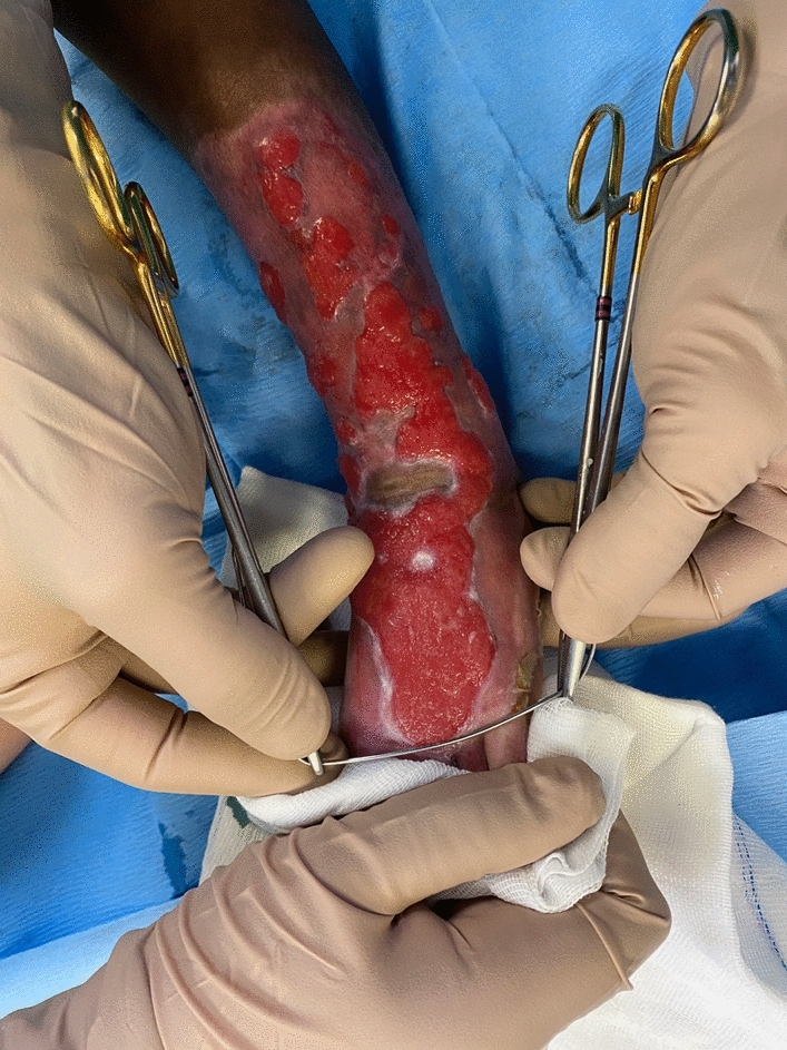
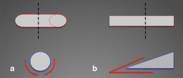
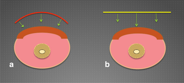
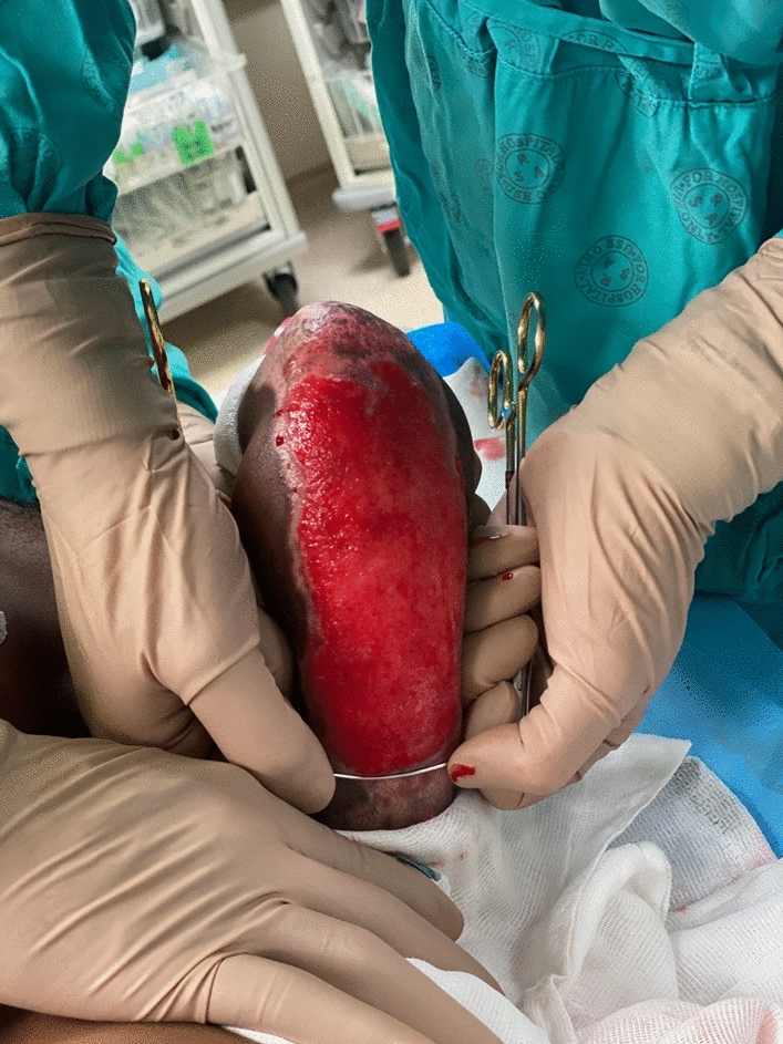
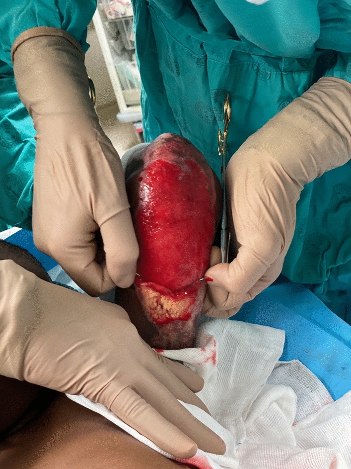
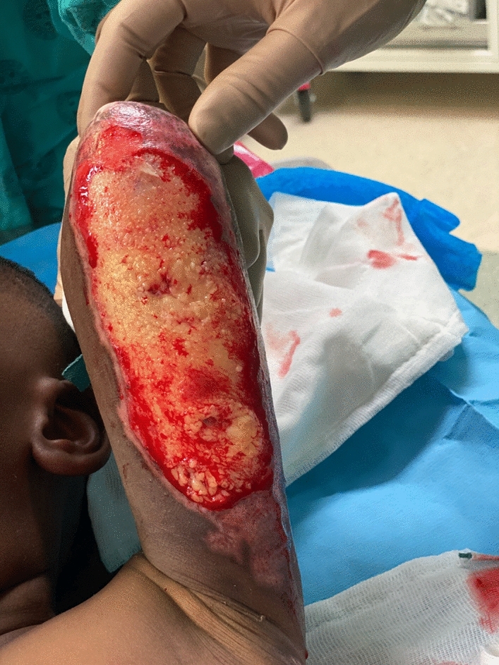
The burn wound healing process can be protracted due to various factors, including burn depth, infection and hypergranulation. Hypergranulation impedes epithelialisation macroscopically by preventing cellular migration across the wound bed and microscopically through cell-to-cell signal interferences. Debridement, which is the act of removing necrotic tissue, hypergranulation, slough and foreign debris from the wound in order to expose the underlying viable bed, can be achieved using various techniques. This aids with wound bed preparation to facilitate and expedite healing. In this article, we present a novel surgical debridement technique using a malleable orthopaedic cerclage wire for the management of a hypergranulated burn wound.
© 2023. The Author(s).
Conflict of interest statement
The authors (mentioned above) declare that they have no known competing financial interests or personal relationships that could have appeared to influence the work reported in this paper.
Figures
Similar articles
-
[Debridement of burn wounds using a hydrosurgery system].Zhonghua Shao Shang Za Zhi. 2015 Dec;31(6):470-2. Zhonghua Shao Shang Za Zhi. 2015. PMID: 26837254 Chinese.
-
Enzymatic wound debridement.J Wound Ostomy Continence Nurs. 2008 May-Jun;35(3):273-80. doi: 10.1097/01.WON.0000319125.21854.78. J Wound Ostomy Continence Nurs. 2008. PMID: 18496083 Review.
-
[Effects of minimally invasive tangential excision in treating deep partial-thickness burn wounds on trunk and limbs in pediatric patients in the early stage post burn].Zhonghua Shao Shang Za Zhi. 2018 Oct 20;34(10):714-718. doi: 10.3760/cma.j.issn.1009-2587.2018.10.012. Zhonghua Shao Shang Za Zhi. 2018. PMID: 30369140 Chinese.
-
Clinical performance and safety of a debridement pad with abrasive and non-abrasive fibres.J Wound Care. 2024 Jun 2;33(6):408-416. doi: 10.12968/jowc.2024.0162. J Wound Care. 2024. PMID: 38843013
-
Understanding wound bed preparation and wound debridement.Br J Community Nurs. 2010 Mar;15(3):S22, S24, S26, passim. doi: 10.12968/bjcn.2010.15.Sup12.S22. Br J Community Nurs. 2010. PMID: 20220634 Review.
Cited by
-
Invited Commentary: Management of Hypergranulation Requires a Multimodal Approach.World J Surg. 2023 Dec;47(12):3105-3106. doi: 10.1007/s00268-023-07227-7. Epub 2023 Nov 25. World J Surg. 2023. PMID: 38006424 Free PMC article. No abstract available.
References
-
- D’avignon LC, Saffle JR, Chung KK, et al. Prevention and management of infections associated with burns in the combat casualty. J Trauma Inj Infect Crit Care. 2008;64:S277–S286. - PubMed
MeSH terms
LinkOut - more resources
Full Text Sources
Medical
