Review
. 2024 Feb;22(2):89-104.
doi: 10.1038/s41579-023-00963-6. Epub 2023 Sep 12.

The oral microbiome: diversity, biogeography and human health

Affiliations
Review

The oral microbiome: diversity, biogeography and human health

Jonathon L Baker et al. Nat Rev Microbiol. 2024 Feb.

Abstract

The human oral microbiota is highly diverse and has a complex ecology, comprising bacteria, microeukaryotes, archaea and viruses. These communities have elaborate and highly structured biogeography that shapes metabolic exchange on a local scale and results from the diverse microenvironments present in the oral cavity. The oral microbiota also interfaces with the immune system of the human host and has an important role in not only the health of the oral cavity but also systemic health. In this Review, we highlight recent advances including novel insights into the biogeography of several oral niches at the species level, as well as the ecological role of candidate phyla radiation bacteria and non-bacterial members of the oral microbiome. In addition, we summarize the relationship between the oral microbiota and the pathology of oral diseases and systemic diseases. Together, these advances move the field towards a more holistic understanding of the oral microbiota and its role in health, which in turn opens the door to the study of novel preventive and therapeutic strategies.


Conflict of interest statement

Competing interests

The authors declare no competing interests.

Figures
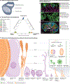
Fig. 1 |
Fig. 1 |. Biogeography of the oral microbiome and relative sizes of its members.
a–d, Microbial communities with disparate structure and composition colonize different surfaces in the mouth. a, Distinctive habitats within the oral cavity host a diversity of resident taxa, whose biogeography can be visualized via combinatorial labelling and spectral imaging fluorescence in situ hybridization microscopy of the supragingival plaque (part b), the buccal mucosa (part c) and the dorsum of the tongue (part d). Shown are bacterial members of the oral microbiome and the characteristic structures they form at each site. Until recently, most human microbiome studies analysed the bacterial distribution at the genus level or even at the phylum level. With the increasing availability of whole-genome metagenomic sequence data, distribution patterns of closely related species can be distinguished. e, A recent study analysed the distribution of species of the abundant oral genus Streptococcus using short-read metagenomic sequence data from the human mouth and found that each species was found primarily at one oral site. In the figure, species are indicated by coloured dots corresponding to the colours in the legend; the size of species dots corresponds to their abundance. These whole-genome data could differentiate closely related taxa such as Streptococcus mitis, found primarily on the buccal mucosa; Streptococcus oralis found in dental plaque; and Streptococcus infantis, found on the tongue dorsum. f, Together, the sizes of oral microorganisms and microbial structures span four orders of magnitude, from nanoscale viruses to bacterial aggregates of hundreds of microns. Bacterial cells range in size from 200–300 nm (the diminutive Saccharibacteria) up to 10 μm (such as the long spirilliform Treponema), with the majority around 1 μm (for example, 0.8-μm diameter Streptococcus spp.). Bacterial aggregates and consortia comprise the largest component of oral microbial biomass (up to hundreds of microns) and include ordered polymicrobial structures, distinctively named to suggest their features. ‘Hedgehog’ aggregates are found abundantly in healthy oral microbiomes and are composed of long filaments of multiple cells of Corynebacterium decorated with Streptococcus and other cocci at their periphery (forming ‘corncobs’ up to 50 μm in length) and creating densely packed environments that facilitate the growth of anaerobe species including Leptotrichia spp., Fusobacterium spp. and Actinomyces spp. within. ‘Rotund’ aggregates have been identified in association with caries and comprise an inner mass of Streptococcus mutans and associated exopolysaccharide matrix, a surrounding layer of S. oralis or other non-mutans streptococci and an outer layer of non-streptococci. Oral eukaryotes include the rare motile protists Trichomonas and Entamoeba, as well as the non-motile fungal genera Candida and Malassezia, and they are all around the same order of magnitude in size as human neutrophils (around 5–15 μm), which are abundant in the gingival crevicular fluid during inflammation. The smallest members of the oral microbiome are the viruses, which are known to include human-infecting viruses (for example, the abundant anelloviruses), viruses infecting bacteria (bacteriophages) and likely also viruses infecting oral micro-eukaryotes. Part b reprinted with permission from ref. , PNAS. Photos in parts c and d courtesy of J.M.W. Part e adapted with permission from ref. , Wiley.
Fig. 2 |
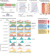
Fig. 2 |. Experimental gingivitis in humans reveals three distinct response types.
a, Human experimental gingivitis model study design (Supplementary Box 1). A typical experimental gingivitis model with healthy subjects includes the following phases: hygiene phase for 2 weeks before baseline (days −14 to 0), gingivitis induction phase lasting for 3 weeks (days 0–21) and resolution phase for 2 weeks (days 21–35). Subgingival plaque and gingival crevicular fluid (GCF) are taken from unbrushed test teeth (test teeth) as well as the teeth that had maintained oral hygiene (control teeth). This model has allowed tracking of changes in the oral microbiome as well as variation in the inflammatory response of the subjects. Although all subjects typically respond to oral plaque accumulation in experimental gingivitis studies, the rate and severity of the inflammatory response has been shown to vary. A recent study investigating healthy subjects (age 18–35) has stratified these responses on the basis of the combined analyses and clustering of temporal trajectories in clinical measures of inflammation. This analysis resulted in three inflammatory responder types: high and low responders (previously recognized in the literature) and a novel slow responder each with distinct microbial and host signatures. b, Plaque growth rate variation between inflammatory responder types. Slow responders display a delayed plaque growth rate, whereas high and low responders have the same growth rate. c, Inflammation variation over time measured by the percent of unbrushed test sites with bleeding on probing. The low responders do not reach a high level of inflammation and the newly described slow responders have delayed inflammation. d, Heatmap illustrating variation in a panel of host mediators (cytokines and chemokines) in GCF. When comparing mean values for chemokine expression over time for responders (row normalized data, red represents high values, white is low value and each column is a visit), the low responder phenotype exhibits lower overall mediator concentrations than the other types. Relative inflammation changes measured by the gingival index (a standard clinical measure of gingivitis severity based on tissue redness) shown as a row-normalized heatmap across the bottom of the plot. e, Dynamic changes in relative abundance across the seven major phyla and three candidate phyla radiation group members in responder type across the phases. Variation is observed across subgingival plaque microbial compositions as well as the rate of change in the relative abundance of certain genera over the induction phase. Coloured areas represent the different genera that make up each phylum, and the plots show the mean relative abundance of different genera by responder group. Consistently across experimental gingivitis studies that have relied on culturing or culture-independent methods, the two most abundant Gram-positive phyla, Firmicutes and Actinobacteria, decrease in relative abundance as the plaque community grows and matures. With 16S rRNA sequencing across early and late time points, additional resolution has been gained recently. For example, Selenomonas becomes a higher proportion of the total Firmicutes by day 21 across responders. By contrast, members of the Prevotella genus predominantly contribute to this increase of the Bacteroidetes. Porphyromonas also increase over time but represent a smaller relative proportion of this total. Within the phylum Spirochaetes, Treponema genus members in gingivitis here tend to show a large enrichment that occurs after a week or more of plaque growth. A defining feature of the slow responder phenotype is the higher abundance of Streptococcus at the time of inclusion in the study (day −14), which is then restored after the induction phase inflammation is resolved. In addition, the newly recognized ultrasmall reduced genome epibionts belonging to the CPR, ‘Candidatus Absconditabacteria’, ‘Candidatus Gracilibacteria’ and Saccharibacteria also show increases during gingival inflammation with variation across responder types. f, Overview of the prevalence and defining features of the newly characterized responder types. Data in parts bd from ref. . Parts b–d adapted from ref. , PNAS.
Fig. 3 |
Fig. 3 |. Links between the oral microbiota and systemic diseases.
The oral microbiota, and particularly periodontal pathogens, have been increasingly linked to a number of systemic diseases, either directly through translocation of oral pathogens to other sites or indirectly through modulation of the host immune system and inflammatory response. This illustration highlights a number of these links with the insets highlighting the mechanism(s). a, Cardiovascular disease: in vitro and ex vivo studies have suggested that Porphyromonas gingivalis bacteraemia alters dendritic cell behaviour, causing pro-inflammatory accumulations at atherosclerotic plaques. b, Inflammatory bowel disease: animal studies have illustrated that when T cells primed and reactive to oral pathobionts encounter ectopic periodontal pathogens in the gut, they increase inflammation (particularly via IL-1B) and contribute to colitis,. c, Colorectal cancer: mouse models and human studies have established that Fusobacterium nucleatum outgrowth synergizes with tumour growth via multiple mechanisms. The F. nucleatum surface protein Fab2 binds to cancer cells, inducing pro-metastatic chemokines, and activates TIGIT, allowing for evasion of immune surveillance by the tumour,. Another F. nucleatum surface protein, FadA, stimulates cancer cell proliferation by inducing E-cadherin-mediated Wnt-β-catenin signalling. d, Alzheimer disease: animal models and observational studies in humans have shown that P. gingivalis, on reaching the brain via bacteraemia, causes neuronal inflammation, degrades tau protein causing neurofibrillary tangles and contributes to the formation of amyloid-β plaques, reviewed elsewhere. e, Aspiration pneumonia: human studies have shown that the translocation of oral bacteria to the lung can lead to pneumonia, reviewed elsewhere. f, Rheumatoid arthritis: in mice, P. gingivalis that reaches the bone marrow skews immune cell development via IL-6, promoting production of osteoclasts and contributing to bone loss. Meanwhile, animal and human studies have illustrated that P. gingivalis and Aggregatibacter actinomycetemcomitans can citrullinate host proteins, which triggers autoimmunity,.

Similar articles

Cited by

References

    1. Dobell C Antony van Leeuwenhoek and His ‘Little Animals’: Being Some Account of the Father of Protozoology and Bacteriology and His Multifarious Discoveries in these Disciplines (Harcourt, Brace and Company, 1932).
    1. Mukherjee C et al. Acquisition of oral microbiota is driven by environment, not host genetics. Microbiome 9, 54 (2021). - PMC - PubMed
    1. Sulyanto RM, Thompson ZA, Beall CJ, Leys EJ & Griffen AL The predominant oral microbiota is acquired early in an organized pattern. Sci. Rep 9, 10550 (2019). - PMC - PubMed
    1. Kaan AMM, Kahharova D & Zaura E Acquisition and establishment of the oral microbiota. Periodontol 2000 86, 123–141 (2021). - PMC - PubMed
    1. Baker JL, Bor B, Agnello M, Shi W & He X Ecology of the oral microbiome: beyond bacteria. Trends Microbiol. 25, 362–374 (2017). - PMC - PubMed