. 2023 Sep 4;24(17):13662.
doi: 10.3390/ijms241713662.

FoxO3 Modulates Circadian Rhythms in Neural Stem Cells

Affiliations

FoxO3 Modulates Circadian Rhythms in Neural Stem Cells

Swip Draijer et al. Int J Mol Sci. .

Abstract

Both FoxO transcription factors and the circadian clock act on the interface of metabolism and cell cycle regulation and are important regulators of cellular stress and stem cell homeostasis. Importantly, FoxO3 preserves the adult neural stem cell population by regulating cell cycle and cellular metabolism and has been shown to regulate circadian rhythms in the liver. However, whether FoxO3 is a regulator of circadian rhythms in neural stem cells remains unknown. Here, we show that loss of FoxO3 disrupts circadian rhythmicity in cultures of neural stem cells, an effect that is mediated via regulation of Clock transcriptional levels. Using Rev-Erbα-VNP as a reporter, we then demonstrate that loss of FoxO3 does not disrupt circadian rhythmicity at the single cell level. A meta-analysis of published data revealed dynamic co-occupancy of multiple circadian clock components within FoxO3 regulatory regions, indicating that FoxO3 is a Clock-controlled gene. Finally, we examined proliferation in the hippocampus of FoxO3-deficient mice and found that loss of FoxO3 delayed the circadian phase of hippocampal proliferation, indicating that FoxO3 regulates correct timing of NSC proliferation. Taken together, our data suggest that FoxO3 is an integral part of circadian regulation of neural stem cell homeostasis.

Keywords: FoxO3; cell cycle; circadian rhythms; liver; metabolism; neural stem cell.


Conflict of interest statement

The authors declare no conflict of interest.

Figures

Figure 1
Figure 1
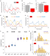
Differential effect of FoxO transcription factors on circadian oscillations in neural stem cells in vitro. (A) Representative examples of bioluminescence rhythms in cells co-transfected with a mBmal1::luciferase reporter construct and either empty vector (grey line), FOXO1-EGFP (left, yellow line), FOXO3-EGFP (middle, red line), or FOXO6-EGFP (right, blue line). (B) Representative examples of bioluminescence rhythms in cells co-transfected with a mBmal1::luciferase reporter construct and control siRNA (grey line), FoxO1 siRNA (left, yellow line), FoxO3 siRNA (middle, red line), or FoxO6 siRNA (right, blue line). (C,D) Graphical representation of the amplitude and period length under FOXO overexpression (C) or FoxO knockdown (D). Values are averages of independent experiments (n = 3) performed in triplicate. Error bars represent SEM. One-way ANOVA with Dunnett’s multiple comparison test. * p < 0.05; ** p < 0.01; *** p < 0.001.
Figure 2
Figure 2
Clock rescues the FoxO3 siRNA phenotype in neural stem cells. (A) Clock mRNA expression in adult neural stem cells following 24 and 48 h after transfection of siRNA control or FoxO3 siRNA. (B) Clock expression in adult neural stem cells following siRNA control or FoxO3 siRNA during 24 h following circadian synchronization with dexamethasone. (C) FoxO3 expression in adult neural stem cells following siRNA control or FoxO3 siRNA during 24 h following circadian synchronization with dexamethasone. (D,E) Representative examples of bioluminescence rhythms in cells co-transfected with a mBmal1::luciferase reporter construct and either siRNA control plus empty vector (light blue), siRNA control plus pFlag-CLOCK (dark blue), FoxO3 siRNA plus empty vector (red) or FoxO3 siRNA plus pFlag-CLOCK (orange). (F) Quantification of amplitudes of bioluminescence rhythms shown in D and E. Values are averages of biological replicates (n = 3) performed in triplicate. One-way ANOVA with Tukey’s multiple comparison test. Error bars represent SD. * p < 0.05, n.s. is “not significant”.
Figure 3
Figure 3
Loss of FoxO3 does not disrupt circadian rhythmicity in single cells. (A) Single cell recordings over multiple days of Rev-ERbα-VNP fluorescence levels in NIH3T33C transfected with FoxO3 siRNA (siO3, red) or control siRNA (siC, grey). Representative examples. (B) Magnitude of circadian oscillations (mesor) in Rev-ERbα-VNP fluorescence levels shown in (A). siC, n = 13; siO3, n = 20. * p < 0.025. (C) Amplitude of circadian oscillation in Rev-ERbα-VNP fluorescence levels shown in (A). siC, n = 13; siO3, n = 20. n.s., not significant. (D,E) Representative examples of single cell recordings of fluorescence marker expression for circadian clock oscillations (Rev-ERbα-VNP, yellow), G1 phase of the cell cycle (hCdt1-mKOrange fusion protein, red) and combined S/G2/M phases (hGeminin-CFP fusion protein, blue) in NIH3T33C cells transfected with FoxO3 siRNA (D) or siRNA control (E). (F) Comparison of the average length of cell cycle phase S/G2/M (blue), G1 (red) and circadian cycle (yellow) of data shown in (DG). siC, n = 13; siO3, n = 20. n.s., not significant. (G) Frequency histograms of circadian cycle length (Clock, yellow) and total cell cycle length (purple) in single NIH3T33C cells transfected with FoxO3 siRNA (bottom) or siRNA control (top). siC, n = 13; siO3, n = 20.
Figure 4
Figure 4
Circadian landscape of the FoxO3 gene. (A) University of California Santa Cruz (UCSC) genome browser track view of the FoxO3 gene showing ChIP-seq binding peaks for circadian clock components Clock, Bmal1, Per1, Per2, Npas2, Cry1 and Cry2, as well as transcriptional regulators CBP, p300 and recruitment and initiation of RNA polymerase II (8WG16 and Ser5P, respectively). Data from [36]. (B,C) Circadian landscape of the second intron (B) and promoter (C) within the FoxO3 gene showing phase distributions of Bmal1, Clock, Per1, Per2, Npas2, Cry1 and Cry2, CBP and RNAPII recruitment and initiation. The mean circadian phase of peak binding is indicated under the name. The black line denotes circadian expression of FoxO3 mRNA.
Figure 5
Figure 5
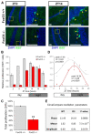
Diurnal proliferation of neural stem cells in the dentate gyrus of adult FOXO3-deficient mice. (A) Representative examples of Ki67-positive cells (yellow arrow heads) in the subgranular zone of the dentate gyrus of FoxO3−/− mice and littermate controls at CT2 and CT14. (B) Diurnal proliferation of neural stem cells in the dentate gyrus of 6-month-old FoxO3+/+ (grey) and FoxO3−/− mice (red). Values are averages per genotype and normalized to CT2 of controls. Error bars represent SEM. (C) Data in (B) shown as total proliferation over 24 h in 6-month-old FoxO3+/+ and FoxO3−/− mice (n ≥ 14). Values are average sum of all time points per genotype. Error bars represent SEM. (** p < 0.01). (D) Data in (B) fitted to a linear harmonic regression model to determine circadian rhythmicity (CircWave). (E) Parameter comparison of circadian oscillation in Ki67-positive cells in the subgranular zone of the dentate gyrus of FoxO3+/+ and FoxO3−/− mice, calculated with CircaCompare [40].

Similar articles

References

    1. Dibner C., Schibler U., Albrecht U. The mammalian circadian timing system: Organization and coordination of central and peripheral clocks. Annu. Rev. Physiol. 2010;72:517–549. doi: 10.1146/annurev-physiol-021909-135821. - DOI - PubMed
    1. Zhang R., Lahens N.F., Ballance H.I., Hughes M.E., Hogenesch J.B. A circadian gene expression atlas in mammals: Implications for biology and medicine. Proc. Natl. Acad. Sci. USA. 2014;111:16219–16224. doi: 10.1073/pnas.1408886111. - DOI - PMC - PubMed
    1. Buhr E.D., Takahashi J.S. Molecular components of the mammalian circadian clock. Handb. Exp. Pharmacol. 2013;217:3–27. doi: 10.1007/978-3-642-25950-0_1. - DOI - PMC - PubMed
    1. Takahashi J.S. Transcriptional architecture of the mammalian circadian clock. Nat. Rev. Genet. 2016;18:164–179. doi: 10.1038/nrg.2016.150. - DOI - PMC - PubMed
    1. Brown S.A. Circadian clock-mediated control of stem cell division and differentiation: Beyond night and day. Development. 2014;141:3105–3111. doi: 10.1242/dev.104851. - DOI - PubMed

Substances

Grants and funding

This research received no external funding.