Review
. 2023 Sep 4;24(17):13659.
doi: 10.3390/ijms241713659.

Meiotic Cell Cycle Progression in Mouse Oocytes: Role of Cyclins

Affiliations
Review

Meiotic Cell Cycle Progression in Mouse Oocytes: Role of Cyclins

Hye Min Kim et al. Int J Mol Sci. .

Abstract

All eukaryotic cells, including oocytes, utilize an engine called cyclin-dependent kinase (Cdk) to drive the cell cycle. Cdks are activated by a co-factor called cyclin, which regulates their activity. The key Cdk-cyclin complex that regulates the oocyte cell cycle is known as Cdk1-cyclin B1. Recent studies have elucidated the roles of other cyclins, such as B2, B3, A2, and O, in oocyte cell cycle regulation. This review aims to discuss the recently discovered roles of various cyclins in mouse oocyte cell cycle regulation in accordance with the sequential progression of the cell cycle. In addition, this review addresses the translation and degradation of cyclins to modulate the activity of Cdks. Overall, the literature indicates that each cyclin performs unique and redundant functions at various stages of the cell cycle, while their expression and degradation are tightly regulated. Taken together, this review provides new insights into the regulatory role and function of cyclins in oocyte cell cycle progression.

Keywords: cyclin; maturation promoting factor; oocyte maturation; protein modification; translational regulation.


Conflict of interest statement

The authors declare no conflict of interest.

Figures

Figure 1
Figure 1
Oocyte maturation and impact of cyclin deletion. Mouse oocyte remains arrested in prophase I of the meiotic cell cycle until ovulation. At this stage, a distinctive large nucleus known as the germinal vesicle (GV) is observed in the oocyte, and upon resumption of the cell cycle triggered by luteinizing hormone the nuclear envelope disassembles in a process called germinal vesicle breakdown (GVBD). Subsequently, the oocyte proceeds through the first meiotic division, releasing the first polar body, and then progresses to metaphase II of the second meiotic division, and the oocyte is arrested again until fertilization. Upon fertilization, the second meiotic division is completed, leading to the release of the second polar body, followed by zygote formation and mitotic cell cycle. This process is primarily governed by the Cdk1–cyclin complex, commonly known as MPF. Recently, various cyclins have been identified in mouse oocytes. Deletion of each cyclin at specific stages of oocyte maturation results in defects, underscoring their unique roles in the process.
Figure 2
Figure 2
Structural features of cyclins found in oocytes. (A) Mouse cyclin proteins commonly possess a cyclin-box domain, a characteristic structural motif. Additionally, sequences containing the D box and KEN box motifs, which facilitate protein degradation, are also present. These motifs are crucial for regulating protein levels during the cell cycle. The D box and KEN box motifs were predicted at http://slim.icr.ac.uk/apc/index.php (accessed on 10 August 2023), with a similarity score threshold of 0.8. Recently, a new degradation site (PM motif) has been identified in the cyclin B1 and B2. This motif is involved in regulating the timing of spindle alignment by allowing free cyclin B to be degraded earlier during progression to metaphase I. The cyclin accession numbers utilized for the analysis are indicated on the left. (B) The comparison of D-boxes found in various cyclins. The D box has a highly conserved (especially arginine and leucine at 1 and 4 position) RXALGXIXN motif. Cyclin B3 has a quasi-D box, which contains phenylalanine instead of leucine at the conserved position 4 of the D box.
Figure 3
Figure 3
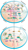
Roles of cyclins in the oocyte maturation process. (Prophase I) Maturation-promoting factor (MPF), composed of Cdk1 kinase and cyclin B, governs cell cycle arrest and resumption. Maternal mRNA is translated selectively. Cyclin B2 expression persists during prophase I arrest, while only the shortest cyclin B1 transcript translates, regulated by cytoplasmic polyadenylation element (CPE) in the 3′ UTR via CPEB. MPF remains inactive due to active Wee2, which is phosphorylated by cAMP-PKA. BubRI, Emi1, Hec1, and securin control APCCdh1 activity and APCcdh1-mediated cyclin degradation maintains levels, ensuring controlled progression without undesired resumption or arrest (Prometaphase I). Cell cycle resumes by inactivating PKA and Wee2. Activated Cdc25B triggers GVBD with mainly cyclin B2-based MPF. Cyclin B1 transcripts, the intermediate and longest, translate, elevating cyclin B1 protein levels. APCCdh1 activity is modulated by Mis12 and CenpH. Cyclin O’s function in Cdk1 inhibitory phosphate removal and MTOC formation is unclear. (Metaphase I and Anaphase I) MPF and SAC inhibit APCcdc20 and separase until proper spindle alignment. Residual APCcdc20 degrades free cyclin B1, extending metaphase I. SAC inhibition activates APCcdc20, degrading cyclin B1 and B2, and lowering MPF activity. Cyclin B3 phosphorylates Emi2, promoting anaphase I (Metaphase II and Fertilization). After meiosis I, S phase-free cell division ensues. Anaphase I-metaphase II transition increases Cdk1-cyclin B1 activity. Cyclin B1 absence leads to interphase re-entry, unique to this stage. Metaphase II oocytes arrest via cytostatic factor (CSF), regulated by MPF and mitogen-activated protein kinase pathway. Fertilization activates APC/C and Wee2 via CaMKII, reducing MPF and enabling zygote’s mitotic cell cycle entry.
Figure 4
Figure 4
Structural motifs in the 3′ untranslated regions (UTRs) of cyclin B transcripts in mouse, human, and Xenopus. The 3′ UTR architecture of Ccnb gene orthologs varies across species, including mice, humans, and Xenopus. Horizontal bars represent sections of the 3′ UTR from each stop codon, with lengths indicated in base pairs (bp). Distinct hexanucleotide motifs within the same transcript generate mRNAs with different 3′ UTR lengths. Currently known examples include mouse ccnb1 with three transcripts. Transcripts exhibit varying lengths of polyadenylation at different time points based on the structure of the 3′ UTR, thereby regulating translation. The structural motifs include hexanucleotide polyadenylation signal, canonical cytoplasmic element (CPE) which has well-conserved sequence, and non-canonical cytoplasmic element (CPE-NC) which has some modification.

Similar articles

References

    1. Masui Y., Markert C.L. Cytoplasmic control of nuclear behavior during meiotic maturation of frog oocytes. J. Exp. Zool. 1971;177:129–145. doi: 10.1002/jez.1401770202. - DOI - PubMed
    1. Evans T., Rosenthal E.T., Youngblom J., Distel D., Hunt T. Cyclin: A protein specified by maternal mRNA in sea urchin eggs that is destroyed at each cleavage division. Cell. 1983;33:389–396. doi: 10.1016/0092-8674(83)90420-8. - DOI - PubMed
    1. Draetta G., Luca F., Westendorf J., Brizuela L., Ruderman J., Beach D. Cdc2 protein kinase is complexed with both cyclin A and B: Evidence for proteolytic inactivation of MPF. Cell. 1989;56:829–838. doi: 10.1016/0092-8674(89)90687-9. - DOI - PubMed
    1. Booher R.N., Alfa C.E., Hyams J.S., Beach D.H. The fission yeast cdc2/cdc13/suc1 protein kinase: Regulation of catalytic activity and nuclear localization. Cell. 1989;58:485–497. doi: 10.1016/0092-8674(89)90429-7. - DOI - PubMed
    1. Nigg E.A. Cyclin-dependent protein kinases: Key regulators of the eukaryotic cell cycle. Bioessays. 1995;17:471–480. doi: 10.1002/bies.950170603. - DOI - PubMed

LinkOut - more resources