. 2023 Dec 1;22(12):1413-1421.
doi: 10.1158/1535-7163.MCT-23-0179.

Microtubule-Targeting Combined with HDAC Inhibition Is a Novel Therapeutic Strategy for Diffuse Intrinsic Pontine Gliomas

Affiliations

Microtubule-Targeting Combined with HDAC Inhibition Is a Novel Therapeutic Strategy for Diffuse Intrinsic Pontine Gliomas

Anahid Ehteda et al. Mol Cancer Ther. .

Abstract

Diffuse intrinsic pontine gliomas (DIPG) are an incurable childhood brain cancer for which novel treatments are needed. DIPGs are characterized by a mutation in the H3 histone (H3K27M), resulting in loss of H3K27 methylation and global gene dysregulation. TRX-E-009-1 is a novel anticancer agent with preclinical activity demonstrated against a range of cancers. We examined the antitumor activity of TRX-E-009-1 against DIPG neurosphere cultures and observed tumor-specific activity with IC50s ranging from 20 to 100 nmol/L, whereas no activity was observed against normal human astrocyte cells. TRX-E-009-1 exerted its anti-proliferative effect through the induction of apoptotic pathways, with marked increases in cleaved caspase 3 and cleaved PARP levels, while also restoring histone H3K27me3 methylation. Co-administration of TRX-E-009-1 and the histone deacetylase (HDAC) inhibitor SAHA extended survival in DIPG orthotopic animal models. This antitumor effect was further enhanced with irradiation. Our findings indicate that TRX-E-009-1, combined with HDAC inhibition, represents a novel, potent therapy for children with DIPG.


Figures

Figure 1. TRX-E-009–1 has anticancer effect against patient-derived DIPG cells. A, Patient-derived DIPG cell lines are sensitive to TRX-E-009–1 compared with normal healthy astrocytes (NHA) and normal lung fibroblast (MRC5) cells. TRX-E-009–1 impairs TRX-E-009–1 impairs clonogenic activity in three DIPG cell lines (B), which is further potentiated in HSJD-DIPG007 cells (C) when combined with irradiation in a dose-dependent manner. B and C, n = 4 independent experiments performed in duplicate. Statistical analysis was calculated using one-way ANOVA between cohorts and for treated and untreated samples (*, P < 0.05; **, P < 0.01; ***, P < 0.001; ****, P < 0.0001). C, Synergy scores were calculated by Calcusyn software.
Figure 1.
TRX-E-009–1 has anticancer effect against patient-derived DIPG cells. A, Patient-derived DIPG cell lines are sensitive to TRX-E-009–1 compared with normal healthy astrocytes (NHA) and normal lung fibroblast (MRC5) cells. TRX-E-009–1 impairs TRX-E-009–1 impairs clonogenic activity in three DIPG cell lines (B), which is further potentiated in HSJD-DIPG007 cells (C) when combined with irradiation in a dose-dependent manner. B and C,n = 4 independent experiments performed in duplicate. Statistical analysis was calculated using one-way ANOVA between cohorts and for treated and untreated samples (*, P < 0.05; **, P < 0.01; ***, P < 0.001; ****, P < 0.0001). C, Synergy scores were calculated by Calcusyn software.
Figure 2. Mechanism of action of TRX-E-009-1 on DIPG cells. A, Treatment of HSJD-DIPG007 cells with 1 and 5 μmol/L TRX-E-009-1 leads to increased protein expression of p21, c-PARP, and c-Caspase 3. Western blot analysis is representative of two independent experiments. B, HSJD-DIPG007 cells were treated with TRX-E-009-1 at 1, 5, and 10 μmol/L to observe the effect on polymerized and depolymerized α-tubulin levels. Western blot analysis is representative of three independent experiments. C, TRX-E-009-1 restores H3K27 trimethylation and increases H3K27 acetylation in treated HSJD-DIPG007 cells. Western blot analysis is representative of two independent experiments. D, TRX-E-009–1 significantly extends survival in the orthotopic mouse model of HSJD-DIPG007. Treatment commenced 4 weeks after intracranial injection of HSJD-DIPG007 cells 50 mg/kg TRX-E-009–1 intravenously three times a week (M, W, F) for 4 weeks. Statistical analysis has been performed using the log-rank (Mantel–Cox) test. E, TRX-E-009–1 drug concentration in the brainstem region of treated mice 2 hours posttreatment. Data are presented as mean values ± SEM from brain samples collected from n = 2 in each cohort.
Figure 2.
Mechanism of action of TRX-E-009-1 on DIPG cells. A, Treatment of HSJD-DIPG007 cells with 1 and 5 μmol/L TRX-E-009-1 leads to increased protein expression of p21, c-PARP, and c-Caspase 3. Western blot analysis is representative of two independent experiments. B, HSJD-DIPG007 cells were treated with TRX-E-009-1 at 1, 5, and 10 μmol/L to observe the effect on polymerized and depolymerized α-tubulin levels. Western blot analysis is representative of three independent experiments. C, TRX-E-009-1 restores H3K27 trimethylation and increases H3K27 acetylation in treated HSJD-DIPG007 cells. Western blot analysis is representative of two independent experiments. D, TRX-E-009–1 significantly extends survival in the orthotopic mouse model of HSJD-DIPG007. Treatment commenced 4 weeks after intracranial injection of HSJD-DIPG007 cells 50 mg/kg TRX-E-009–1 intravenously three times a week (M, W, F) for 4 weeks. Statistical analysis has been performed using the log-rank (Mantel–Cox) test. E, TRX-E-009–1 drug concentration in the brainstem region of treated mice 2 hours posttreatment. Data are presented as mean values ± SEM from brain samples collected from n = 2 in each cohort.
Figure 3. Combination of TRX-E-009–1 and SAHA is synergistic against DIPG. Combination of TRX-E-009–1 and SAHA is synergistic against HSJD-DIPG007 and SU-DIPGXVII neurospheres, resulting in decreased (A) cell survival and (B) fewer colonies formed upon combination treatment. Data are presented as mean values ± SEM. A, Single well examined over five independent experiments. B, n = 4 independent experiments performed in duplicate. Synergy scores were calculated by Calcusyn software.
Figure 3.
Combination of TRX-E-009–1 and SAHA is synergistic against DIPG. Combination of TRX-E-009–1 and SAHA is synergistic against HSJD-DIPG007 and SU-DIPGXVII neurospheres, resulting in decreased (A) cell survival and (B) fewer colonies formed upon combination treatment. Data are presented as mean values ± SEM. A, Single well examined over five independent experiments. B,n = 4 independent experiments performed in duplicate. Synergy scores were calculated by Calcusyn software.
Figure 4. Combination of TRX-E-009–1 and SAHA induces apoptosis and restores H3K27 trimethylation and acetylation in DIPG cells. Protein expression in HSJD-DIPG007 cells when treated with TRX-E-009–1 (1 μmol/L) and SAHA (5 μmol/L) of (A) c-PARP, c-caspase 3, γ-H2AX and (B) H3K27 trimethylation and acetylation. Representative blot from two independent experiments.
Figure 4.
Combination of TRX-E-009–1 and SAHA induces apoptosis and restores H3K27 trimethylation and acetylation in DIPG cells. Protein expression in HSJD-DIPG007 cells when treated with TRX-E-009–1 (1 μmol/L) and SAHA (5 μmol/L) of (A) c-PARP, c-caspase 3, γ-H2AX and (B) H3K27 trimethylation and acetylation. Representative blot from two independent experiments.
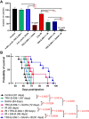
Figure 5. TRX-E-009–1 synergizes with SAHA and irradiation in orthotopic models of DIPG. A, Triple combination treatment of TRX-E-009–1 (0.005 μmol/L), SAHA (0.01 μmol/L), and irradiation (2Gy) significantly reduced the clonogenic activity of HSJD-DIPG007 cells. Data are presented as mean values ±SEM. n = 4 independent experiments performed in duplicate. Synergy scores were calculated by Calcusyn software (*, P < 0.05; ****, P < 0.0001). B, Mice were intracranially injected with DIPG cells and 4 weeks post-injection, treatments commenced. Mice were humanely euthanized when they displayed severe neurological decline and/or weight loss or reached maximum holding time (MHT). Survival curve of HSJD-DIPG007 with TRX-E-009–1/SAHA/Irradiation treatment. Median survival of cohorts is indicated in brackets. Statistical analysis has been performed using the log-rank (Mantel–Cox) test.
Figure 5.
TRX-E-009–1 synergizes with SAHA and irradiation in orthotopic models of DIPG. A, Triple combination treatment of TRX-E-009–1 (0.005 μmol/L), SAHA (0.01 μmol/L), and irradiation (2Gy) significantly reduced the clonogenic activity of HSJD-DIPG007 cells. Data are presented as mean values ±SEM. n = 4 independent experiments performed in duplicate. Synergy scores were calculated by Calcusyn software (*, P < 0.05; ****, P < 0.0001). B, Mice were intracranially injected with DIPG cells and 4 weeks post-injection, treatments commenced. Mice were humanely euthanized when they displayed severe neurological decline and/or weight loss or reached maximum holding time (MHT). Survival curve of HSJD-DIPG007 with TRX-E-009–1/SAHA/Irradiation treatment. Median survival of cohorts is indicated in brackets. Statistical analysis has been performed using the log-rank (Mantel–Cox) test.
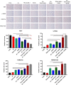
Figure 6. Triple combination of TRX-E-009–1, SAHA, and irradiation inhibits tumour cell proliferation, induces DNA damage response and restores H3K27 trimethylation and acetylation in orthotopic tumors. Ki67, γ-H2AX. H3K27ac and H3K27me3 staining of DIPG tumors after 4 weeks of treatment in HSJD-DIPG007 model. Three images were taken from brain samples collected from two mice in each cohort. Statistical analysis was calculated using one-way ANOVA between cohorts and for treated and untreated samples (**, P < 0.01; ***, P < 0.001; ****, P < 0.0001). Specific P values are listed in Supplementary Table S1.
Figure 6.
Triple combination of TRX-E-009–1, SAHA, and irradiation inhibits tumour cell proliferation, induces DNA damage response and restores H3K27 trimethylation and acetylation in orthotopic tumors. Ki67, γ-H2AX. H3K27ac and H3K27me3 staining of DIPG tumors after 4 weeks of treatment in HSJD-DIPG007 model. Three images were taken from brain samples collected from two mice in each cohort. Statistical analysis was calculated using one-way ANOVA between cohorts and for treated and untreated samples (**, P < 0.01; ***, P < 0.001; ****, P < 0.0001). Specific P values are listed in Supplementary Table S1.

Similar articles

Cited by

References

    1. Lin GL, Wilson KM, Ceribelli M, Stanton BZ, Woo PJ, Kreimer S, et al. . Therapeutic strategies for diffuse midline glioma from high-throughput combination drug screening. Sci Transl Med 2019;11:eaaw0064. - PMC - PubMed
    1. Donaldson SS, Laningham F, Fisher PG. Advances toward an understanding of brainstem gliomas. J Clin Oncol 2006;24:1266–72. - PubMed
    1. Johung TB, Monje M. Diffuse intrinsic pontine glioma: new pathophysiological insights and emerging therapeutic targets. Curr Neuropharmacol 2017;15:88–97. - PMC - PubMed
    1. Jansen MHA, van Vuurden DG, Vandertop WP, Kaspers GJL. Diffuse intrinsic pontine gliomas: a systematic update on clinical trials and biology. Cancer Treat Rev 2012;38:27–35. - PubMed
    1. Upton DH, Ung C, George SM, Tsoli M, Kavallaris M, Ziegler DS. Challenges and opportunities to penetrate the blood-brain barrier for brain cancer therapy. Theranostics 2022;12:4734–52. - PMC - PubMed