. 2023 Sep 28;97(9):e0088123.
doi: 10.1128/jvi.00881-23. Epub 2023 Sep 8.

Epstein-Barr virus-encoded miR-BART11-3p modulates the DUSP6-MAPK axis to promote gastric cancer cell proliferation and metastasis

Affiliations

Epstein-Barr virus-encoded miR-BART11-3p modulates the DUSP6-MAPK axis to promote gastric cancer cell proliferation and metastasis

Mingqian Xu et al. J Virol. .

Abstract

Epstein-Barr virus (EBV)-encoded miRNAs within the BamHI-A rightward transcript (BART) region are abundantly expressed in EBV-associated gastric cancer (EBVaGC), suggesting that they play roles in tumorigenesis. However, how these viral miRNAs contribute to the development of EBVaGC remains largely obscure. In this study, we found that EBV-encoded miR-BART11-3p targets 3' -UTR of dual-specificity phosphatase 6 (DUSP6) mRNA to upregulate ERK phosphorylation and downregulate JNK and p38 phosphorylation. By doing so, miR-BART11-3p promotes gastric cancer (GC) cell proliferation, migration, and invasion in vitro, and facilitates tumor growth in vivo. Restoration of DUSP6 expression reverses the tumor-promoting activity of miR-BART11-3p in AGS GC cells. Consistently, knockdown of DUSP6 ablates the antitumor effects of miR-BART11-3p inhibitors in EBV-positive GC cells. Furthermore, blocking ERK phosphorylation with trametinib inhibited the proliferation, migration, and invasion of miR-BART11-3p-expressing AGS cells. Administration of a miR-BART11-3p antagomir reduced the growth of EBV-positive xenograft tumors. Together, these findings reveal a novel mechanism by which EBV dysregulates MAPK pathways through an EBV-encoded microRNA to promote the development and progression of EBVaGC, which may be harnessed to develop new therapeutics to treat EBVaGC. IMPORTANCE The Epstein-Barr virus (EBV) is the first human tumor virus found to encode miRNAs, which within the BART region have been detected abundantly in EBV-associated gastric cancer (EBVaGC) and play various roles in promoting tumorigenesis. In our study, we observed that EBV-miR-BART11-3p promotes cell proliferation and induces migration and invasion in GC. Interestingly, we showed that miR-BART11-3p upregulates p-ERK and downregulates p-JNK and p-p38 by directly targeting 3'-UTR of dual-specificity phosphatase 6 (DUSP6). Restoration of DUSP6 rescues the effects generated by miR-BART11-3p in GC cells, and blocking ERK phosphorylation with Trametinib augments JNK and p38 phosphorylation and inhibits the effects of miR-BART11-3p-expressing AGS cells, suggesting that miR-BART11-3p promotes cell proliferation, migration, and invasion by modulating DUSP6-MAPK axis in EBVaGC. The findings presented in this study provide new mechanisms into the tumorigenesis in EBVaGC and new avenues for the development of therapeutic strategies to combat EBVaGC targeting miR-BART11-3p or phospho-ERK.

Keywords: EBV-miR-BART11-3p; Epstein-Barr virus; dual-specificity phosphatase 6; gastric cancer; mitogen-activated protein kinases.


Conflict of interest statement

The authors declare no conflict of interest.

Figures

Fig 1
Fig 1
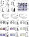
Downregulation of DUSP6 is correlated with increased phosphorylated ERK (p-ERK) in AGS-EBV and associated with worse prognosis in GC patients. (A) Total and phosphorylated forms of ERK, JNK, and p38 were detected by western blotting in AGS and AGS-EBV cells and xenografts. β-Actin was used as a loading control. (B) Heat map of mRNAs of 17 DUSPs that were differentially expressed in AGS vs. AGS-EBV cells (deposited in the online repository: https://ngdc.cncb.ac.cn/search/, HREQ001940). (C) Transcript levels of DUSP6 were examined in AGS and AGS-EBV cell lines and xenografts by RT-qPCR. β-Actin served as the loading controls. (D) Protein levels of DUSP6 in AGS and AGS-EBV cells and xenografts were determined by western blotting. β-Actin served as the loading controls. Transcript levels of DUSP6 were examined in EBV-negative GC cell lines (AGS, HGC27) and EBV-positive GC cell lines (AGS-EBV, HGC27-EBV, SNU719) (E) and GC cell line-based xenografts (F) by RT-qPCR. β-Actin was used for normalizing the expression of DUSP6. Western blotting was used to detect the expression of DUSP6, total and phosphorylated ERK in GC cell lines (G), and GC cell line-based xenografts (H). β-Actin was used as a loading control. “SE” and “LE” represent short exposure and long exposure, respectively. Data were shown as the mean ± SD (*P < 0.05, **P < 0.01, and ***P < 0.001). (I) Relationship between expression of DUSP6 and prognosis of GC patients was analyzed by the Kaplan-Meier method.
Fig 2
Fig 2
EBV-encoded miR-BART11-3p expressed in AGS-EBV cells directly targets the 3′-UTR of DUSP6. (A) The expression levels of EBV-miR-BART11-3p (BART11-3p) in AGS and AGS-EBV cells and xenografts were determined by stem-loop RT-qPCR, and amplified products were further verified by gel electrophoresis. RNU6B (U6) was used as an internal control. (B) The expression of BART11-3p in EBV-negative GC cell lines (AGS, HGC27) and EBV-positive GC cell lines (AGS-EBV, HGC27-EBV, SNU719) was determined by stem-loop real-time PCR. U6 was used for normalizing the expression of BART11-3p and the lowest expression level in each group was set as “1”. (C) The expression of BART11-3p was determined in xenografts as in (B). (D) Bioinformatics predictions of one binding site by BART11-3p in the wild-type (wt) DUSP6 3′-UTR. Mutant sequence of DUSP6 3′-UTR (mut) generated in the complementary site that binds to the seed region of BART11-3p is indicated. (E) Luciferase reporter assays. HEK293T cells were co-transfected with a luciferase reporter vector containing wt or mut DUSP6 3′-UTR and the BART11-3p mimic or scramble control (nc). Luciferase activities were measured 48 h post-transfection. “ND” indicates “not detectable.” Data are shown as the mean ± SD, **P < 0.01 and ***P < 0.001.
Fig 3
Fig 3
EBV-miR-BART11-3p dysregulates the phosphorylation of ERK, JNK, and p38 by reducing the level of DUSP6, and enhances cell proliferation, migration, and invasion of GC cells. (A) Expression of EBV-miR-BART11-3p (BART11-3p) in AGS cells transduced by a lentiviral vector expressing miR-BART11-3p (AGS-B11-3p) or its control vector (AGS-Mock) was verified by stem-loop RT-qPCR. AGS-EBV cells served as controls. “ND” indicates the expression of BART11-3p in AGS-Mock is “not detectable.” (B) Expression of DUSP6 mRNA in AGS-B11-3p cells or AGS-Mock cells was examined by RT-qPCR. (C and D) AGS-EBV or SNU719 cells were transiently transfected with BART11-3p inhibitor (In-B11-3p) or its nonspecific control inhibitor (In-NC). BART11-3p levels in cells were quantified by stem-loop RT-qPCR. (E and F) Transcript levels of DUSP6 in AGS-EBV or SNU719 cells were quantified by RT-qPCR. (G) Protein levels of DUSP6, total and the phosphorylation of ERK, JNK, and p38 were determined by western blotting in the cells mentioned previously. CCK-8 (H), colony formation (I), transwell migration (J), and transwell chamber invasion (K) assays were performed with the cells mentioned previously to examine the potential of cell proliferation, colony formation, migration, and invasion. Original magnification of (J) and (K), ×200. The data are shown as the mean ± SD, N = 3, *P < 0.05, **P < 0.01, ***P < 0.001.
Fig 4
Fig 4
Reconstitution of DUSP6 offsets the effects of EBV-miR-BART11-3p in GC cells. (A) Transcript levels of DUSP6 were quantified by RT-qPCR in AGS-B11-3p cells transiently transfected with either pcDNA3.1-DUSP6 plasmid or the empty vector (NC). (B) Expressions of DUSP6, total and phosphorylated ERK, JNK, and p38 in AGS-B11-3p cells transfected with the pcDNA3.1-DUSP6 plasmid or its empty vector (NC) were detected by western blotting. β-Actin was used as the protein loading control. “SE” and “LE” represent short exposure and long exposure, respectively. Proliferation, colony formation, migration, and invasion of AGS-B11-3p cells transfected with pcDNA3.1-DUSP6 plasmid or the empty vector (NC) were examined by CCK-8 (C), colony formation (D), transwell migration (E), and transwell chamber invasion (F) assays. Original magnification of (E) and (F) ×200. The data are shown as the mean ± SD, N = 3, *P < 0.05, **P < 0.01, ***P < 0.001.
Fig 5
Fig 5
Knockdown of DUSP6 offsets the effects of EBV-miR-BART11-3p inhibitor in AGS-EBV cells. AGS-EBV cells were co-transfected with a miR-BART11-3p inhibitor (B11-3p inhibitor) and DUSP6 siRNA (siRNA-1 and siRNA-2) or their corresponding nonspecific controls. (A) Expression of DUSP6, total and phosphorylated ERK, JNK, and p38 was detected by western blotting in AGS-EBV cells after co-transfection of the B11-3p inhibitor and DUSP6 siRNA or their corresponding negative controls. β-Actin was used as the protein loading control. Proliferation, colony formation, migration, and invasion of AGS-EBV cells after co-transfection of a miR-BART11-3p inhibitor (In-B11-3p or B11-3p inhibitor) and DUSP6 siRNA (siRNA-1 and siRNA-2) or their corresponding negative controls were examined by CCK-8 (B), colony formation (C), transwell migration (D), and Boyden chamber invasion (E) assays. Original magnification of (F) and (G) ×200. Data are shown as the mean ± SD, N = 3, *P < 0.05, **P < 0.01, ***P < 0.001.
Fig 6
Fig 6
Blocking ERK phosphorylation results in activation of JNK and p38, and inhibition of proliferation, migration, and invasion in EBV-miR-BART11-3p-expressing GC cells. (A) AGS-B11-3p cells were incubated with 0, 1, and 5 nM trametinib for 1 h and immunoblotted for the expression of total and phosphorylated ERK, JNK, and p38. β-Actin was used as the protein loading control. Trametinib (5 nM) treatment was applied to conduct (B) CCK-8, (C) colony formation, (D) transwell migration, and (E) transwell chamber invasion assays. Original magnification of (D) and (E) ×200. Data are shown as the mean ± SD, N = 3, *P < 0.05, **P < 0.01, ***P < 0.001.
Fig 7
Fig 7
Inhibition of EBV-miR-BART11-3p decreases the growth of EBV-positive GC cell-derived xenografts in NSG mice. (A through B) Ectopic expression of miR-BART11-3p in AGS cells enhanced the growth of xenografts in NSG mice. AGS-Mock or AGS-B11-3p cells were subcutaneously inoculated into the armpit of the right forelimb of NSG mice (n = 4). Tumor volume was periodically measured for each mouse and tumor growth curves were plotted. Xenograft tumors were collected on Day 45 following subcutaneous implantation. Tumor growth curves (A), and images of tumor xenografts and tumor weight (B) are shown. (C through F) MiR-BART11-3p antagomir decreased the growth of EBV-positive GC cell-bearing xenografts in NSG mice. AGS-EBV or SNU719 cells were subcutaneously inoculated into the armpit of the right forelimb of NSG mice (n = 8). Arrows indicate the first administration of miR-BART11-3p antagomir or its corresponding negative control antagomir (NC) in mice (n = 4). Xenograft tumors were collected on Day 60 post subcutaneous implantation of AGS-EBV cells or on Day 30 post subcutaneous implantation of SNU719 cells. Tumor growth curves (C and E), and images of tumor xenografts and tumor weights (D and F) are shown. Data are summarized as mean ± SD. (G through I) The protein levels of DUSP6, total and phosphorylated ERK in tumor samples from NSG mice in the groups studied earlier were detected by western blotting and normalized to the level of β-actin.
Fig 8
Fig 8
Model for miR-BART11-3p-mediated enhancement of proliferation and migration by targeting a DUSP6/MAPK pathway in EBVaGC. EBV-encoded miR-BART11-3p downregulates the expression of DUSP6 by directly targeting the 3′-UTR of DUSP6, which consequently leads to increased phosphorylation of ERK coupled with decreased phosphorylation of JNK and p38. The increased phospho-ERK signaling pathway ultimately enhances EBVaGC progression by facilitating proliferation, migration, and invasion of tumor cells.

Similar articles

Cited by

References

    1. Young LS, Yap LF, Murray PG. 2016. Epstein-Barr virus: more than 50 years old and still providing surprises. Nat Rev Cancer 16:789–802. doi:10.1038/nrc.2016.92 - DOI - PubMed
    1. Cohen JI, Fauci AS, Varmus H, Nabel GJ. 2011. Epstein-Barr virus: an important vaccine target for cancer prevention. Sci Transl Med 3:107fs7. doi:10.1126/scitranslmed.3002878 - DOI - PMC - PubMed
    1. Cancer Genome Atlas Research Network . 2014. Comprehensive molecular characterization of gastric adenocarcinoma. Nature 513:202–209. doi:10.1038/nature13480 - DOI - PMC - PubMed
    1. Yang J, Liu Z, Zeng B, Hu G, Gan R. 2020. Epstein-Barr virus-associated gastric cancer: a distinct subtype. Cancer Lett 495:191–199. doi:10.1016/j.canlet.2020.09.019 - DOI - PubMed
    1. Shinozaki-Ushiku A, Kunita A, Isogai M, Hibiya T, Ushiku T, Takada K, Fukayama M. 2015. Profiling of virus-encoded microRNAs in Epstein-Barr virus-associated gastric carcinoma and their roles in gastric carcinogenesis. J Virol 89:5581–5591. doi:10.1128/JVI.03639-14 - DOI - PMC - PubMed