

Deletion of a 7-amino-acid region in the porcine epidemic diarrhea virus envelope protein induces higher type I and III interferon responses and results in attenuation in vivo
- PMID: 37681956
- PMCID: PMC10537754
- DOI: 10.1128/jvi.00847-23
Deletion of a 7-amino-acid region in the porcine epidemic diarrhea virus envelope protein induces higher type I and III interferon responses and results in attenuation in vivo
Abstract
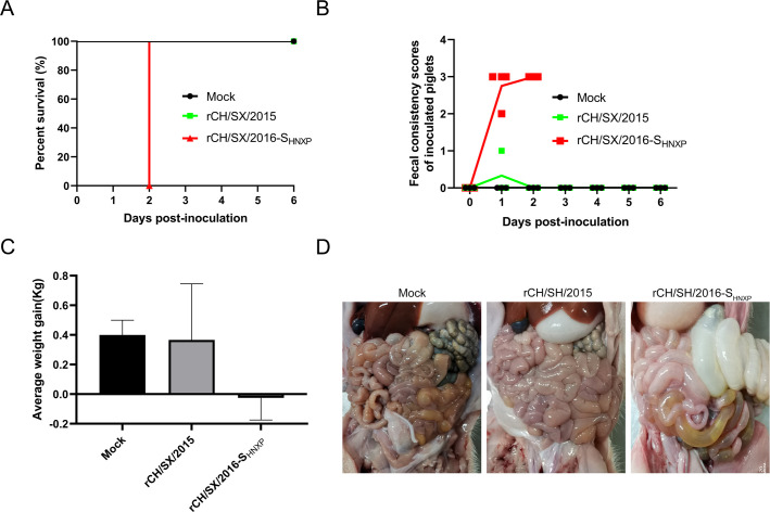
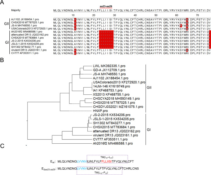
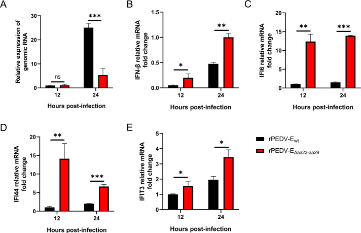
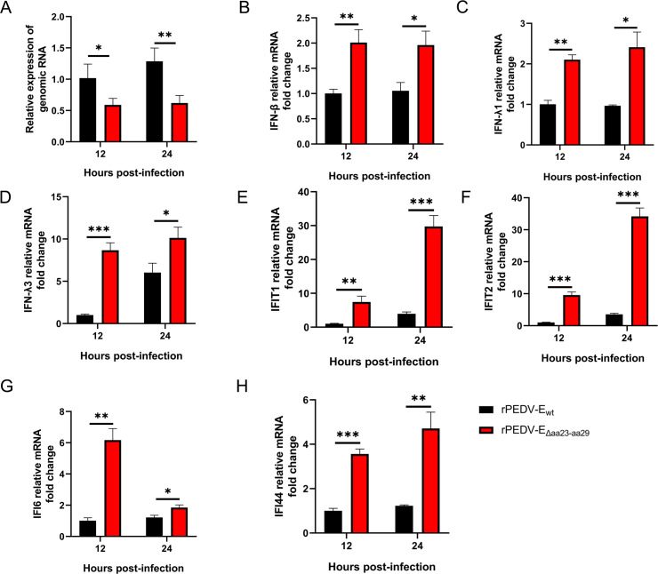
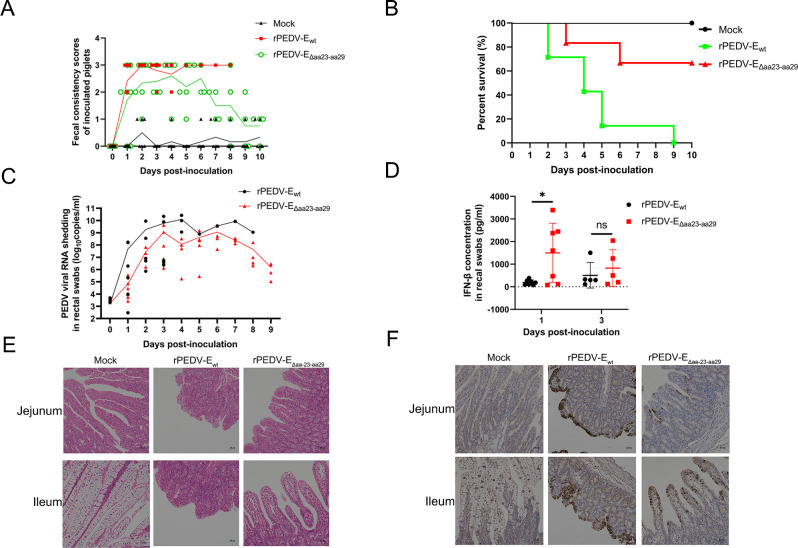
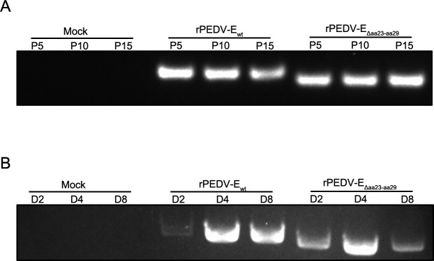
Porcine epidemic diarrhea virus (PEDV) leads to enormous economic losses for the pork industry. However, the commercial vaccines failed to fully protect against the epidemic strains. Previously, the rCH/SX/2016-SHNXP strain with the entire E protein and the rCH/SX/2015 strain with the deletion of 7-amino-acid (7-aa) at positions 23-29 in E protein were constructed and rescued. The pathogenicity assay indicated that rCH/SX/2015 is an attenuated strain, but rCH/SX/2016-SHNXP belongs to the virulent strains. Then, the recombination PEDV (rPEDV-EΔaa23-aa29)strain with a 7-aa deletion in the E protein was generated, using the highly virulent rCH/SX/2016-SHNXP strain (rPEDV-Ewt) as the backbone. Compared with the rPEDV-Ewt strain, the release and infectivity of the rPEDV-EΔaa23-aa29 strain were significantly reduced in vitro, but stronger interferon (IFN) responses were triggered both in vitro and in vivo. The pathogenicity assay showed that the parental strain resulted in severe diarrhea (100%) and death (100%) in all piglets. Compared with the parental strain group, rPEDV-EΔaa23-aa29 caused lower mortality (33%) and diminished fecal PEDV RNA shedding. At 21 days, all surviving pigs were challenged orally with rPEDV-Ewt. No pigs died in the two groups. Compared with the mock group, significantly delayed and milder diarrhea and reduced fecal PEDV RNA shedding were detected in the rPEDV-EΔaa23-aa29 group. In conclusion, the deletion of a 7-aa fragment in the E protein (EΔaa23-aa29) attenuated PEDV but retained its immunogenicity, which can offer new ideas for the design of live attenuated vaccines and provide new insights into the attenuated mechanism of PEDV. IMPORTANCE Porcine epidemic diarrhea virus (PEDV) causes high mortality in neonatal piglets and remains a large challenge to the pork industry. Unfortunately, no safe and effective vaccines are available yet. The pathogenesis and molecular basis of the attenuation of PEDV remain unclear, which seriously hinders the development of PEDV vaccines. This study found that the rPEDV carrying EΔaa23-aa29 mutation in the E protein induced significantly higher IFN responses than the parental virus, partially attenuated, and remained immunogenic in piglets. For the first time, PEDV E was verified as an IFN antagonist in the infection context and identified as a virulence factor of PEDV. Our data also suggested that EΔaa23-aa29 mutation can be a good target for the development of live attenuated vaccines for PEDV and also provide new perspectives for the attenuated mechanism of PEDV.
Keywords: PEDV; coronavirus; envelope; interferon; reverse genetic analysis; the molecular basis of the attenuation of PEDV; virulence factor.
Conflict of interest statement
The authors declare no conflict of interest.
Figures
Similar articles
-
Mutations in Porcine Epidemic Diarrhea Virus nsp1 Cause Increased Viral Sensitivity to Host Interferon Responses and Attenuation In Vivo.J Virol. 2022 Jun 8;96(11):e0046922. doi: 10.1128/jvi.00469-22. Epub 2022 May 18. J Virol. 2022. PMID: 35583324 Free PMC article.
-
Deletion of a 197-Amino-Acid Region in the N-Terminal Domain of Spike Protein Attenuates Porcine Epidemic Diarrhea Virus in Piglets.J Virol. 2017 Jun 26;91(14):e00227-17. doi: 10.1128/JVI.00227-17. Print 2017 Jul 15. J Virol. 2017. PMID: 28490591 Free PMC article.
-
Engineering a Live Attenuated Porcine Epidemic Diarrhea Virus Vaccine Candidate via Inactivation of the Viral 2'-O-Methyltransferase and the Endocytosis Signal of the Spike Protein.J Virol. 2019 Jul 17;93(15):e00406-19. doi: 10.1128/JVI.00406-19. Print 2019 Aug 1. J Virol. 2019. PMID: 31118255 Free PMC article.
-
Emerging Highly Virulent Porcine Epidemic Diarrhea Virus: Molecular Mechanisms of Attenuation and Rational Design of Live Attenuated Vaccines.Int J Mol Sci. 2019 Nov 4;20(21):5478. doi: 10.3390/ijms20215478. Int J Mol Sci. 2019. PMID: 31689903 Free PMC article. Review.
-
Porcine epidemic diarrhea virus: Molecular mechanisms of attenuation and vaccines.Microb Pathog. 2020 Dec;149:104553. doi: 10.1016/j.micpath.2020.104553. Epub 2020 Oct 1. Microb Pathog. 2020. PMID: 33011361 Free PMC article. Review.
Cited by
-
Reverse Genetics Systems for Emerging and Re-Emerging Swine Coronaviruses and Applications.Viruses. 2023 Sep 26;15(10):2003. doi: 10.3390/v15102003. Viruses. 2023. PMID: 37896780 Free PMC article. Review.
-
Developing Next-Generation Live Attenuated Vaccines for Porcine Epidemic Diarrhea Using Reverse Genetic Techniques.Vaccines (Basel). 2024 May 19;12(5):557. doi: 10.3390/vaccines12050557. Vaccines (Basel). 2024. PMID: 38793808 Free PMC article. Review.
-
A Comprehensive View on the Protein Functions of Porcine Epidemic Diarrhea Virus.Genes (Basel). 2024 Jan 26;15(2):165. doi: 10.3390/genes15020165. Genes (Basel). 2024. PMID: 38397155 Free PMC article. Review.
References
-
- Hou Y, Ke H, Kim J, Yoo D, Su Y, Boley P, Chepngeno J, Vlasova AN, Saif LJ, Wang Q. 2019. Engineering a live attenuated porcine epidemic diarrhea virus vaccine candidate via inactivation of the viral 2'-O-methyltransferase and the Endocytosis signal of the spike protein. J Virol 93:e00406-19. doi:10.1128/JVI.00406-19 - DOI - PMC - PubMed
Publication types
MeSH terms
Substances
LinkOut - more resources
Full Text Sources
